Radiological pathology of brain to brain metastasis: Pattern of spread of pri...Professor Yasser Metwally
Radiological pathology of brain to brain metastasis: Pattern of spread of primary brain tumors within the neural axis
http://yassermetwally.com
http://yassermetwally.net
The document discusses medulloblastoma (MB), the most common malignant brain tumor in children. MB originates in the cerebellum and projects into the fourth ventricle. It is thought to arise from primitive neuroepithelial cells. On imaging, MB typically appears as a large lobulated mass in the posterior fossa that compresses the fourth ventricle and causes hydrocephalus. Molecular analysis has classified MB into four subgroups: SHH, WNT, Group III, and Group IV, which have different characteristics and prognoses. Prognostic factors include age at diagnosis, extent of disease, extent of resection, histology, and biological markers.
A large malignant peripheral nerve sheath tumour (mpsnt) of radial nervedhanabir thangjam
This case report describes a rare case of a large malignant peripheral nerve sheath tumor (MPNST) arising from the radial nerve in a 50-year-old woman. Imaging and biopsy initially suggested a benign nerve sheath tumor. However, surgical excision and histopathological examination confirmed a low-grade MPNST. MPNSTs are aggressive sarcomas that typically arise from large nerve trunks in the extremities. While rare, this case highlights that MPNSTs can arise from the radial nerve and emphasizes the importance of thorough histological examination for accurate diagnosis and treatment planning.
Leptomeningeal carcinomatosis is the infiltration of malignant cells into the leptomeninges, which is the protective layers surrounding the brain and spinal cord. It occurs in 5% of cancer patients and most commonly spreads from primary cancers of the breast and lung. Clinical presentation involves neurological symptoms affecting the brain, cranial nerves, and spinal cord. Diagnosis is made through lumbar puncture and imaging of the craniospinal axis. Treatment aims to palliate symptoms and improve survival through radiotherapy targeting symptomatic areas and CSF flow pathways. Craniospinal irradiation provides wider treatment but is associated with acute and late toxicities. Localized radiotherapy to specific sites offers additional palliation with reduced side effects.
Thermo-Elastography is a new method using ultrasound to quantitatively measure tissue elasticity. Elastography works by measuring tissue displacement after compression to estimate strain, with higher strain indicating softer tissue. Several applications are discussed, including detecting breast and prostate cancers, monitoring HIFU therapy, and evaluating arteries. Limitations include difficulty distinguishing tissue types by ultrasound alone. New techniques like intravascular elastography using arterial pulsation or balloons aim to characterize plaque vulnerability for guiding interventions. Overall, elastography provides complementary information to conventional ultrasound for visualizing tissue mechanical properties.
The document discusses craniospinal irradiation (CSI) for treating tumors that can spread through the cerebrospinal fluid (CSF), such as medulloblastoma. It describes how CSI was developed to treat the entire nervous system by irradiating the brain, spinal cord, and CSF spaces. Key points covered include tumor types treated with CSI, techniques for delivering CSI safely and uniformly, matching fields at junctions, and dose schedules for average and high-risk patients.
Thesis section: The role of neuroimaging in muscle and peripheral nerve disor...Professor Yasser Metwally
This document discusses the role of neuroimaging in muscle and peripheral nerve diseases. It begins by explaining how MRI is commonly used to evaluate neuromuscular disorders by detecting signal changes in muscles or nerves. It then classifies peripheral neuropathies and myopathies. The document discusses the pathology and pathogenesis of several muscle and nerve diseases like GBS, CMT, muscular dystrophies and inflammatory myopathies. It provides examples of characteristic patterns of muscle involvement seen on MRI for different conditions.
Tractography uses diffusion MRI to visually represent nerve tracts in the brain through 3D modeling. It can reveal both long tracts connecting different brain regions and more complex short circuits within the brain. However, nerve tracts are not directly visible through standard imaging techniques. Diffusion MRI measures the directionality of water diffusion within tissues to infer the orientation of fiber tracts. Software then maps the diffusion data to generate color-coded 3D representations of tracts called tractograms. Tractography provides insights into the structure and integrity of white matter pathways in the living brain.
This document summarizes various pathologies that can affect the eye and orbit. It discusses congenital anomalies, infections and inflammations, tumors, trauma, and miscellaneous conditions. For each condition, it provides a brief description and highlights relevant imaging findings on modalities such as CT, MRI, and plain films. Key features that help characterize many lesions include enhancement pattern, presence of calcification, and signal characteristics on different MRI sequences.
This document provides guidelines for the radiotherapy treatment of medulloblastoma. It describes the target volumes that should be treated with craniospinal irradiation (CSI), including the entire brain, spinal cord, and cerebrospinal fluid spaces. It recommends a dose of 30-36 Gy in 18-21 fractions to the cranium and spine during CSI, followed by a posterior fossa boost of 18-20 Gy in 10-11 fractions. The document discusses various techniques for achieving adequate coverage of the treatment volumes during CSI, including the use of opposed lateral fields for whole brain treatment and direct posterior fields for the spine, with adjustments like collimator and couch rotation needed to match field borders.
This case discusses a 40-year-old male patient who presented with gradual paraplegia and sensory loss. MRI revealed an intramedullary ependymoma in the dorsal spinal cord that was cystic with bands, causing cord expansion. Ependymoma is the most common intramedullary spinal tumor in adults. The patient was diagnosed with a spinal ependymoma based on the radiological and clinical findings. Complete surgical resection of intramedullary ependymomas offers the best chance for reduced recurrence and improved prognosis.
This document describes the case of an 8-year-old boy who presented with 6 months of headaches and 1 month of gait disturbance. After examination, MRI revealed a posterior fossa midline neoplastic lesion involving the right cerebellum and compressing the fourth ventricle, causing hydrocephalus. The diagnosis was medulloblastoma with hydrocephalus. The patient underwent VP shunt placement for hydrocephalus and then midline suboccipital craniectomy for tumor resection. Outcomes and complications of the surgery are discussed.
Metastasis to the brain parenchyma is common, accounting for about half of all brain tumors. Parenchymal metastases are often located near gray-white junctions supplied by arteries. They enhance with contrast and can be solitary or multiple. Survival is dictated by size, number and location of lesions.
Glioblastoma multiforme is the most common primary brain tumor. It appears as an irregular, heterogeneously enhancing mass usually with areas of necrosis. Location is commonly the cerebral hemispheres with involvement of white matter tracts. Surrounding edema and mass effect are usually present.
Low-grade astrocytoma is a slow growing tumor that most commonly occurs in younger adults. It appears as
Case series: Diffuse Leptomeningeal Glioneuronal Tumor of ChildhoodDhairya A. Lakhani
DL-GNT is a rare low-grade tumor seen in children that involves diffuse leptomeningeal spread, with or without a small parenchymal component. It is characterized by numerous small T2 hyperintense pseudocystic lesions along the brain and spinal cord surface. Histologically, it shows a monomorphic population of round cells with synaptophysin and S100 immunoreactivity. Key diagnostic features include the diffuse leptomeningeal enhancement and tiny cystic lesions, unlike other pediatric tumors that typically present with a dominant mass.
Charting the human thalamus - basic contepts and recent developmentsDr. Jakab András
This document summarizes a study on developing a probabilistic tractography and segmentation method to chart the human thalamus. The study used diffusion tensor imaging and probabilistic tractography to visualize cortico-thalamic connections. It then developed a statistical shape model of the mean thalamus atlas incorporating these connectivity maps. The method was able to align the atlas to individual subjects' geometry with sub-millimeter accuracy, outperforming conventional alignment methods. This individualized target mapping method could help guide image-guided neurosurgery of the thalamus.
This document presents a case study of a 40-year-old female patient diagnosed with a cerebellopontine angle meningioma. The patient presented with symptoms of increased intracranial pressure, long tract signs, cranial nerve palsies, and cerebellar signs. MRI images showed a densely enhanced tumor with a wide base attachment and compression of the brain stem. The case study discusses the MRI appearance of different types of meningiomas and characteristics used to identify meningiomas. It also describes dural tail enhancement commonly seen with meningiomas.
Intracranial chordomas are rare, slow-growing tumors that arise from remnants of the embryonic notochord in the skull base. They typically present as soft tissue masses originating in the clivus with associated bone destruction. MR imaging and CT are effective in diagnosing and monitoring these tumors. Treatment involves surgical resection followed by radiation therapy, which provides the best outcomes for patients with intracranial chordomas.
Multiple Meningiomas and Meningiomatosis. Molecular Pathogenesis NeuroAcademy
The document discusses the molecular pathogenesis of multiple meningiomas and meningiomatosis. It begins with the embryonic development of the meninges, the membranes surrounding the brain and spinal cord. It then describes normal meningothelial cells and examples of multiple meningiomas in different patients. Neurofibromatosis type II is discussed as a common genetic cause associated with multiple meningiomas. The Merlin gene is identified as encoding the tumor suppressor protein involved in NF2. Finally, some potential drug treatments for NF2-associated meningiomas are mentioned.
The document describes an interactive microscopy session on deep soft tissue spindle cell tumors. It presents two cases of monophasic synovial sarcoma in the first case, discussing the histology, immunoprofile and genetics of this tumor. The second case presents two examples of malignant peripheral nerve sheath tumor (MPNST), noting their characteristic histological features and association with neurofibromatosis type 1 or prior radiation therapy. Key differential diagnoses and diagnostic markers are also mentioned.
This document summarizes a case report of a congenital myofibroma tumor in a newborn male. Histopathological examination revealed a biphasic tumor composed of round and spindle-shaped cells. Immunohistochemistry showed the tumor cells were positive for CD34 and smooth muscle markers. Ultrastructural examination demonstrated cytoplasmic intermediate filaments, indicating smooth muscle differentiation. The findings support that this tumor is a true hemangiopericytoma, arising from pericytes and undergoing myofibroblastic differentiation, rather than a solitary fibrous tumor.
The document summarizes two cases of Kimura disease seen on CT and MR imaging. In Case 1, the lesion showed slightly high intensity on T2-weighted MR and low to intermediate intensity on T1-weighted images. It enhanced intermediately. In Case 2, the lesion showed very high intensity on T2-weighted images and marked enhancement on gadolinium-enhanced T1-weighted images. Kimura disease typically presents as an asymptomatic mass and lymphadenopathy in the head and neck region, especially the parotid and submandibular glands, in Asian patients in their second to third decades of life.
A radiological insight into various musculoskeletal complications in patients suffering from AIDS and how it'll affect the management of the patient. A must know for all Radiologists.
This case report describes a 40-year-old man who was previously treated for borderline leprosy and presented with a painful lump on his left ulnar nerve one year after treatment. Differential diagnoses included a post-treatment reaction nerve abscess versus a peripheral nerve tumor. Ultrasound revealed a nodule on the ulnar nerve that was surgically removed and found to be a benign schwannoma rather than a nerve abscess from leprosy. The report emphasizes that differential diagnoses including neural tumors should be considered for peripheral neuropathies in patients with a history of leprosy.
1) Brainmapping provides a quantitative analysis of EEG data by measuring frequency bands and topographic distribution, allowing objective characterization beyond visual analysis.
2) Studies have found focal increases in beta band energy can localize epileptic foci, while decreased alpha/theta with increased beta may indicate surrounding gliosis.
3) Brainmapping uncovered covert epilepsy and subclinical foci, with patterns of diminished and augmented activity suggesting regions of atrophy surrounded by epileptogenic cortex.
A 32-year-old male patient presented with a single grand mal seizure. Imaging showed extradural masses in the left parietal and frontal regions. Biopsy revealed non-Hodgkin B-cell lymphoma. The CNS manifestations were initially diagnosed as secondary CNS lymphoma. The patient was referred for oncology treatment. The document discusses epidural lymphomatous deposits and provides information on accessing additional case publications and archives on the author's website.
This document discusses a case of subarachnoid hemorrhage and bilateral anterior cerebral artery infarctions caused by a ruptured anterior communicating artery aneurysm in a 40-year-old male patient found unconscious. It provides radiological images showing the location of the aneurysm and resulting hemorrhage. The diagnosis is given as a ruptured anterior communicating artery aneurysm. The discussion section covers complications of cerebral aneurysms including hematoma formation and patterns associated with different aneurysm locations. It describes how aneurysm ruptures can lead to subarachnoid or intracerebral hemorrhage and secondary effects like vasospasm and infarction.
Research section...The role of MR imaging in the management of parkinson diseaseProfessor Yasser Metwally
The document discusses a study of 20 patients with idiopathic Parkinson's disease who underwent MRI brain scans and clinical evaluations. Patients were divided into two groups: Group A had a short duration of symptoms and good response to levodopa treatment, while Group B had a longer duration and fluctuating levodopa response. MRI showed changes in the substantia nigra and red nucleus in both groups. Additionally, Group B showed signal changes in the putamen, suggesting more advanced disease involving both nigral and striatal structures.
A 40-year-old patient presented with relapsing-remitting spinal multiple sclerosis. MRI revealed asymptomatic brain involvement and recent progression of myelopathy. The document discusses the diagnosis of spinal MS and differentiating it from transverse myelitis using MRI characteristics such as lesion location and contrast enhancement. Figures show examples of MS plaques and diffuse abnormalities on MRI of the spinal cord.
Issues in brainmapping...EEG and brainmap spectral profiles in cortical lesio...Professor Yasser Metwally
Issues in brainmapping...EEG and brainmap spectral profiles in cortical lesions, subcortical white matter lesions and subcortical gray matter (diencephalic) lesions
This document discusses neurological complications that can occur during hematopoietic cell transplantation (HCT). It describes complications that occur during different stages of HCT: 1) During conditioning regimens, complications include encephalopathy, seizures, and cerebral infarction caused by chemotherapy drugs or medical procedures. 2) During bone marrow depletion, complications include encephalopathy, seizures, cerebral infarctions, and hemorrhages due to metabolic issues, drugs, or infections. 3) During chronic immunosuppression in allogeneic HCT, complications include infections by viruses and opportunistic organisms. 4) Late complications include relapse of the original disease, neurological graft-versus-host disease, and second neoplasms.
This document summarizes the pathology of pituitary tumors, including:
1) Pituitary adenomas are benign neoplasms originating from pituitary cells and are the most common tumors of the sella region.
2) Pituitary tumors are currently classified based on their hormonal content and ultrastructural morphology, with 14 recognized subtypes.
3) Growth hormone-secreting adenomas are associated with acromegaly or gigantism and can have various histologic appearances, most commonly densely or sparsely granulated somatotroph adenomas.
A 66-year-old male patient presented with non-specific lower back pain. MRI images showed signs of lumbar spondylosis including disc degeneration and bulging in the lower lumbar spine. The document provides figures showing disc herniation and details on accessing additional materials on the author's website, including updates versions of the case publication.
This document is a case publication from January 2008 edited by Professor Yasser Metwally about acute disseminated encephalomyelitis (ADEM). It describes an 18-year-old female patient who presented with disturbed consciousness, seizures, optic neuritis and meningism. MRI images show lesions in the brain consistent with ADEM. The publication discusses differentiating ADEM from other conditions like multiple sclerosis, infections and metabolic disorders based on clinical features and test results. It provides guidance on the diagnosis and management of ADEM cases.
This document is a short case publication about a 60-year-old male patient presenting with symptoms of cerebellopontine angle syndrome affecting the 7th, 5th, and bulbar cranial nerves, as well as cerebellar deficits and right ear tinnitus. Imaging revealed a fusiform aneurysm of the vertebrobasilar system with mural thrombosis. Fusiform aneurysms commonly involve the vertebrobasilar system, causing arteries to be diffusely dilated, tortuous, and prolonged with frequent mural thrombosis. They rarely rupture but can cause ischemic manifestations or pressure effects from mass effect. The publication is periodically updated on the author's website.
A 40-year-old female presented with headache, seizures, and altered mental status. MRI showed widespread cerebral venous thrombosis resulting in venous hypertension and congestion. This led to dilated veins and edema in the brain, seen as hyperintensities on T2-weighted MRI. The document discusses the pathophysiology of cerebral venous thrombosis, noting that increased venous pressure can cause edema, hemorrhage, and potentially infarction if pressure exceeds arterial pressure long enough. It describes three stages: initial sinus changes only, early encephalopathy with reversible edema on MRI, and later irreversible infarction if not resolved.
This document provides an overview of psychiatry disorders in pregnancy. It discusses the prevalence and course of several disorders including anxiety disorders like panic disorder and PTSD, mood disorders like major depression and bipolar disorder, schizophrenia and psychosis, and eating disorders. For many disorders, data on incidence and course during pregnancy is limited. The document highlights that pregnancy does not necessarily improve psychiatric conditions. It also notes potential risks to pregnancy outcomes from conditions like depression, including preterm birth and low birth weight. The importance of understanding psychiatric illnesses and treatments during pregnancy is emphasized to guide clinical decision making.
This document summarizes different types of generalized epilepsies based on their EEG patterns and clinical presentations. It discusses primary generalized epilepsies, which have no identifiable cause and normal brain imaging. It also discusses more serious secondary generalized epilepsies, which are caused by diffuse brain injury and often associated with developmental delays. Specific syndromes like Lennox-Gastaut syndrome and West syndrome are examined in detail based on their characteristic EEG patterns including slow spike wave discharges, hypsarrhythmia, and independent multifocal spike discharges.
A 40-year-old female patient presented with a history of migraines. MRI scans revealed two meningioma tumors, one in the parasellar region and one in the cerebellopontine angle. The tumors appeared hyperintense on T2-weighted MRI and showed dense contrast enhancement. Based on the MRI characteristics and location of the tumors, they were diagnosed as multiple syncytial meningiomas. Syncytial meningiomas tend to appear hyperintense on T2-weighted MRI due to high cell count and microcysts.
A 30-year-old male presented with bilateral pyramidal signs and pseudobulbar palsy. MRI showed hyperintense lesions in the crus cerebri and basis pontis, involving the corticospinal tract, more on the left side. This represents demyelination and edema along the corticospinal tract fibers, mainly located in the midbrain/thalamus with extension to the posterior limb of the internal capsule, basis pontis, and medulla. The diagnosis is neuro-Behcet disease.
This document summarizes the classification and treatment of headaches. Headaches are classified as either primary/idiopathic or secondary/symptomatic. Primary headaches include tension headaches, migraines, and cluster headaches and have no underlying cause, while secondary headaches are a symptom of another condition like hypertension or meningitis. The document outlines diagnostic criteria and treatment approaches for different headache types, including acute treatment with medications and preventative treatment.
This document discusses a case of cortical dysplasia in a 9-year-old female patient presenting with Lennox-Gastaut syndrome. Radiological findings showed lissencephaly, pachygyria, and subependymal nodular heterotopia. The diagnosis was cortical dysplasia. The discussion section covers malformations of abnormal cell proliferation including hemimegalencephaly, focal cortical dysplasia, tuberous sclerosis, and neoplastic conditions. Disorders of cellular migration such as band heterotopia, lissencephaly, and periventricular nodular heterotopia are also discussed. Imaging features of these conditions are presented.
This document summarizes the case of a 67-year-old woman presenting with chronic headache and right-sided weakness. Imaging revealed a heterogeneous mass centered in the splenium of the corpus callosum, with significant edema. The differential diagnosis includes glioblastoma, lymphoma, metastasis, demyelination, and toxoplasmosis. Histopathology found features consistent with glioblastoma such as cellular atypia, necrosis, endothelial proliferation, and GFAP positivity.
Desmoplastic Non-Infantile Ganglioglioma; An Incidental Finding with Unusual ...CrimsonPublishersTNN
Desmoplastic non-infantile ganglioglioma (DNIG) is a rare tumor entity and only a small number of cases have been published until now in the literature. Although its infantile counterpart (desmoplastic infantile ganglioglioma [DIG]) has been already recognized and included together with desmoplastic infantile astrocytoma (DIA) in the WHO Classification of CNS tumors, DNIG has not been accepted as a distinct tumor entity. The reason for this is obviously the scarcity of the neoplasm and subsequently the absence of substantial information regarding its pathogenesis and its association to the infantile form.
HemangioEndotelioma epiteloide de la silla turcaCarlos Casallo
This document describes a case report of a 53-year-old man who presented with headaches and loss of libido due to an epithelioid hemangioendothelioma (EHE) tumor located in his infundibular-hypothalamic region. MRI scans showed a suprasellar mass that enhanced with contrast. A biopsy was performed via transnasal surgery which revealed EHE, a rare vascular tumor. The patient underwent radiotherapy after interferon treatment caused intolerance. On follow-up MRI the cyst portion of the tumor had regressed while the solid portion remained stable. The report reviews 23 prior cases of intracranial EHE and discusses the pathology and clinical aspects of this uncommon condition
This document discusses the evaluation and diagnosis of neck masses in children through imaging. It begins by stating that most neck masses in children are benign and congenital or inflammatory in origin. It then describes the various imaging modalities used to evaluate neck masses in children, including ultrasound, CT, MRI, and nuclear medicine imaging. The rest of the document focuses on discussing the imaging appearance and characteristics of common cystic and solid neck masses seen in children, including thyroglossal duct cysts, branchial apparatus cysts, dermoid cysts, lipomas, and lymphatic malformations. The goal of imaging is to determine the extent of disease and suggest a differential diagnosis to guide appropriate patient management.
This case report documents a 28 month old girl presenting with progressive ataxia and lower extremity weakness. Imaging reveals a heterogeneous posterior fossa mass with calcifications, hydrocephalus, and mass effect. Histopathology of the tumor shows a "small blue cell" neoplasm positive for synaptophysin and GFAP. The proliferation index varies within the tumor. The findings are consistent with medulloblastoma, the most common pediatric brain tumor of the posterior fossa.
A 40-year-old female presented with a single grand mal seizure. MRI images showed a parasagittal meningioma that was hypointense on T1 images, surrounded by CSF, and causing mild mass effect and edema. Based on the hyperintensity on T2 images and lack of calcification, the meningioma was diagnosed as a syncytial subtype. Meningiomas are common, often located in the parasagittal region, and can cause symptoms by compressing brain structures. Histological subtypes include fibroblastic, transitional, and syncytial, which were determined based on imaging and pathology in this case.
Dr. Pooja discusses various types of spinal dysraphism seen on imaging. She describes open spinal dysraphisms like myelomeningocele and myelocele which involve defects in closure of the neural tube. Closed spinal dysraphisms include lipomas, meningoceles, myelocystoceles, tight filum terminale, and dermal sinuses. Complex dysraphic states arise from disorders of midline notochordal development and include dorsal enteric fistulas, neurenteric cysts, and diastematomyelia where the spinal cord is split. Imaging plays a key role in characterizing these lesions preoperatively.
- A 6-month-old male patient presented with macrocephaly, West syndrome, right-sided hemiplegia and severe developmental delay. MRI showed abnormal enlargement of the left cerebral hemisphere with signs of lissencephaly, pachygyria and heterotopias as well as white matter changes.
- The diagnosis was hemimegalencephaly, a rare congenital brain malformation where one hemisphere is abnormally enlarged. It involves neuronal and glial abnormalities and commonly causes seizures, hemiplegia and developmental delays.
- Hemimegalencephaly is characterized on imaging as asymmetric skull and brain enlargement, cortical dysplasias, and white matter signal changes correlated to poor
This document summarizes various pediatric white matter diseases seen on imaging. It begins by describing normal myelination patterns in the neonatal brain. It then divides white matter diseases into dysmyelinating, demyelinating, and hypomyelinating categories. Several specific diseases are described in detail, including their characteristic imaging findings. Metachromatic leukodystrophy, Krabbe disease, mucopolysaccharidoses, peroxisomal disorders, adrenoleukodystrophy, Zellweger syndrome, and mitochondrial diseases like MELAS are discussed. Common imaging patterns include abnormal white matter signal, involvement of specific tracts, sparing of U-fibers, and enhancement patterns.
This document summarizes key information about a tectal plate glioma case study. It describes the patient's clinical presentation with Parinaud syndrome since age 12. MRI findings showed an isointense mass in the tectal plate with ring enhancement and central hypointensities. The tumor was diagnosed as a tectal plate glioma (focal midbrain glioma). The discussion section provides an overview of brainstem gliomas, including epidemiology, anatomy, pathology, clinical presentations, neuroimaging characteristics, and the importance of MRI for diagnosis and monitoring of these tumors.
Presentation1, radiological imaging of intra cranial dermoid tumours.Abdellah Nazeer
This document provides an overview of radiological imaging features of intra-cranial dermoid tumors. It discusses that dermoid tumors are benign inclusion cysts composed of ectodermal elements that are typically located in the midline. On imaging, dermoid tumors often appear as well-defined lobulated masses with low attenuation on CT and high signal intensity on T1-weighted MRI, and they typically do not enhance with contrast. The document outlines characteristics, locations, epidemiology, clinical presentations, pathology, and radiographic features of dermoid tumors.
This patient presented with chronic headache and right-sided weakness. Imaging revealed a heterogeneous mass centered in the splenium of the corpus callosum, with significant edema. Histopathology of a biopsy showed features of a high-grade glioma such as cellular atypia, endothelial proliferation, and a high Ki-67 proliferation index. Immunohistochemistry was positive for GFAP. Features were consistent with glioblastoma multiforme.
This document discusses several types of primary brain tumors that can occur in the posterior fossa of adult patients. It first provides an overview of the posterior fossa anatomy. It then describes several specific tumor types:
1) Hemangioblastoma, the most common primary posterior fossa tumor in adults. They are highly vascular grade I meningeal tumors that often present as a cyst with an enhancing mural nodule.
2) Medulloblastoma, an embryonal tumor that is more common in children but can occur in adults. They often appear hyperattenuating on CT and hypointense on T2-weighted MRI with circumscribed margins.
3) Pilocytic astrocyt
1. The document discusses spinal dysraphisms, which are congenital lesions of the spine. The most common are diverse forms of spinal dysraphism and caudal spinal anomalies.
2. Imaging techniques like radiography, CT, MRI, ultrasound, and nuclear imaging can be used to diagnose spinal dysraphisms prenatally, at birth, in early childhood or adulthood. The causes are multifactorial involving genetic and environmental factors like folic acid deficiency.
3. Spinal dysraphisms can be open like myelocele or myelomeningocele, or occult. Chiari malformations are also discussed, in particular Chiari type I and II which are commonly associated with spinal dysraph
The document provides information on pilocytic astrocytomas including:
- Pilocytic astrocytomas most commonly occur in the cerebellum in children between 5-15 years old. They present as well-circumscribed yet non-encapsulated masses that are often cystic with a projecting nodule.
- Microscopically, they have a biphasic pattern of dense, elongated astrocytes alternating with loose, microcystic regions. They frequently contain Rosenthal fibers and abnormal, thick-walled blood vessels.
- Pilocytic astrocytomas are considered low-grade tumors that grow slowly by expansion rather than infiltration. They have a benign
This document summarizes various pathologies that can present as pineal region masses. It describes signs and symptoms such as Parinaud syndrome, precocious puberty, and hydrocephalus. It then discusses tumors of pineal parenchymal origin including pineocytoma, pineal parenchymal tumor of intermediate differentiation, and pineoblastoma. It also covers germ cell tumors, pineal cysts, astrocytoma, meningioma, and metastasis that can occur in the pineal region. Imaging findings for each condition are provided with examples.
This document summarizes various pathologies that can present as pineal region masses. It describes signs and symptoms such as Parinaud syndrome, hydrocephalus, precocious puberty, and pineal apoplexy. It then discusses tumors of pineal parenchymal origin including pineocytoma, pineal parenchymal tumor of intermediate differentiation, and pineoblastoma. It also covers germ cell tumors, pineal cysts, astrocytoma, and meningioma that can occur in the pineal region. Imaging findings for each condition are provided with examples.
Spinal cord lesions and its radiological imaging finding.Navneet Ranjan
1. The document discusses imaging approaches for evaluating intramedullary spinal cord lesions. It outlines a systematic approach including assessing the lesion length, extent of cord involvement, location within the cord, cord swelling, and enhancement characteristics.
2. Differential diagnoses discussed include demyelinating diseases, tumors, vascular causes, and infections. Specific conditions like multiple sclerosis, neuromyelitis optica, transverse myelitis, and various tumor types are described.
3. Imaging features of different pathologies are provided to help differentiate between conditions like ependymoma, astrocytoma, ganglioglioma, and hemangioblastoma.
Similar to Case record...Dysembryoplastic neuroepithelial tumor (20)
The document discusses the benefits of exercise for mental health. It states that regular physical activity can help reduce anxiety and depression and improve mood and cognitive function. Exercise causes chemical changes in the brain that may help alleviate symptoms of mental illness.
The document discusses the benefits of exercise for mental health. Regular physical activity can help reduce anxiety and depression and improve mood and cognitive functioning. Exercise causes chemical changes in the brain that may help protect against mental illness and improve symptoms.
The document discusses the benefits of exercise for mental health. Regular physical activity can help reduce anxiety and depression and improve mood and cognitive function. Exercise causes chemical changes in the brain that may help protect against mental illness and improve symptoms.
The document discusses the benefits of exercise for mental health. Regular physical activity can help reduce anxiety and depression and improve mood and cognitive function. Exercise causes chemical changes in the brain that may help protect against mental illness and improve symptoms for those who already suffer from conditions like depression and anxiety.
The document discusses the benefits of exercise for mental health. Regular physical activity can help reduce anxiety and depression and improve mood and cognitive function. Exercise causes chemical changes in the brain that may help protect against mental illness and improve symptoms.
This document discusses the radiological pathology of seizure disorders. It describes various developmental anomalies, neoplasms, infections, immune-mediated disorders, cerebrovascular diseases, and trauma that can cause seizures. Specific conditions mentioned include cortical dysplasia, tuberous sclerosis, Sturge-Weber syndrome, neuronal migration disorders, vascular malformations, infections, and immune-mediated Rasmussen's encephalitis. The document provides detailed descriptions of the histopathological findings and MRI/CT appearances of different lesions that can underlie seizure disorders.
This document discusses cerebral haemorrhage (ICH), which accounts for 10-15% of strokes. ICH can result from several mechanisms, including hypertension (47-66% of cases), cerebral amyloid angiopathy (CAA), and vascular malformations. CAA typically affects the elderly and causes lobar ICH that is often recurrent or involves multiple simultaneous haemorrhages. Vascular malformations like arteriovenous malformations (AVMs) and cavernous angiomas are a common cause of ICH in young, non-hypertensive patients. Imaging techniques like CT and MRI can identify vascular malformations and help determine the underlying cause of ICH.
Cerebral amyloid angiopathy (CAA) refers to the deposition of β-amyloid in the arteries of the cerebral cortex. It is commonly seen in Alzheimer's disease but can also occur in healthy elderly individuals. CAA can cause intracerebral hemorrhage, dementia, or transient neurological symptoms. The deposition damages blood vessels and increases the risk of hemorrhage. Imaging such as CT scans can detect hemorrhages characteristic of CAA, which are often lobar and cortical. Genetic factors like the ApoE genotype can influence the severity and presentation of CAA.
Cerebral microbleeds are small brain hemorrhages detected by MRI that are caused by leakage of blood from damaged small vessel walls. They are increasingly recognized in patients with cerebrovascular disease, Alzheimer's disease, vascular cognitive impairment, and normal elderly populations. Microbleeds in lobar regions may indicate cerebral amyloid angiopathy and link vascular and amyloid neuropathologies, while deep or infratentorial microbleeds often reflect hypertensive vasculopathy. Detection of microbleeds provides insight into cerebral small vessel disease and its relationship to cognitive impairment and dementia.
The document discusses the benefits of exercise for mental health. Regular physical activity can help reduce anxiety and depression and improve mood and cognitive functioning. Exercise causes chemical changes in the brain that may help protect against mental illness and improve symptoms.
A 57-year-old male patient presented with left lower limb weakness that had progressed over three months. MRI images showed bilateral, symmetrical lesions in the posterior parieto-occipital white matter, which had scalloped margins and did not enhance or cause mass effect. Based on the clinical presentation and MRI findings, the patient was diagnosed with progressive multifocal leukoencephalopathy (PML), a demyelinating disease caused by JC virus reactivation that predominantly affects immunocompromised individuals. PML lesions are typically multifocal and located in the white matter of the brain, most often in the parieto-occipital region.
Issues in radiological pathology: Radiological pathology of watershed infarct...Professor Yasser Metwally
The document discusses border zone or watershed infarcts, which occur at the junction between two main arterial territories and constitute approximately 10% of all brain infarcts. There are two types - external (cortical) and internal (subcortical). External infarcts are often embolic in nature while internal infarcts are mainly caused by hemodynamic compromise. Advanced imaging can help identify areas of low perfusion and distinguish the two types. The document then examines the classification, imaging appearance, causal mechanisms, and clinical course of both external and internal border zone infarcts in more detail.
TEST BANK For Community and Public Health Nursing: Evidence for Practice, 3rd...Donc Test
TEST BANK For Community and Public Health Nursing: Evidence for Practice, 3rd Edition by DeMarco, Walsh, Verified Chapters 1 - 25, Complete Newest Version TEST BANK For Community and Public Health Nursing: Evidence for Practice, 3rd Edition by DeMarco, Walsh, Verified Chapters 1 - 25, Complete Newest Version TEST BANK For Community and Public Health Nursing: Evidence for Practice, 3rd Edition by DeMarco, Walsh, Verified Chapters 1 - 25, Complete Newest Version Test Bank For Community and Public Health Nursing: Evidence for Practice 3rd Edition Pdf Chapters Download Test Bank For Community and Public Health Nursing: Evidence for Practice 3rd Edition Pdf Download Stuvia Test Bank For Community and Public Health Nursing: Evidence for Practice 3rd Edition Study Guide Test Bank For Community and Public Health Nursing: Evidence for Practice 3rd Edition Ebook Download Stuvia Test Bank For Community and Public Health Nursing: Evidence for Practice 3rd Edition Questions and Answers Quizlet Test Bank For Community and Public Health Nursing: Evidence for Practice 3rd Edition Studocu Test Bank For Community and Public Health Nursing: Evidence for Practice 3rd Edition Quizlet Test Bank For Community and Public Health Nursing: Evidence for Practice 3rd Edition Stuvia Community and Public Health Nursing: Evidence for Practice 3rd Edition Pdf Chapters Download Community and Public Health Nursing: Evidence for Practice 3rd Edition Pdf Download Course Hero Community and Public Health Nursing: Evidence for Practice 3rd Edition Answers Quizlet Community and Public Health Nursing: Evidence for Practice 3rd Edition Ebook Download Course hero Community and Public Health Nursing: Evidence for Practice 3rd Edition Questions and Answers Community and Public Health Nursing: Evidence for Practice 3rd Edition Studocu Community and Public Health Nursing: Evidence for Practice 3rd Edition Quizlet Community and Public Health Nursing: Evidence for Practice 3rd Edition Stuvia Community and Public Health Nursing: Evidence for Practice 3rd Edition Test Bank Pdf Chapters Download Community and Public Health Nursing: Evidence for Practice 3rd Edition Test Bank Pdf Download Stuvia Community and Public Health Nursing: Evidence for Practice 3rd Edition Test Bank Study Guide Questions and Answers Community and Public Health Nursing: Evidence for Practice 3rd Edition Test Bank Ebook Download Stuvia Community and Public Health Nursing: Evidence for Practice 3rd Edition Test Bank Questions Quizlet Community and Public Health Nursing: Evidence for Practice 3rd Edition Test Bank Studocu Community and Public Health Nursing: Evidence for Practice 3rd Edition Test Bank Quizlet Community and Public Health Nursing: Evidence for Practice 3rd Edition Test Bank Stuvia
TEST BANK For Community Health Nursing A Canadian Perspective, 5th Edition by...Donc Test
TEST BANK For Community Health Nursing A Canadian Perspective, 5th Edition by Stamler, Verified Chapters 1 - 33, Complete Newest Version Community Health Nursing A Canadian Perspective, 5th Edition by Stamler, Verified Chapters 1 - 33, Complete Newest Version Community Health Nursing A Canadian Perspective, 5th Edition by Stamler Community Health Nursing A Canadian Perspective, 5th Edition TEST BANK by Stamler Test Bank For Community Health Nursing A Canadian Perspective, 5th Edition Pdf Chapters Download Test Bank For Community Health Nursing A Canadian Perspective, 5th Edition Pdf Download Stuvia Test Bank For Community Health Nursing A Canadian Perspective, 5th Edition Study Guide Test Bank For Community Health Nursing A Canadian Perspective, 5th Edition Ebook Download Stuvia Test Bank For Community Health Nursing A Canadian Perspective, 5th Edition Questions and Answers Quizlet Test Bank For Community Health Nursing A Canadian Perspective, 5th Edition Studocu Test Bank For Community Health Nursing A Canadian Perspective, 5th Edition Quizlet Test Bank For Community Health Nursing A Canadian Perspective, 5th Edition Stuvia Community Health Nursing A Canadian Perspective, 5th Edition Pdf Chapters Download Community Health Nursing A Canadian Perspective, 5th Edition Pdf Download Course Hero Community Health Nursing A Canadian Perspective, 5th Edition Answers Quizlet Community Health Nursing A Canadian Perspective, 5th Edition Ebook Download Course hero Community Health Nursing A Canadian Perspective, 5th Edition Questions and Answers Community Health Nursing A Canadian Perspective, 5th Edition Studocu Community Health Nursing A Canadian Perspective, 5th Edition Quizlet Community Health Nursing A Canadian Perspective, 5th Edition Stuvia Community Health Nursing A Canadian Perspective, 5th Edition Test Bank Pdf Chapters Download Community Health Nursing A Canadian Perspective, 5th Edition Test Bank Pdf Download Stuvia Community Health Nursing A Canadian Perspective, 5th Edition Test Bank Study Guide Questions and Answers Community Health Nursing A Canadian Perspective, 5th Edition Test Bank Ebook Download Stuvia Community Health Nursing A Canadian Perspective, 5th Edition Test Bank Questions Quizlet Community Health Nursing A Canadian Perspective, 5th Edition Test Bank Studocu Community Health Nursing A Canadian Perspective, 5th Edition Test Bank Quizlet Community Health Nursing A Canadian Perspective, 5th Edition Test Bank Stuvia
Muktapishti is a traditional Ayurvedic preparation made from Shoditha Mukta (Purified Pearl), is believed to help regulate thyroid function and reduce symptoms of hyperthyroidism due to its cooling and balancing properties. Clinical evidence on its efficacy remains limited, necessitating further research to validate its therapeutic benefits.
Promoting Wellbeing - Applied Social Psychology - Psychology SuperNotesPsychoTech Services
A proprietary approach developed by bringing together the best of learning theories from Psychology, design principles from the world of visualization, and pedagogical methods from over a decade of training experience, that enables you to: Learn better, faster!
Case record...Dysembryoplastic neuroepithelial tumor
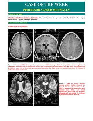
1. CASE OF THE WEEK
PROFESSOR YASSER METWALLY
CLINICAL PICTURE
CLINICAL PICTURE: CLINICAL PICTURE: A 6 years old male patient presented clinically with intractable complex
partial seizure. The child is mentally subnormal.
RADIOLOGICAL FINDINGS
RADIOLOGICAL FINDINGS:
Figure 1. Precontrast MRI T1 image (A) and postcontrast MRI T1 images (B,C) showing evidence of lissencephaly and
pachygyria. Also observed an intracortical mass in the left posterior parieto-occipital; area. The mass is hypointense on
precontrast scans with no observable postcontrast enhancement. The mass has a very minimal mas effect. No evidence of
perilesional edema is observed.
Figure 2. MRI T2 images showing
showing similar finding observed in
figure 1. The mass is hyperintense on the
MRI T2 images, purely intracortical with
minimal mass effect. Also noted
lissencephaly, pachygyria with probable
subependymal heterotopia. No evidence
of perilesional edema is observed.
2. Figure 3. MRI FLAIR images. The left parieto-occipital mass is hyperintense on FLAIR images, purely intracortical with mild
mass effect. Also observed lissencephaly, polymicrogyria and subependymal nodular heterotopia. No evidence of perilesional
edema is observed.
Stereotactic biopsy of the cortical mass revealed a Dysembryoplastic neuroepithelial tumor [DNET]. SEE figure 4.
Figure 4. Photomicrograph (original magnification, x160;
hematoxylineosin stain) of a dysembryoplastic
neuroepithelial tumor with cystic degeneration shows a
trabecular pattern (long arrows) of glial elements, including
astrocytes and oligodendrocytes. Oligodendroglial cells
(arrowhead) contain small dark nuclei, whereas the
astrocytic cells (short arrow) are somewhat larger with pink
cytoplasm.
Dysembryoplastic neuroepithelial tumor [DNET] is a relatively newly described benign tumor arising within the
supratentorial cortex and almost always associated with partial complex seizures. These lesions may occasionally appear
cystic and show one of the three characteristics which include: a) specific glioneuronal element, b) nodular component, or c)
association with cortical dysplasia.
MR scan usually demonstrates a focal cortical lesion most commonly in the temporal lobe that is hypodense on T1 and
hyperintense on T2 weighted studies. It is not uncommon for a small subset of these tumors to resemble benign cysts with
slightly increased signal on proton density-weighted and FLAIR sequences. Post-contrast enhancement and calcification may
also occur occasionally.
Imaging Findings for Dysembryoplastic neuroepithelial tumor
Imaging characteristics of dysembryoplastic neuroepithelial tumor is similar to those of other low-grade glial tumors and may
not be possible to distinguish this tumor from diffuse astrocytoma, ganglioglioma, oligodendroglioma, or other low-grade
neoplasms.
At CT, the tumor manifests as a hypoattenuating mass.
Calcification may be seen.
3. Remodeling of the adjacent inner table of the skull may also be seen.
At MR imaging, dysembryoplastic neuroepithelial tumors most commonly manifest as cortical masses that are
hypointense on T1-weighted images and hyperintense on T2-weighted images without surrounding vasogenic edema.
Some lesions may appear as an enlarged gyrus, producing a soap bubble appearance at the cortical margin.
Approximately one-third of dysembryoplastic neuroepithelial tumors enhance following intravenous administration of
contrast material.
DIAGNOSIS:
DIAGNOSIS: DYSEMBRYOPLASTIC NEUROEPITHELIAL TUMOUR ASSOCIATED WITH CORTICAL DYSPLASIA
DISCUSSION
DISCUSSION:
Dysembryoplastic neuroepithelial tumor [DNET] is a benign tumor of neuroepithelial origin arising from the cortical or deep
gray matter. Since the description of 39 cases in 1988 by investigators from Paris and the Mayo Clinic, there are now more
than 300 cases on record (1,2). These tumors virtually always manifest in patients with medically refractory partial seizures.
The vast majority of patients are younger than 20 years, and males are more commonly affected (3). In contrast to other brain
neoplasms, neurologic deficits are not common with dysembryoplastic neuroepithelial tumors unless they occur in the
presence of complex congenital abnormalities (3). The temporal lobe is the most common site (62%), followed by the frontal
lobe (31%) (3). Although the vast majority of dysembryoplastic neuroepithelial tumors are confined to the cortical gray
matter, they may also arise within the caudate nucleus, cerebellum, or pons (4,5). Patients with cerebellar dysembryoplastic
neuroepithelial tumors present with symptoms (eg, ataxia, vertigo, gait problems) related to this location rather than seizure
activity (2). The locations of these tumors support the hypothesis that these lesions are derived from secondary germinal
layers (1).
Figure 1. Dysembryoplastic
neuroepithelial tumor
Dysembryoplastic neuroepithelial tumors appear to be remarkably stable in terms of biologic behavior. Despite only partial
resection of many of these lesions, complete cessation of all seizure activity is a common result following neurosurgical
intervention (1). Recurrence is also very rare (3). However, there is one recent report in the literature of malignant
transformation of a dysembryoplastic neuroepithelial tumor that showed a higher degree of mitotic activity than is commonly
seen in these tumors (6). The tumor in this case recurred 11 years after the initial surgical resection (6). This case emphasizes
4. the need for long-term follow-up of patients with atypical dysembryoplastic neuroepithelial tumors.
Figure 2. Dysembyroplastic neuroepithelial tumor. Coronal proton density-weighted image (A) and postcontrast (B) images
demonstrate a peripherally based lesion (arrow) with trabeculated enhancement. Based solely on these images, it appears that
the lesion is extra-axial because gray matter can be seen surrounding the lesion, especially in (A). However, it is not
uncommon for an intra-axial neoplasm causing chronic epilepsy to appear extra-axial because it is situated within or replaces
the cortex and has the appearance of being outside the cortex in some cross-sectional planes. Thin section imaging allowed
visualization of the multicystic nature of this lesion on the enhanced image (B).
The original description of 39 dysembryoplastic neuroepithelial tumors in 1988 resulted from a retrospective review of nearly
300 tumors initially diagnosed as low-grade astrocytomas in patients with medically refractory seizures (1). These 39
specimens shared common histologic features: a multinodular architecture, a "specific glioneuronal element" in a columnar
pattern oriented perpendicular to the cortical surface, and focal areas of cortical dysplasia (1). Since that initial description,
neuropathologists have noted that these three characteristics may not always be present. This observation has led to the latest
classification scheme, which divides these tumors into a "simple form" (without a nodular architecture) and a "complex
form" (with a multinodular appearance) (3). They are characterized by an admixture of astrocytes and oligodendroglial
elements, in association with "floating neurons" and mucinous degeneration. The tumors generally have a low growth
potential as measured by certain metabolic indexes (3). There are several reports of composite neoplasms with both
ganglioglioma and dysembryoplastic neuroepithelial tumor components. Since cortical dysplasia is commonly seen with both
of these tumors, perhaps they represent the tumoral form of cortical dysplasia or neoplastic transformation of a dysplastic
area (7,8,9,10,11).
Figure 3. Dysembryoplastic neuroepithelial tumor in a 14-year-old girl who experienced a seizure while sleeping. (a) Axial CT
image shows a hypoattenuating mass of the right parietal lobe. Note the remodeling of the adjacent inner table of the skull. (b)
Axial T2-weighted MR image shows marked high signal intensity of the mass, which extends beyond the normal cortical
margin ("soap bubble" appearance) and directly remodels the skull. There is no evidence of vasogenic edema associated with
the mass. (c) Contrast-enhanced axial T1-weighted MR image shows no evidence of enhancement within the mass.
5. Figure 4. Photomicrograph (original magnification, x120; hematoxylineosin stain) of a dysembryoplastic neuroepithelial
tumor shows neuronal elements surrounded by prominent vacuoles, which represent so-called floating neurons (arrows).
SUMMARY
SUMMARY
Dysembryoplastic Neuroepithelial Tumor (DNT) is a recently recognized, benign tumor associated with medically intractable,
partial complex seizures. Mean age of onset of symptoms is nine years (range 1-19 years). All reported DNT's have been
supratentorial, most often involving the temporal lobe (approximately 2/3) followed in frequency by the frontal lobe (1/3). The
tumors are primarily cortical in location, although they may extend to involve the subcortical white matter. On CT scans,
DNT's are well-defined, low-attenuation lesions which may be mistaken for cysts. The tumors tend to be low signal on T1-
weighted MR images and high signal on T2-weighted images, i.e., similar to CSF, but on proton-density images, they are
slightly higher in signal than CSF, allowing them to be differentiated from simple cysts. Less than 25% calcify or enhance.
There is associated calvarial remodeling in approximately 1/3 of cases.
Differential diagnoses include ganglioglioma and low-grade astrocytoma. A ganglioglioma, however, is more likely to be
located within the white matter. Calvarial remodeling, if present, is a helpful differentiating feature in that it would be
unlikely for either a ganglioglioma or a low-grade astrocytoma to cause such changes.
The prognosis for patients with DNT is excellent. 70-81% of patients are seizure-free following surgery, and the tumor does
not recur, even in cases where resection is considered incomplete.
Addendum
A new version of this PDF file (with a new case) is uploaded in my web site every week (every Saturday and remains
available till Friday.)
To download the current version follow the link "http://pdf.yassermetwally.com/case.pdf".
You can also download the current version from my web site at "http://yassermetwally.com".
To download the software version of the publication (crow.exe) follow the link:
http://neurology.yassermetwally.com/crow.zip
The case is also presented as a short case in PDF format, to download the short case follow the link:
http://pdf.yassermetwally.com/short.pdf
At the end of each year, all the publications are compiled on a single CD-ROM, please contact the author to know more
details.
Screen resolution is better set at 1024*768 pixel screen area for optimum display.
6. For an archive of the previously reported cases go to www.yassermetwally.net, then under pages in the right panel, scroll
down and click on the text entry "downloadable case records in PDF format"
Also to view a list of the previously published case records follow the following link (http://wordpress.com/tag/case-
record/) or click on it if it appears as a link in your PDF reader
REFERENCES
References
1.Daumas-Duport C, Scheithauer BW, Chodkiewicz JP, Laws ER, Jr, Vedrenne C. Dysembryoplastic neuroepithelial tumour:
a surgically curable tumour of young patients with intractable partial seizures. Neurosurgery 1988; 23:545-556.
2.Fujimoto K, Ohnishi H, Tsujimoto M, Hoshida T, Nakazato Y. Dysembryoplastic neuroepithelial tumor of the cerebellum
and brainstem. J Neurosurg 2000; 93:487-489.
3.Daumas-Duport C, Pietsch T, Lantos PL. Dysembryoplastic neuroepithelial tumour. In: Kleihues P, Cavenee WK, eds.
Pathology and genetics of tumours of the nervous system. Lyon, France: IARC, 2000; 103-106.
4.Cervera-Pierot P, Varlet P, Chodkiewicz JP, Daumas-Duport C. Dysembryoplastic neuroepithelial tumors located in the
caudate nucleus area: report of four cases. Neurosurgery 1997; 40:1065-1070.
5.Kuchelmeister K, Demirel T, Schlorer E, Bergmann M, Gullota F. Dysembryoplastic neuroepithelial tumour of the
cerebellum. Acta Neuropathol (Berl) 1995; 89:385-390.
6.Hammond RR, Duggal N, Woulfe JMJ, Girvin JP. Malignant transformation of a dysembryoplastic neuroepithelial tumor.
J Neurosurg 2000; 92:722-725.
7.Prayson RA. Composite ganglioglioma and dysembryoplastic neuroepithelial tumor. Arch Pathol Lab Med 1999; 123:247-
250.
8.Koeller KK, Dillon WP. MR appearance of dysembryoplastic neuroepithelial tumors (DNT). AJNR Am J Neuroradiol 1992;
13:1319-1325.
9.Kuroiwa T, Kishikawa T, Kato A, Ueno M, Kudo S, Tabuci K. Dysembryoplastic neuroepithelial tumors: MR findings. J
Comput Assist Tomogr 1994; 18:352-356.
10.Ostertun B, Wolf HK, Campos MG, et al. Dysembryoplastic neuroepithelial tumors: MR and CT evaluation. AJNR Am J
Neuroradiol 1996; 17:419-430.
11.Abe M, Tabuchi K, Tsuji T, Shiraishi T, Koga H, Takagi M. Dysembryoplastic neuroepithelial tumor: report of three
cases. Surg Neurol 1995; 43:240-245.
12. Metwally, MYM: Textbook of neuroimaging, A CD-ROM publication, (Metwally, MYM editor) WEB-CD agency for
electronic publication, version 10.4a October 2009