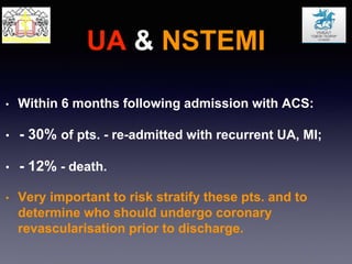
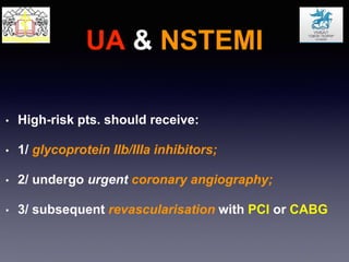
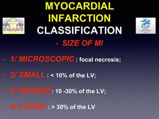
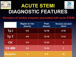
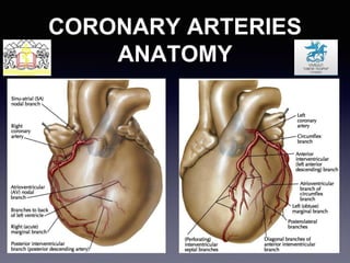
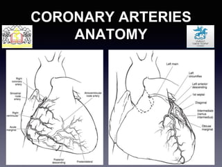
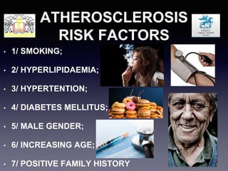
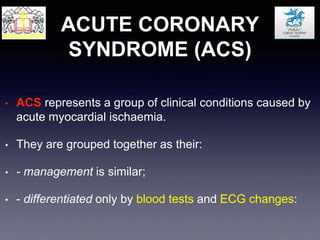
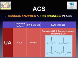
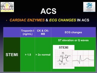
CORONARY
ARTERY DISEASE
Hristo A. Rahman
Department of CardioVascular surgery
Medical University Plovdiv
University General Hospital Saint George-Plovdiv
CORONARY ARTERIES
ANATOMY
CORONARY ARTERIES
ANATOMY
PATHOPHYSIOLOGY
CAD
• CAD:caused by atherosclerosis, developing in 3 stages:
• a) linear fatty streak: focal intimal thickening caused by
deposition of lipid-filled macrophages and smooth
muscle cells;
• b) fibroid plaques: consisting of these lipid-laden cells,
an inflammatory core and cholesterol deposits, encased
by a fibrous cap, resulting in luminal narrowing;
PATHOPHYSIOLOGY
CAD
PATHOPHYSIOLOGY
CAD
• c) evolution of plaques: plaque rupture and exposure of blood
to the contents of plaque can result in luminal thrombosis or a
“major plaque event”.
• - with smaller amounts of thrombosis, luminal occlusion does not
occur but subsequent embolisation of thrombi may trigger
arrhythmias or plaque growth by the release of platelet-derived
growth factors which stimulate smooth muscle division.
• - plaque rupture - more likely in soft plaques with low fibrin
content than hard plaques.
• - soft plaques - potentially more lethal even if they are
smaller than hard plaques which have a thick fibrous cap.
PATHOPHYSIOLOGY
CAD
ATHEROSCLEROSIS
RISK FACTORS
• 1/ SMOKING;
• 2/ HYPERLIPIDAEMIA;
• 3/ HYPERTENTION;
• 4/ DIABETES MELLITUS;
• 5/ MALE GENDER;
• 6/ INCREASING AGE;
• 7/ POSITIVE FAMILY HISTORY
DEFINITIONS
• MYOCARDIAL ISCHAEMIA:
IMBALANCE BETWEEN MYOCARDIAL OXYGEN SUPPLY
AND DEMAND
• MYOCARDIAL INFARCTION:
ISCHAEMIA-INDUCED CARDIOMYOCYTE LOSS
(NECROSIS), CAUSED BY CORONARY ARTERY
OCCLUSION SECONDARY TO:
• 1/ PROGRESSIVE ATHEROSCLEROSIS;
• 2/ DISRUPTION OF UNSTABLE PLAQUE WITH ACUTE
THROMBOSIS
ANGINA PECTORIS
TREATMENT PRINCIPLES
• CONSERVATIVE: cessation of smoking, healthy diet,
BP control, diabetic control.
• MEDICAL: antiplatelet, statin, nitrate, B-blocker, calcium
channel blocker, nicorandil.
• PERCUTANEOUS CORONARY INTERVENTION
(PCI): balloon angioplasty, bare metal stent, drug-eluting
stent.
• SURGERY: coronary artery bypass grafting (CABG),
transmyocardial revascularisation.
CANADIAN
CARDIOVASCULAR SOCIETY
CLASSIFICATION
• Functional classification that relates patient’s
symptoms of angina pectoris with the ability to perform
activities.
• I: no limitations of physical activity and no symptoms
with ordinary activity;
• II: slight limitation of physical activity with angina
precipitated by vigorous activity;
• III: marked limitation of physical activity with angina
precipitated by routine activity;
• IV: inability to perform any activities without angina or
symptoms of angina at rest
ACUTE CORONARY
SYNDROME (ACS)
• ACS represents a group of clinical conditions caused by
acute myocardial ischaemia.
• They are grouped together as their:
• - management is similar;
• - differentiated only by blood tests and ECG changes:
ACUTE CORONARY
SYNDROME (ACS)
• 1/ UNSTABLE ANGINA PECTORIS (UA) - defined as
angina:
• - 1.1: occurring with increasing frequency and severity;
• - 1.2: occurring at rest or more frequently at night;
• - 1.3: not relieved quickly with sublingual glyceryl trinitrate
(GTN).
• 2/ non-ST elevation MI (NSTEMI)
• 3/ ST elevation MI (STEMI)
ACS
• CARDIAC ENZYMES & ECG CHANGES IN ACS
Troponin I
(ng/mL)
CK & CK-MB ECG changes
UA < 0.6 Normal
Transient ST & T-wave changes
or normal ECG
ACS
• CARDIAC ENZYMES & ECG CHANGES IN ACS
Troponin I
(ng/mL)
CK & CK-
MB
ECG changes
NSTEMI 0.6 - 1.5 < 2x normal
Transient ST & T-wave changes
or normal ECG
ACS
• CARDIAC ENZYMES & ECG CHANGES IN ACS
Troponin I
(ng/mL)
CK & CK-
MB
ECG changes
STEMI > 1.5 > 2x normal
ST elevation or Q waves
UA & NSTEMI
MANAGEMENT
• Main principles of managing pts. with UA & NSTEMI
include:
• 1/ Stabilisation of the atheromatous plaque;
• 2/ Restoration of coronary blood flow;
• 3/ Alleviation of the flow-limiting stenosis.
UA & NSTEMI
MANAGEMENT
• Initial medical management ot pts. with ACS includes:
• 1/ analgesia (fentanyl, diamorphine);
• 2/ anti-anginal therapy (B-blocker, nitrate, calcium
channel blocker);
• 3/ dual anti platelet therapy (aspirin & clopidogrel);
• 4/ low-molecular weight heparin (LMWH);
• 5/ statin
UA & NSTEMI
• Within 6 months following admission with ACS:
• - 30% of pts. - re-admitted with recurrent UA, MI;
• - 12% - death.
• Very important to risk stratify these pts. and to
determine who should undergo coronary
revascularisation prior to discharge.
UA & NSTEMI
• High-risk pts. should receive:
• 1/ glycoprotein IIb/IIIa inhibitors;
• 2/ undergo urgent coronary angiography;
• 3/ subsequent revascularisation with PCI or CABG
UA & NSTEMI
• HIGH-RISK FACTORS INCLUDE:
• 1/ PATIENT FACTORS;
• 2/ ECG CHANGES;
• 3/ ANY ELEVATION IN CARDIAC ENZYMES.
UA & NSTEMI
• PATIENT FACTORS:
• 1/ AGE > 65;
• 2/ HYPERTENSION;
• 3/ DIABETES MELLITUS;
• 4/ PREVIOUS MI;
• 5/ LV DYSFUNCTION;
• 6/ ONGOING PAIN DESPITE MEDICAL THERAPY;
• 7/ CARDIOGENIC SHOCK
UA & NSTEMI
• ECG CHANGES:
• 1/ TRANSIENT ST ELEVATION OR DEPRESSION
DURING PAIN;
• 2/ PERSISTENT ST DEPRESSION;
• 3/ DEEP T-WAVE INVERSION;
• 4/ LBBB;
• 5/ VENTRICULAR TACHYCARDIA
UA & NSTEMI
• REMAINING PTS. SHOULD UNDERGO
INVESTIGATION WITH:
• 1/ EXERCISE TOLERANCE TEST;
• 2/ STRESS NUCLEAR MEDICINE SCANNING
• 3/ IF 1/ +/- 2/ ARE POSITIVE, CORONARY
ANGIOGRAPHY
MYOCARDIAL
INFARCTION
CLASSIFICATION
• CLASSIFICATION, BASED ON:
• 1/ SIZE;
• 2/ LOCATION;
• 3/ TIME FROM ONSET;
• 4/ ST ELEVATION
MYOCARDIAL
INFARCTION
CLASSIFICATION
• SIZE OF MI
• 1/ MICROSCOPIC : focal necrosis;
• 2/ SMALL : < 10% of the LV;
• 3/ MEDIUM : 10 -30% of the LV;
• 4/ LARGE : > 30% of the LV
MYOCARDIAL
INFARCTION
CLASSIFICATION
• LOCATION OF MI
• 1/ ANTERIOR;
• 2/ SEPTAL;
• 3/ INFERIOR;
• 4/ LATERAL;
• 5/ POSTERIOR.
MYOCARDIAL
INFARCTION
CLASSIFICATION
• TIME FROM ONSET OF MI
• 1/ ACUTE : 6 hours to 7 days
(polymorphonuclear leukocytes);
• 2/ HEALING : 7 to 28 days
(mononuclear cells and fibroblasts);
• 3/ HEALED : > 28 days
(scar tissue without infiltration)
MYOCARDIAL
INFARCTION
CLASSIFICATION
• ST ELEVATION
• 1/ non-ST elevation MI : no transmurality;
• 2/ ST elevation MI : transmural
ACUTE STEMI
DIAGNOSTIC FEATURES
• According to ESC & ACC guidelines:
• 1/ Typical rise & fall of cardiac enzymes
(troponin or CK-MB) associated with one of the
following:
• 2.1/ ST elevation with peaked T wave;
• 2.2/ Pathological Q waves;
• 2.3/ Ischaemic type of chest pain lasting > 20 min.
ACUTE STEMI
DIAGNOSTIC FEATURES
• Elevation of cardiac enzymes associated with acute STEMI
Begins to rise
(hours)
Peak
(hours)
Remains elevated
(days)
Tp I 4-6 12-16 5-10
Tp T 4-6 12-16 5-14
CK 4-8 12-24 3
CK-MB 2-6 12-24 2
Myoglobin 2 6-8 24
ACUTE STEMI
DIAGNOSTIC FEATURES
• Elevation of cardiac enzymes associated with acute STEMI
• 1/ Creatinine kinase
(normal <140 IU/L for women and <170 IU/L for men)
(normal <3.0 ng/L)
(normal <0.1 ng/L)
• 4/ Troponin I
(normal <0.3 ng/L) may also be raised with renal impairment
ACUTE STEMI
MANAGEMENT
• Main principles of managing pts. with STEMI (similar to
those for UA & NSTEMI) include:
• 1/ Stabilisation of the atheromatous plaque;
• 2/ Restoration of coronary blood flow;
• 3/ Alleviation of the flow-limiting stenosis.
ACUTE STEMI
MANAGEMENT
• Initial medical management ot pts. with STEMI includes:
• 1/ oxygen;
• 2/ analgesia (diamorphine) & anti-emetic
(metoclopramide);
• 3/ antiplatelet therapy (aspirin);
• 4/ B-blocker;
• 5/ statin
ACUTE STEMI
MANAGEMENT
• Nitrates, diuretics, atropine, lidocaine may be
required for complications of MI including:
• 1/ Ongoing chest pain;
• 2/ Pulmonary oedema;
• 3/ Bradycardia;
• 4/ Ventricular arrythmia
ACUTE STEMI
MANAGEMENT
• Restoration of coronary blood flow is achieved by:
• 1/ Primary PCI;
• 2/ Thrombolysis.
• High-risk rescue PCI or CABG may be required for
pts. with:
• 1/ Ongoing chest pain;
• 2/ ECG changes following thrombolysis
CABG guidelines
AHA / ACC/ ESC / ЕACTS
CABG
• INDICATIONS ACCORDING TO AHA GUIDELINES:
• 1/ For asymptomatic pts. or pts. with mild angina;
• 2/ For pts. with chronic stable angina;
• 3/ For pts. with UA or NSTEMI;
• 4/ For pts. with STEMI;
• 5/ For pts. with poor LV function;
• 6/ For pts. with life-threatening ventricular arrhythmias;
• 7/ CABG after failed PCI;
• 8/ For pts. with previous CABG
Asymptomatic CAD
• LMCAD >/= 50%
• Left main equivalent
• 3-VCAD (especially if LVEF<50%)
• 1-/2-CAD with large area at risk +/- LVEF<50%
CLASSES OF
RECOMMENDATION DEFINITION
SUGGESTED
WORDING TO USE
CLASS I
1. Significant LMS stenosis (>50%);
2. LMS equivalent disease;
3. 3-VCAD (survival benefit is greater in pts. with
LVEF <50%;
4. 1-/2-VCAD (incl. pLAD stenosis and LVEF <50%);
5. 1-/2-VCAD (without pLAD stenosis) but with large
area of myocardium at risk
RECOMMENDED
INDICATED
CLASS II
CLASS IIa
1. 1-/2-VCAD (incl. pLAD stenosis and LVEF >50%;
2. 1-/2 VCAD (without pLAD stenosis) and moderate
area of myocardium at risk
SHOULD BE
CONSIDERED
CLASS IIb
MAY BE
CONSIDERED
CLASS III
1. 1-/2 VCAD (without pLAD stenosis) and no
myocardium at risk;
2. Borderline stenosis 50-60% with no myocardium at
risk;
3. Insignificant stenosis (<50%)
NOT
RECOMMENDED
CONTRAINDICATED
CABG for chronic stable angina
Chronic stable angina
• LMCAD >/= 50%
• Left main equivalent (prox.LAD + prox.LCx)
• 3-VCAD (especially if LVEF<50%)
• 1-/2-CAD with large area at risk +/- LVEF<50%
• Disabling angina (if low operative risk)
CLASSES OF
RECOMMENDATION DEFINITION
SUGGESTED
WORDING TO USE
CLASS I
1. Significant LMS stenosis (>50%);
2. LMS equivalent disease;
3. Other CAD with ongoing ischaemia unresponsive
to max. non-surgical treatment
RECOMMENDED
INDICATED
CLASS II
CLASS IIa 1. 1-/2-VCAD (incl. pLAD stenosis) and LVEF >50%
SHOULD BE
CONSIDERED
CLASS IIb
1. 1-/2-VCAD (without pLAD stenosis) when PCI is not
an option but a large area of myocardium is at risk
MAY BE
CONSIDERED
CLASS III
NOT
RECOMMENDED
CONTRAINDICATED
CABG for UA & NSTEMI
Unstable angina & NSTEMI
• LMCAD >/= 50%
• Left main equivalent (prox.LAD + prox.LCx)
• 3-VCAD (especially if LVEF<50%)
• 1-/2-CAD with large area at risk +/- LVEF<50%
• Ongoing ischaemia
CLASSES OF
RECOMMENDATION DEFINITION
SUGGESTED
WORDING TO USE
CLASS I
1. Emergency or urgent CABG indicated in pts. with
suitable coronary anatomy and:
2. Failed PCI with haemodynamic instability;
3. Persistent or recurrent ischaemia with large area
of myocardium at risk but not suitable for PCI;
4. Mechanical complications of MI including: VSR,
IMR, LVR;
5. Cariogenic shock within 36 h. of MI, unless further
intervention is futile;
6. Life-threatening ventricular arrhythmias with
significant LMCAD or 3-VCAD
RECOMMENDED
INDICATED
CLASS II
CLASS IIa
1. Primary reperfusion within 6-12 h. of MI, in pts. not
suitable for, or following failed, PCI and thrombolysis
SHOULD BE
CONSIDERED
CLASS IIb
MAY BE
CONSIDERED
CLASS III
1. Haemodynamically stable pt. with small area of
myocardium at risk
NOT
RECOMMENDED
CONTRAINDICATED
CABG for STEMI
STEMI >12h.
• LMCAD >/= 50%
• Left main equivalent
• 3-VCAD (especially if LVEF<50%)
• 1-/2-CAD with large area at risk +/- LVEF<50%
• Ongoing ischaemia
• STEMI <12h : No CABG ( CONTRAINDICATED )
CLASSES OF
RECOMMENDATION DEFINITION
SUGGESTED
WORDING TO USE
CLASS I
1. Ongoing ischaemia;
2. Threatened occlusion;
3. Haemodynamic compromise
RECOMMENDED
INDICATED
CLASS II
CLASS IIa
1. Retained foreign body;
2. Haemodynamic compromise with impaired
clotting and no previous median sternotomy
SHOULD BE
CONSIDERED
CLASS IIb
1. Haemodynamic compromise with impaired
clotting and previous median sternotomy
MAY BE
CONSIDERED
CLASS III
1. No evidence of ischaemia
2. No suitable targets for grafting
NOT
RECOMMENDED
CONTRAINDICATED
CABG after failed PCI
CABG
CONVENTIONAL TECHNIQUE ON
CPB
• 1/ STERNOTOMY;
• 2/ GRAFT (CONDUIT) HARVESTING;
• 3/ CPB ESTABLISHMENT;
• 4/ CARDIOPLEGIC ARREST;
• 5/ DISTAL ANASTOMOSES;
• 6/ PROXIMAL ANASTOMOSES;
• 7/ CPB WEANING-OFF;
• 8/ HAEMOSTASIS;
• 9/ CLOSURE
FULL STERNOTOMY
GRAFTS / CONDUITS
• ARTERIAL:
• - Left/Right Internal Thoracic Arteries ( LIMA/RIMA );
• - Radial artery ( RA );
• - Right Gastroepiploic Artery ( rGEA ).
• VENOUS:
• - Greater Saphenous Vein (GSV);
• - Smaller Saphenous Vein (SSV)
GREATER SAPHENOUS
VEIN HARVESTING
GREATER SAPHENOUS
VEIN HARVESTING
GREATER SAPHENOUS VEIN
TECHNIQUES & FINAL
RESULTS
Conventional
Bridge Technique
Endoscopic
LEFT INTERNAL THORACIC
ARTERY HARVESTING
RADIAL ARTERY
HARVESTING
RIGHT GASTROEPIPLOIC
ARTERY HARVESTING
RIGHT GASTROEPIPLOIC
ARTERY HARVESTING
CABG
RCA
LAD
LIMA
RA
CABG x 3
all arterial
CABG
RCA
LAD
LIMA
SVG
CABG x 3
LAD-LIMA
RCA,OM- sequential SVG
OM
CABG
LAD
LIMA
RIMA
CABG x 2
all arterial
LAD-RIMA
OM-LIMA
OM
CABG
LAD
LIMA
RIMA
CABG x 2
all arterial
LAD-LIMA
RCA-RIMA
RCA
CABG
RCx
OM
SVG
CABG x 3
RCA,RCx,OM- sequential SVG
RCA
CABG
RCA/PD/
SVG
CABG x 2
RCA,RCA/PD/- sequential SVG
RCA
CABG
LIMA
SVG
CABG x 3
LAD-LIMA
RCA, RDg-SVG
SVG

CAD & CABG

  • 1.
    CORONARY ARTERY DISEASE Hristo A.Rahman Department of CardioVascular surgery Medical University Plovdiv University General Hospital Saint George-Plovdiv
  • 2.
  • 3.
  • 4.
    PATHOPHYSIOLOGY CAD • CAD:caused byatherosclerosis, developing in 3 stages: • a) linear fatty streak: focal intimal thickening caused by deposition of lipid-filled macrophages and smooth muscle cells; • b) fibroid plaques: consisting of these lipid-laden cells, an inflammatory core and cholesterol deposits, encased by a fibrous cap, resulting in luminal narrowing;
  • 5.
  • 6.
    PATHOPHYSIOLOGY CAD • c) evolutionof plaques: plaque rupture and exposure of blood to the contents of plaque can result in luminal thrombosis or a “major plaque event”. • - with smaller amounts of thrombosis, luminal occlusion does not occur but subsequent embolisation of thrombi may trigger arrhythmias or plaque growth by the release of platelet-derived growth factors which stimulate smooth muscle division. • - plaque rupture - more likely in soft plaques with low fibrin content than hard plaques. • - soft plaques - potentially more lethal even if they are smaller than hard plaques which have a thick fibrous cap.
  • 7.
  • 8.
    ATHEROSCLEROSIS RISK FACTORS • 1/SMOKING; • 2/ HYPERLIPIDAEMIA; • 3/ HYPERTENTION; • 4/ DIABETES MELLITUS; • 5/ MALE GENDER; • 6/ INCREASING AGE; • 7/ POSITIVE FAMILY HISTORY
  • 9.
    DEFINITIONS • MYOCARDIAL ISCHAEMIA: IMBALANCEBETWEEN MYOCARDIAL OXYGEN SUPPLY AND DEMAND • MYOCARDIAL INFARCTION: ISCHAEMIA-INDUCED CARDIOMYOCYTE LOSS (NECROSIS), CAUSED BY CORONARY ARTERY OCCLUSION SECONDARY TO: • 1/ PROGRESSIVE ATHEROSCLEROSIS; • 2/ DISRUPTION OF UNSTABLE PLAQUE WITH ACUTE THROMBOSIS
  • 10.
    ANGINA PECTORIS TREATMENT PRINCIPLES •CONSERVATIVE: cessation of smoking, healthy diet, BP control, diabetic control. • MEDICAL: antiplatelet, statin, nitrate, B-blocker, calcium channel blocker, nicorandil. • PERCUTANEOUS CORONARY INTERVENTION (PCI): balloon angioplasty, bare metal stent, drug-eluting stent. • SURGERY: coronary artery bypass grafting (CABG), transmyocardial revascularisation.
  • 11.
    CANADIAN CARDIOVASCULAR SOCIETY CLASSIFICATION • Functionalclassification that relates patient’s symptoms of angina pectoris with the ability to perform activities.
  • 12.
    • I: nolimitations of physical activity and no symptoms with ordinary activity; • II: slight limitation of physical activity with angina precipitated by vigorous activity; • III: marked limitation of physical activity with angina precipitated by routine activity; • IV: inability to perform any activities without angina or symptoms of angina at rest
  • 13.
    ACUTE CORONARY SYNDROME (ACS) •ACS represents a group of clinical conditions caused by acute myocardial ischaemia. • They are grouped together as their: • - management is similar; • - differentiated only by blood tests and ECG changes:
  • 14.
    ACUTE CORONARY SYNDROME (ACS) •1/ UNSTABLE ANGINA PECTORIS (UA) - defined as angina: • - 1.1: occurring with increasing frequency and severity; • - 1.2: occurring at rest or more frequently at night; • - 1.3: not relieved quickly with sublingual glyceryl trinitrate (GTN). • 2/ non-ST elevation MI (NSTEMI) • 3/ ST elevation MI (STEMI)
  • 15.
    ACS • CARDIAC ENZYMES& ECG CHANGES IN ACS Troponin I (ng/mL) CK & CK-MB ECG changes UA < 0.6 Normal Transient ST & T-wave changes or normal ECG
  • 16.
    ACS • CARDIAC ENZYMES& ECG CHANGES IN ACS Troponin I (ng/mL) CK & CK- MB ECG changes NSTEMI 0.6 - 1.5 < 2x normal Transient ST & T-wave changes or normal ECG
  • 17.
    ACS • CARDIAC ENZYMES& ECG CHANGES IN ACS Troponin I (ng/mL) CK & CK- MB ECG changes STEMI > 1.5 > 2x normal ST elevation or Q waves
  • 18.
    UA & NSTEMI MANAGEMENT •Main principles of managing pts. with UA & NSTEMI include: • 1/ Stabilisation of the atheromatous plaque; • 2/ Restoration of coronary blood flow; • 3/ Alleviation of the flow-limiting stenosis.
  • 19.
    UA & NSTEMI MANAGEMENT •Initial medical management ot pts. with ACS includes: • 1/ analgesia (fentanyl, diamorphine); • 2/ anti-anginal therapy (B-blocker, nitrate, calcium channel blocker); • 3/ dual anti platelet therapy (aspirin & clopidogrel); • 4/ low-molecular weight heparin (LMWH); • 5/ statin
  • 20.
    UA & NSTEMI •Within 6 months following admission with ACS: • - 30% of pts. - re-admitted with recurrent UA, MI; • - 12% - death. • Very important to risk stratify these pts. and to determine who should undergo coronary revascularisation prior to discharge.
  • 21.
    UA & NSTEMI •High-risk pts. should receive: • 1/ glycoprotein IIb/IIIa inhibitors; • 2/ undergo urgent coronary angiography; • 3/ subsequent revascularisation with PCI or CABG
  • 22.
    UA & NSTEMI •HIGH-RISK FACTORS INCLUDE: • 1/ PATIENT FACTORS; • 2/ ECG CHANGES; • 3/ ANY ELEVATION IN CARDIAC ENZYMES.
  • 23.
    UA & NSTEMI •PATIENT FACTORS: • 1/ AGE > 65; • 2/ HYPERTENSION; • 3/ DIABETES MELLITUS; • 4/ PREVIOUS MI; • 5/ LV DYSFUNCTION; • 6/ ONGOING PAIN DESPITE MEDICAL THERAPY; • 7/ CARDIOGENIC SHOCK
  • 24.
    UA & NSTEMI •ECG CHANGES: • 1/ TRANSIENT ST ELEVATION OR DEPRESSION DURING PAIN; • 2/ PERSISTENT ST DEPRESSION; • 3/ DEEP T-WAVE INVERSION; • 4/ LBBB; • 5/ VENTRICULAR TACHYCARDIA
  • 25.
    UA & NSTEMI •REMAINING PTS. SHOULD UNDERGO INVESTIGATION WITH: • 1/ EXERCISE TOLERANCE TEST; • 2/ STRESS NUCLEAR MEDICINE SCANNING • 3/ IF 1/ +/- 2/ ARE POSITIVE, CORONARY ANGIOGRAPHY
  • 26.
    MYOCARDIAL INFARCTION CLASSIFICATION • CLASSIFICATION, BASEDON: • 1/ SIZE; • 2/ LOCATION; • 3/ TIME FROM ONSET; • 4/ ST ELEVATION
  • 27.
    MYOCARDIAL INFARCTION CLASSIFICATION • SIZE OFMI • 1/ MICROSCOPIC : focal necrosis; • 2/ SMALL : < 10% of the LV; • 3/ MEDIUM : 10 -30% of the LV; • 4/ LARGE : > 30% of the LV
  • 28.
    MYOCARDIAL INFARCTION CLASSIFICATION • LOCATION OFMI • 1/ ANTERIOR; • 2/ SEPTAL; • 3/ INFERIOR; • 4/ LATERAL; • 5/ POSTERIOR.
  • 29.
    MYOCARDIAL INFARCTION CLASSIFICATION • TIME FROMONSET OF MI • 1/ ACUTE : 6 hours to 7 days (polymorphonuclear leukocytes); • 2/ HEALING : 7 to 28 days (mononuclear cells and fibroblasts); • 3/ HEALED : > 28 days (scar tissue without infiltration)
  • 30.
    MYOCARDIAL INFARCTION CLASSIFICATION • ST ELEVATION •1/ non-ST elevation MI : no transmurality; • 2/ ST elevation MI : transmural
  • 31.
    ACUTE STEMI DIAGNOSTIC FEATURES •According to ESC & ACC guidelines: • 1/ Typical rise & fall of cardiac enzymes (troponin or CK-MB) associated with one of the following: • 2.1/ ST elevation with peaked T wave; • 2.2/ Pathological Q waves; • 2.3/ Ischaemic type of chest pain lasting > 20 min.
  • 32.
    ACUTE STEMI DIAGNOSTIC FEATURES •Elevation of cardiac enzymes associated with acute STEMI Begins to rise (hours) Peak (hours) Remains elevated (days) Tp I 4-6 12-16 5-10 Tp T 4-6 12-16 5-14 CK 4-8 12-24 3 CK-MB 2-6 12-24 2 Myoglobin 2 6-8 24
  • 33.
    ACUTE STEMI DIAGNOSTIC FEATURES •Elevation of cardiac enzymes associated with acute STEMI • 1/ Creatinine kinase (normal <140 IU/L for women and <170 IU/L for men) (normal <3.0 ng/L) (normal <0.1 ng/L) • 4/ Troponin I (normal <0.3 ng/L) may also be raised with renal impairment
  • 34.
    ACUTE STEMI MANAGEMENT • Mainprinciples of managing pts. with STEMI (similar to those for UA & NSTEMI) include: • 1/ Stabilisation of the atheromatous plaque; • 2/ Restoration of coronary blood flow; • 3/ Alleviation of the flow-limiting stenosis.
  • 35.
    ACUTE STEMI MANAGEMENT • Initialmedical management ot pts. with STEMI includes: • 1/ oxygen; • 2/ analgesia (diamorphine) & anti-emetic (metoclopramide); • 3/ antiplatelet therapy (aspirin); • 4/ B-blocker; • 5/ statin
  • 36.
    ACUTE STEMI MANAGEMENT • Nitrates,diuretics, atropine, lidocaine may be required for complications of MI including: • 1/ Ongoing chest pain; • 2/ Pulmonary oedema; • 3/ Bradycardia; • 4/ Ventricular arrythmia
  • 37.
    ACUTE STEMI MANAGEMENT • Restorationof coronary blood flow is achieved by: • 1/ Primary PCI; • 2/ Thrombolysis. • High-risk rescue PCI or CABG may be required for pts. with: • 1/ Ongoing chest pain; • 2/ ECG changes following thrombolysis
  • 38.
    CABG guidelines AHA /ACC/ ESC / ЕACTS
  • 39.
    CABG • INDICATIONS ACCORDINGTO AHA GUIDELINES: • 1/ For asymptomatic pts. or pts. with mild angina; • 2/ For pts. with chronic stable angina; • 3/ For pts. with UA or NSTEMI; • 4/ For pts. with STEMI; • 5/ For pts. with poor LV function; • 6/ For pts. with life-threatening ventricular arrhythmias; • 7/ CABG after failed PCI; • 8/ For pts. with previous CABG
  • 40.
    Asymptomatic CAD • LMCAD>/= 50% • Left main equivalent • 3-VCAD (especially if LVEF<50%) • 1-/2-CAD with large area at risk +/- LVEF<50%
  • 41.
    CLASSES OF RECOMMENDATION DEFINITION SUGGESTED WORDINGTO USE CLASS I 1. Significant LMS stenosis (>50%); 2. LMS equivalent disease; 3. 3-VCAD (survival benefit is greater in pts. with LVEF <50%; 4. 1-/2-VCAD (incl. pLAD stenosis and LVEF <50%); 5. 1-/2-VCAD (without pLAD stenosis) but with large area of myocardium at risk RECOMMENDED INDICATED CLASS II CLASS IIa 1. 1-/2-VCAD (incl. pLAD stenosis and LVEF >50%; 2. 1-/2 VCAD (without pLAD stenosis) and moderate area of myocardium at risk SHOULD BE CONSIDERED CLASS IIb MAY BE CONSIDERED CLASS III 1. 1-/2 VCAD (without pLAD stenosis) and no myocardium at risk; 2. Borderline stenosis 50-60% with no myocardium at risk; 3. Insignificant stenosis (<50%) NOT RECOMMENDED CONTRAINDICATED CABG for chronic stable angina
  • 42.
    Chronic stable angina •LMCAD >/= 50% • Left main equivalent (prox.LAD + prox.LCx) • 3-VCAD (especially if LVEF<50%) • 1-/2-CAD with large area at risk +/- LVEF<50% • Disabling angina (if low operative risk)
  • 43.
    CLASSES OF RECOMMENDATION DEFINITION SUGGESTED WORDINGTO USE CLASS I 1. Significant LMS stenosis (>50%); 2. LMS equivalent disease; 3. Other CAD with ongoing ischaemia unresponsive to max. non-surgical treatment RECOMMENDED INDICATED CLASS II CLASS IIa 1. 1-/2-VCAD (incl. pLAD stenosis) and LVEF >50% SHOULD BE CONSIDERED CLASS IIb 1. 1-/2-VCAD (without pLAD stenosis) when PCI is not an option but a large area of myocardium is at risk MAY BE CONSIDERED CLASS III NOT RECOMMENDED CONTRAINDICATED CABG for UA & NSTEMI
  • 44.
    Unstable angina &NSTEMI • LMCAD >/= 50% • Left main equivalent (prox.LAD + prox.LCx) • 3-VCAD (especially if LVEF<50%) • 1-/2-CAD with large area at risk +/- LVEF<50% • Ongoing ischaemia
  • 45.
    CLASSES OF RECOMMENDATION DEFINITION SUGGESTED WORDINGTO USE CLASS I 1. Emergency or urgent CABG indicated in pts. with suitable coronary anatomy and: 2. Failed PCI with haemodynamic instability; 3. Persistent or recurrent ischaemia with large area of myocardium at risk but not suitable for PCI; 4. Mechanical complications of MI including: VSR, IMR, LVR; 5. Cariogenic shock within 36 h. of MI, unless further intervention is futile; 6. Life-threatening ventricular arrhythmias with significant LMCAD or 3-VCAD RECOMMENDED INDICATED CLASS II CLASS IIa 1. Primary reperfusion within 6-12 h. of MI, in pts. not suitable for, or following failed, PCI and thrombolysis SHOULD BE CONSIDERED CLASS IIb MAY BE CONSIDERED CLASS III 1. Haemodynamically stable pt. with small area of myocardium at risk NOT RECOMMENDED CONTRAINDICATED CABG for STEMI
  • 46.
    STEMI >12h. • LMCAD>/= 50% • Left main equivalent • 3-VCAD (especially if LVEF<50%) • 1-/2-CAD with large area at risk +/- LVEF<50% • Ongoing ischaemia • STEMI <12h : No CABG ( CONTRAINDICATED )
  • 47.
    CLASSES OF RECOMMENDATION DEFINITION SUGGESTED WORDINGTO USE CLASS I 1. Ongoing ischaemia; 2. Threatened occlusion; 3. Haemodynamic compromise RECOMMENDED INDICATED CLASS II CLASS IIa 1. Retained foreign body; 2. Haemodynamic compromise with impaired clotting and no previous median sternotomy SHOULD BE CONSIDERED CLASS IIb 1. Haemodynamic compromise with impaired clotting and previous median sternotomy MAY BE CONSIDERED CLASS III 1. No evidence of ischaemia 2. No suitable targets for grafting NOT RECOMMENDED CONTRAINDICATED CABG after failed PCI
  • 48.
    CABG CONVENTIONAL TECHNIQUE ON CPB •1/ STERNOTOMY; • 2/ GRAFT (CONDUIT) HARVESTING; • 3/ CPB ESTABLISHMENT; • 4/ CARDIOPLEGIC ARREST; • 5/ DISTAL ANASTOMOSES; • 6/ PROXIMAL ANASTOMOSES; • 7/ CPB WEANING-OFF; • 8/ HAEMOSTASIS; • 9/ CLOSURE
  • 49.
  • 50.
    GRAFTS / CONDUITS •ARTERIAL: • - Left/Right Internal Thoracic Arteries ( LIMA/RIMA ); • - Radial artery ( RA ); • - Right Gastroepiploic Artery ( rGEA ). • VENOUS: • - Greater Saphenous Vein (GSV); • - Smaller Saphenous Vein (SSV)
  • 51.
  • 52.
  • 53.
    GREATER SAPHENOUS VEIN TECHNIQUES& FINAL RESULTS Conventional Bridge Technique Endoscopic
  • 54.
  • 55.
  • 56.
  • 57.
  • 58.
  • 59.
  • 60.
    CABG LAD LIMA RIMA CABG x 2 allarterial LAD-RIMA OM-LIMA OM
  • 61.
    CABG LAD LIMA RIMA CABG x 2 allarterial LAD-LIMA RCA-RIMA RCA
  • 62.
  • 63.
  • 64.