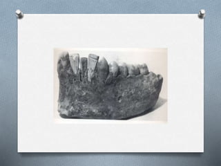
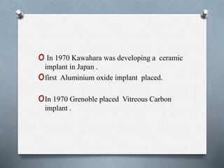
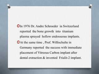
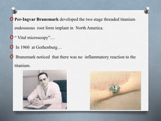
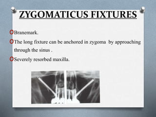
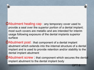
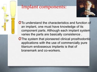
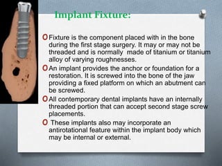
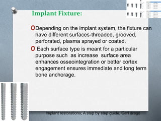
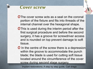
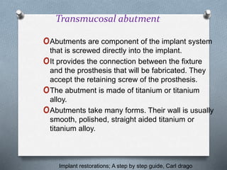
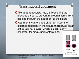
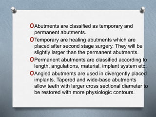
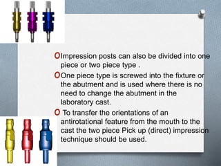
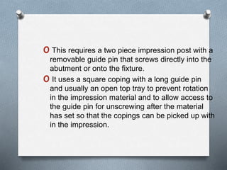
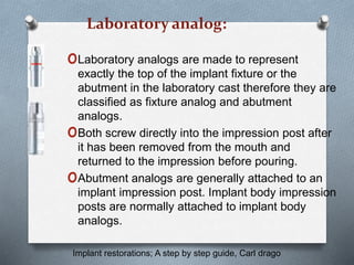
The document provides a history of dental implants from ancient times to the modern era. Some key points discussed include:
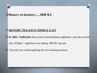
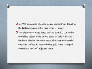
- Ancient implants dating back to 400 BC made of materials like ivory and animal teeth.
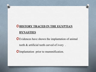
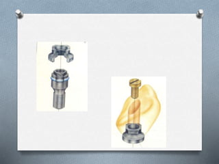
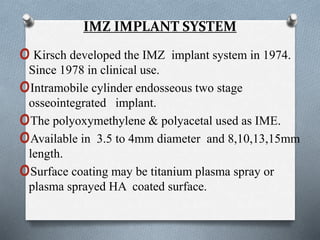
- Medieval period saw allotransplantation of animal teeth and use of materials like bone and ivory.
- Foundational period in the 1800s included early modern style root-form implants made of gold and other materials.
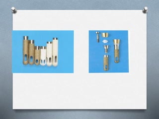
- Premodern era from 1910-1930 saw advances like Payne's silver capsule implant and Greenfield's hollow basket implant design.
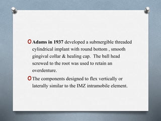
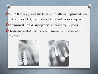
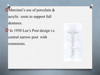
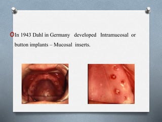
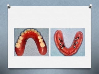
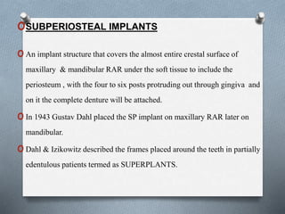
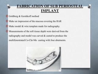
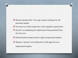
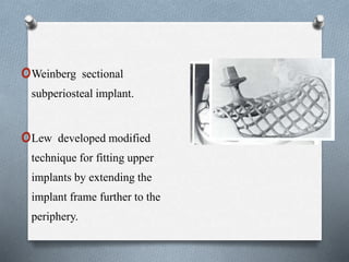
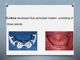
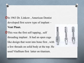
- Dawn of modern era from 1935 included developments like Strock's threaded vitallium implant and subperiosteal implant frames
History and evolution of implants /certified fixed orthodontic courses by Ind...Indian dental academy
Welcome to Indian Dental Academy
The Indian Dental Academy is the Leader in continuing dental education , training dentists in all aspects of dentistry and offering a wide range of dental certified courses in different formats.
Indian dental academy has a unique training program & curriculum that provides students with exceptional clinical skills and enabling them to return to their office with high level confidence and start treating patients
State of the art comprehensive training-Faculty of world wide repute &Very affordable
The Indian Dental Academy is the Leader in continuing dental education , training dentists in all aspects of dentistry and
offering a wide range of dental certified courses in different formats.for more details please visit
www.indiandentalacademy.com
This document provides an overview of the history and evolution of dental implants from ancient times to the modern era. It discusses early attempts at implant dentistry dating back thousands of years, including the use of animal teeth, carved ivory, and other materials as implants. The document then outlines several key periods in the more recent history and development of dental implants, including pioneers who advanced implant techniques and materials in the 18th century through the early 20th century. It focuses on the foundational work done in the late 1930s and 1940s that marked the beginning of modern implant dentistry.
Indian Dental Academy: will be one of the most relevant and exciting training
center with best faculty and flexible training programs for dental
professionals who wish to advance in their dental practice,Offers certified
courses in Dental implants,Orthodontics,Endodontics,Cosmetic Dentistry,
Prosthetic Dentistry, Periodontics and General Dentistry.
History and evolution of implants raju /orthodontic courses by Indian dental ...Indian dental academy
This document provides a history of implantology from ancient times to the present. It begins with examples of early dental implants from ancient Egypt and other civilizations dating back to 600 AD. Key developments are then discussed chronologically, including the medieval use of ivory and bone implants, early modern experimentation with materials like gold and porcelain in the 1800s-1900s, and pioneering studies on osseointegration by Branemark in the 1950s. The modern era saw advancement of endosteal and subperiosteal implant designs, culminating in the establishment of contemporary implant systems like Nobel Biocare in the late 1970s to the present.
Description :
The Indian Dental Academy is the Leader in continuing dental education , training dentists in all aspects of dentistry and
offering a wide range of dental certified courses in different formats.for more details please visit
www.indiandentalacademy.com
2 newhistory and evolution of implants1/ oral surgery courses /endodontic co...Indian dental academy
Description :
The Indian Dental Academy is the Leader in continuing dental education , training dentists in all aspects of dentistry and
offering a wide range of dental certified courses in different formats.for more details please visit
www.indiandentalacademy.com
The document provides a history of the field of prosthodontics, including historical dates and developments in complete dentures, facebows, surveyors, fixed partial dentures, crowns, bridges, implants, articulators, and dental materials. It traces developments from early carved wood and bone dentures in 700 BC to modern materials like acrylics, metals, and lasers. Key developments mentioned include the first facebow in 1889, the first surveyor in 1917, and advances in dental materials in the 20th century.
Evolution of implant designs, Dr Justin Ninan JustinNinan2
The document provides an overview of the evolution of dental implant designs from early attempts at tooth transplantation to modern root form and cylindrical implants. It discusses various classifications of implant designs including endosteal, subperiosteal, blade, and ramus implants. The document also covers considerations in implant design like threads, surfaces, and materials that have been developed to improve osseointegration and long term success of dental implants.
The Indian Dental Academy is the Leader in continuing dental education , training dentists in all aspects of dentistry and
offering a wide range of dental certified courses in different formats.for more details please visit
www.indiandentalacademy.com
Indian Dental Academy: will be one of the most relevant and exciting training
center with best faculty and flexible training programs for dental
professionals who wish to advance in their dental practice,Offers certified
courses in Dental implants,Orthodontics,Endodontics,Cosmetic Dentistry,
Prosthetic Dentistry, Periodontics and General Dentistry.
DENTAL IMPLANTS-1 HISTORY- Dr MEENU MERRY C PAULMeenuMerryCPaul
Dental implants are artificial titanium fixtures surgically placed into the jawbone to replace missing teeth and their roots. Pioneered by Per-Ingvar Branemark in the 1950s, who discovered that titanium forms a biological bond with bone called osseointegration. Since then, titanium has become the material of choice for dental implants. Implants can replace single teeth, multiple teeth, or serve as anchors for dentures, offering improved function, aesthetics, and oral health compared to other tooth replacement options.
History and evolution of dental implants / academy of fixed orthodonticsIndian dental academy
Indian Dental Academy: will be one of the most relevant and exciting training center with best faculty and flexible training programs for dental professionals who wish to advance in their dental practice,Offers certified courses in Dental implants,Orthodontics,Endodontics,Cosmetic Dentistry, Prosthetic Dentistry, Periodontics and General Dentistry.
This document provides an introduction and history of orthodontics. It discusses early concepts and treatments from ancient civilizations through the 14th-17th centuries. Key figures who advanced orthodontic understanding and techniques are highlighted from the 18th century onward, including Fauchard, Hunter, Greenwood, Kingsley, Harris, Farrar, Angell, Baker, and Angle. Angle is described as revolutionizing orthodontics in the late 19th/early 20th century by developing his classification system and innovative appliances like the edgewise bracket. The document traces the evolution of orthodontics to become a recognized specialty.
The document provides a historical overview of orthodontics from ancient times to the late 19th century. Some key points summarized:
- Ancient civilizations like the Greeks and Romans practiced early forms of orthodontics by binding teeth with wires and catgut.
- Major figures like Hippocrates, Aristotle, and Galen contributed early dental anatomy knowledge and descriptions of malocclusions.
- During the Renaissance, da Vinci, Vesalius, and Paré advanced understanding of dental and facial anatomy.
- In the 18th-19th centuries, pioneers like Fauchard, Fox, Greenwood, and the Americans Harris and Angell developed new appliances and treatment techniques, establishing the
I will discuss history of dentures.....
please do like, share and subscribe my channel Dr Aaryas Vlogs
please click the link to view in youtube:
https://youtu.be/8owHut1KJrA
The document discusses the history and evolution of dental implants from ancient times to modern day. Some key points covered include:
- The earliest evidence of dental implants dates back to 600 AD in Mayan civilization in South America using animal teeth and carved ivory.
- In the late 19th century, experimentation began with various materials like gold, lead, and porcelain being used for root forms and capsules placed into extraction sites.
- In the 1950s-60s, designs incorporated threaded fixtures and subperiosteal frames. Peri-implant bone integration was observed but not well understood.
- In 1965, Professor Branemark began pioneering research demonstrating osseointegration of
2 newhistory and evolution of implants1/ dental implant courses by Indian den...Indian dental academy
Description :
The Indian Dental Academy is the Leader in continuing dental education , training dentists in all aspects of dentistry and
offering a wide range of dental certified courses in different formats.for more details please visit
www.indiandentalacademy.com
Description :
The Indian Dental Academy is the Leader in continuing dental education , training dentists in all aspects of dentistry and
offering a wide range of dental certified courses in different formats.for more details please visit
www.indiandentalacademy.com
The document summarizes dentistry in 16th and 17th century England during the Renaissance period. It discusses dental hygiene practices at the time which involved using rosemary or sage tooth whitening scrubs. It also notes that the wealthy could afford sugary foods leading to higher rates of tooth decay among nobility compared to peasants. Lay barbers performed tooth extractions and tools included pelicans, forceps, speculums and mouth gags. Key figures who advanced dental anatomy knowledge included Leonardo Da Vinci, Fallope, Vesalius, Pare, Eustachius, Fauchard and Van Leeuwenhoek.
This document summarizes the evolution of intramedullary nails for long bone fracture fixation from the 16th century to modern times. It describes the early use of wooden sticks and ivory implants, the introduction of metallic rods during WWI, and the development of modern locked intramedullary nails in the mid-20th century. Key figures who advanced nail design include Kuntscher, who introduced reamed nailing in 1940, and Russell and Taylor, who developed the first closed section interlocking nail in the 1980s. The document outlines the progression from first to fourth generation nails, incorporating improvements in materials, locking mechanisms, and designs to optimize stability and healing.
The evolution of oral and maxillofacial surgery began in ancient Egypt and Greece with early treatments involving bandages soaked in honey and egg white for mandibular fractures and tooth extractions using loosening agents. Significant developments occurred during the Middle Ages with the separation of medicine and surgery and the Renaissance period which saw the expansion of procedures like cleft lip repairs. The 17th century saw the first textbook on oral surgery and the 19th century established oral surgery as a distinct field with leaders performing the first orthognathic surgery case and developing fixation techniques. Recent advances include minimally invasive endoscopy, improved imaging like CBCT, advancements in dental extractions using platelet-rich fibrin and atraumatic techniques, and applications of nanome
The Indian Dental Academy is the Leader in continuing dental education , training dentists in all aspects of dentistry and
offering a wide range of dental certified courses in different formats.for more details please visit
www.indiandentalacademy.com
The first comprehensive studies on root canal anatomy were published in the 1840s, with further contributions in the late 19th century using techniques like sectioning teeth and injecting materials into canals. In the early 20th century, improved methods like injecting celluloid or paraffin allowed for more detailed studies of variations. Landmark publications in the 1950s and 1960s further characterized the complex and variable nature of root canal anatomy using wax models and clearing techniques.
History of orthodontics post graduate seminarAboli Muley
This document provides a history of orthodontics from ancient civilizations to modern techniques. It discusses early practitioners from ancient Egypt, Greece, and Rome and their crude early attempts at orthodontic treatment. It then outlines major developments in the 18th-19th centuries with pioneers like Fauchard, Fox, and Kingsley experimenting with new appliances. It highlights Edward Angle as the "Father of Modern Orthodontics" for developing the classification system still used today and standardized appliances. The document traces the evolution of orthodontics through pioneers of the late 19th century to modern techniques.
The document discusses types of dental implants and their components. It defines dental implants and describes their purpose to replace missing teeth. It categorizes implants based on their placement (e.g. endosteal, transosteal), materials (e.g. metallic, non-metallic), and ability to bond with bone (e.g. bioactive, bioinert). Key implant components are also defined, including fixtures, abutments, cover screws, and impression posts. Standardized terminology is important for effective communication among dental professionals regarding different implant systems and their specific parts.
The document discusses restorative options for completely edentulous maxilla patients using dental implants, including maxillary fixed-detachable prostheses and maxillary overdenture prostheses. It provides definitions and considerations for each option, outlining advantages like predictability, fixedness and retrievability for fixed prostheses, and advantages like improved speech and esthetics for overdentures. Patient factors like resorption and needs are important to consider when determining the best treatment option.
This document provides information on epithelial tissue and cell junctions. It discusses the general features of epithelial tissue, including that epithelial cells are closely packed with many cell junctions. It also describes the different types of epithelial tissue (simple vs stratified), the cell shapes (squamous, cuboidal, columnar), and locations in the body. The document further explains the structure and functions of the basement membrane and cell junctions, including occluding junctions, anchoring junctions, and communicating junctions. Key cellular adhesion molecules and proteins involved in different junction types are also outlined.
This document provides information about lymph nodes and the lymphatic system. It discusses the anatomy, embryology, histology, and physiology of lymph nodes and lymphatic drainage. Key points include:
- Lymph nodes act as filters for the lymphatic system and help fight infection. They are located along lymphatic vessels.
- The primary lymphoid organs are the bone marrow and thymus, where lymphocytes develop. Secondary lymphoid organs include the spleen, lymph nodes, tonsils, and skin.
- Lymph nodes have an outer cortex and inner medulla. Lymph enters through afferent vessels and exits through efferent vessels. High endothelial venules are found
More Related Content
Similar to HISTORY AND EVOLUTION OF IMPLANTS1.pptx
Evolution of implant designs, Dr Justin Ninan JustinNinan2
The document provides an overview of the evolution of dental implant designs from early attempts at tooth transplantation to modern root form and cylindrical implants. It discusses various classifications of implant designs including endosteal, subperiosteal, blade, and ramus implants. The document also covers considerations in implant design like threads, surfaces, and materials that have been developed to improve osseointegration and long term success of dental implants.
The Indian Dental Academy is the Leader in continuing dental education , training dentists in all aspects of dentistry and
offering a wide range of dental certified courses in different formats.for more details please visit
www.indiandentalacademy.com
Indian Dental Academy: will be one of the most relevant and exciting training
center with best faculty and flexible training programs for dental
professionals who wish to advance in their dental practice,Offers certified
courses in Dental implants,Orthodontics,Endodontics,Cosmetic Dentistry,
Prosthetic Dentistry, Periodontics and General Dentistry.
DENTAL IMPLANTS-1 HISTORY- Dr MEENU MERRY C PAULMeenuMerryCPaul
Dental implants are artificial titanium fixtures surgically placed into the jawbone to replace missing teeth and their roots. Pioneered by Per-Ingvar Branemark in the 1950s, who discovered that titanium forms a biological bond with bone called osseointegration. Since then, titanium has become the material of choice for dental implants. Implants can replace single teeth, multiple teeth, or serve as anchors for dentures, offering improved function, aesthetics, and oral health compared to other tooth replacement options.
History and evolution of dental implants / academy of fixed orthodonticsIndian dental academy
Indian Dental Academy: will be one of the most relevant and exciting training center with best faculty and flexible training programs for dental professionals who wish to advance in their dental practice,Offers certified courses in Dental implants,Orthodontics,Endodontics,Cosmetic Dentistry, Prosthetic Dentistry, Periodontics and General Dentistry.
This document provides an introduction and history of orthodontics. It discusses early concepts and treatments from ancient civilizations through the 14th-17th centuries. Key figures who advanced orthodontic understanding and techniques are highlighted from the 18th century onward, including Fauchard, Hunter, Greenwood, Kingsley, Harris, Farrar, Angell, Baker, and Angle. Angle is described as revolutionizing orthodontics in the late 19th/early 20th century by developing his classification system and innovative appliances like the edgewise bracket. The document traces the evolution of orthodontics to become a recognized specialty.
The document provides a historical overview of orthodontics from ancient times to the late 19th century. Some key points summarized:
- Ancient civilizations like the Greeks and Romans practiced early forms of orthodontics by binding teeth with wires and catgut.
- Major figures like Hippocrates, Aristotle, and Galen contributed early dental anatomy knowledge and descriptions of malocclusions.
- During the Renaissance, da Vinci, Vesalius, and Paré advanced understanding of dental and facial anatomy.
- In the 18th-19th centuries, pioneers like Fauchard, Fox, Greenwood, and the Americans Harris and Angell developed new appliances and treatment techniques, establishing the
I will discuss history of dentures.....
please do like, share and subscribe my channel Dr Aaryas Vlogs
please click the link to view in youtube:
https://youtu.be/8owHut1KJrA
The document discusses the history and evolution of dental implants from ancient times to modern day. Some key points covered include:
- The earliest evidence of dental implants dates back to 600 AD in Mayan civilization in South America using animal teeth and carved ivory.
- In the late 19th century, experimentation began with various materials like gold, lead, and porcelain being used for root forms and capsules placed into extraction sites.
- In the 1950s-60s, designs incorporated threaded fixtures and subperiosteal frames. Peri-implant bone integration was observed but not well understood.
- In 1965, Professor Branemark began pioneering research demonstrating osseointegration of
2 newhistory and evolution of implants1/ dental implant courses by Indian den...Indian dental academy
Description :
The Indian Dental Academy is the Leader in continuing dental education , training dentists in all aspects of dentistry and
offering a wide range of dental certified courses in different formats.for more details please visit
www.indiandentalacademy.com
Description :
The Indian Dental Academy is the Leader in continuing dental education , training dentists in all aspects of dentistry and
offering a wide range of dental certified courses in different formats.for more details please visit
www.indiandentalacademy.com
The document summarizes dentistry in 16th and 17th century England during the Renaissance period. It discusses dental hygiene practices at the time which involved using rosemary or sage tooth whitening scrubs. It also notes that the wealthy could afford sugary foods leading to higher rates of tooth decay among nobility compared to peasants. Lay barbers performed tooth extractions and tools included pelicans, forceps, speculums and mouth gags. Key figures who advanced dental anatomy knowledge included Leonardo Da Vinci, Fallope, Vesalius, Pare, Eustachius, Fauchard and Van Leeuwenhoek.
This document summarizes the evolution of intramedullary nails for long bone fracture fixation from the 16th century to modern times. It describes the early use of wooden sticks and ivory implants, the introduction of metallic rods during WWI, and the development of modern locked intramedullary nails in the mid-20th century. Key figures who advanced nail design include Kuntscher, who introduced reamed nailing in 1940, and Russell and Taylor, who developed the first closed section interlocking nail in the 1980s. The document outlines the progression from first to fourth generation nails, incorporating improvements in materials, locking mechanisms, and designs to optimize stability and healing.
The evolution of oral and maxillofacial surgery began in ancient Egypt and Greece with early treatments involving bandages soaked in honey and egg white for mandibular fractures and tooth extractions using loosening agents. Significant developments occurred during the Middle Ages with the separation of medicine and surgery and the Renaissance period which saw the expansion of procedures like cleft lip repairs. The 17th century saw the first textbook on oral surgery and the 19th century established oral surgery as a distinct field with leaders performing the first orthognathic surgery case and developing fixation techniques. Recent advances include minimally invasive endoscopy, improved imaging like CBCT, advancements in dental extractions using platelet-rich fibrin and atraumatic techniques, and applications of nanome
The Indian Dental Academy is the Leader in continuing dental education , training dentists in all aspects of dentistry and
offering a wide range of dental certified courses in different formats.for more details please visit
www.indiandentalacademy.com
The first comprehensive studies on root canal anatomy were published in the 1840s, with further contributions in the late 19th century using techniques like sectioning teeth and injecting materials into canals. In the early 20th century, improved methods like injecting celluloid or paraffin allowed for more detailed studies of variations. Landmark publications in the 1950s and 1960s further characterized the complex and variable nature of root canal anatomy using wax models and clearing techniques.
History of orthodontics post graduate seminarAboli Muley
This document provides a history of orthodontics from ancient civilizations to modern techniques. It discusses early practitioners from ancient Egypt, Greece, and Rome and their crude early attempts at orthodontic treatment. It then outlines major developments in the 18th-19th centuries with pioneers like Fauchard, Fox, and Kingsley experimenting with new appliances. It highlights Edward Angle as the "Father of Modern Orthodontics" for developing the classification system still used today and standardized appliances. The document traces the evolution of orthodontics through pioneers of the late 19th century to modern techniques.
The document discusses types of dental implants and their components. It defines dental implants and describes their purpose to replace missing teeth. It categorizes implants based on their placement (e.g. endosteal, transosteal), materials (e.g. metallic, non-metallic), and ability to bond with bone (e.g. bioactive, bioinert). Key implant components are also defined, including fixtures, abutments, cover screws, and impression posts. Standardized terminology is important for effective communication among dental professionals regarding different implant systems and their specific parts.
The document discusses restorative options for completely edentulous maxilla patients using dental implants, including maxillary fixed-detachable prostheses and maxillary overdenture prostheses. It provides definitions and considerations for each option, outlining advantages like predictability, fixedness and retrievability for fixed prostheses, and advantages like improved speech and esthetics for overdentures. Patient factors like resorption and needs are important to consider when determining the best treatment option.
Similar to HISTORY AND EVOLUTION OF IMPLANTS1.pptx (20)
This document provides information on epithelial tissue and cell junctions. It discusses the general features of epithelial tissue, including that epithelial cells are closely packed with many cell junctions. It also describes the different types of epithelial tissue (simple vs stratified), the cell shapes (squamous, cuboidal, columnar), and locations in the body. The document further explains the structure and functions of the basement membrane and cell junctions, including occluding junctions, anchoring junctions, and communicating junctions. Key cellular adhesion molecules and proteins involved in different junction types are also outlined.
This document provides information about lymph nodes and the lymphatic system. It discusses the anatomy, embryology, histology, and physiology of lymph nodes and lymphatic drainage. Key points include:
- Lymph nodes act as filters for the lymphatic system and help fight infection. They are located along lymphatic vessels.
- The primary lymphoid organs are the bone marrow and thymus, where lymphocytes develop. Secondary lymphoid organs include the spleen, lymph nodes, tonsils, and skin.
- Lymph nodes have an outer cortex and inner medulla. Lymph enters through afferent vessels and exits through efferent vessels. High endothelial venules are found
Dr. Sonam Rani presented on the topic of cementum to several professors and colleagues. Cementum is a calcified tissue that covers tooth roots and provides attachment for periodontal ligaments. It comes in several types classified based on cellularity, presence of fibers, and origin. Cementum is formed by cementoblasts and cementocytes and plays an important role in tooth adaptation and repair. Systemic conditions like Paget's disease and cleidocranial dysplasia can affect cementum formation and structure.
This document provides an overview of pain, including its definition, classification, theories, transmission and modulation pathways, assessment, and management approaches. It begins with definitions of pain from Dorland's Medical Dictionary and Monheim. It then classifies pain according to intensity, temporal relationship, qualities, onset, and localization. Theories of pain discussed include specificity, pattern, and gate control theories. It describes the dual nature of pain and the transduction, transmission, modulation, and perception of pain. It discusses referred pain and neuropathic pain. The document concludes by covering pain assessment tools and pharmacological and non-pharmacological management strategies.
COMMON SEMINAR STERILISATION, INFECTION CONTROL AND HOSPITAL MANAGEMENT.pptxmalti19
This document discusses sterilization, infection control, and hospital management in dentistry. It defines key terms like sterilization, disinfection, and asepsis. It then describes various methods of sterilization including physical methods like heat and radiation, and chemical methods like alcohols, phenols, aldehydes, halogens, and gases. The document provides details on specific sterilization techniques and protocols for sterilizing dental instruments used in different specialties. It emphasizes the importance of proper sterilization to prevent disease transmission between patients.
This document summarizes key concepts in immunology as they relate to periodontal disease. It discusses the epithelial barrier and pattern recognition receptors that detect pathogens. Inflammatory mediators recruit immune cells through chemotaxis. T lymphocytes develop and differentiate into subsets like Th1, Th2, Th17 that activate different immune responses. B cells produce antibodies through somatic hypermutation. Regulatory T cells control self-tolerance. The adaptive response becomes antigen-specific and develops memory. Dendritic cells present antigens to activate T cells. An imbalance in T cell subsets can lead to tissue destruction in periodontal disease.
Thrombosis, embolism, and infarction are related pathological processes involving blood clots. Thrombosis is the formation of a blood clot within a blood vessel, while embolism occurs when a piece of a clot breaks off and travels to another location. Infarction results from obstruction of blood flow by a clot, causing tissue death. The document discusses the mechanisms, types, features, and progression of thrombosis, embolism, and infarction. It also covers related topics like Virchow's triad, hypercoagulable states, fat embolism, and amniotic fluid embolism.
Thrombosis, embolism, and infarction are related pathological processes involving blood clots. Thrombosis is the formation of a blood clot within a blood vessel, while embolism occurs when a piece of a clot breaks off and travels to another location. Infarction results from obstruction of blood flow by a clot, causing tissue death. The document discusses the mechanisms, classifications, and morphological features of thrombosis, embolism, and infarction. It also covers related topics like Virchow's triad, hypercoagulable states, and the development and types of infarcts over time.
Immune responses in periodontal disease final.pptxmalti19
This document discusses the immune responses involved in periodontal disease. It begins by defining periodontitis as an infectious disease caused by anaerobic bacteria. Both bacteria and a susceptible host are required to cause disease. It then describes the pathogenesis which involves environmental and genetic risk factors interacting with the microbial challenge to activate the host immune response, resulting in inflammation and bone/tissue destruction. The document discusses the types of immunity, including innate and adaptive immunity. It covers topics such as dendritic cells, T-cell and B-cell roles, the roles of cytokines and RANKL in linking the immune response to bone loss, and hypotheses about the roles of the Th1 and Th2 responses in periodontitis.
This document provides an overview of antibiotics used in periodontics. It begins with an introduction to antibiotics and their historical background. It then covers classification of antimicrobial agents based on chemical structure, mechanism of action, organisms targeted, and spectrum of activity. Guidelines for antibiotic use in periodontal diseases are presented, along with the diseases where antibiotics can be used. Commonly used antibiotics like tetracycline, doxycycline, metronidazole, penicillin, and amoxicillin-clavulanate are described in detail. The document concludes with a reference to research on systemic antibiotic use in periodontics.
This document discusses evidence-based periodontology and decision making. It defines evidence-based periodontology as applying evidence-based healthcare to periodontology by integrating the best available evidence with clinical practice. The document outlines the steps of evidence-based periodontology, including asking questions using the PICO format, searching for and appraising evidence from systematic reviews and clinical practice guidelines, and applying the evidence to clinical decisions. It also discusses tools for critically assessing evidence, such as considering different levels of evidence and ensuring temporality between causes and effects.
Calcium and Phosphorous metabolism 23-03-23.pptxmalti19
Calcium and phosphorus metabolism is tightly regulated by vitamin D, parathyroid hormone, and calcitonin. Calcium is crucial for bone development, nerve function, and other processes. The recommended daily intake is 800 mg for adults. Dietary sources include dairy products, leafy greens, and fish. Absorption occurs in the small intestine and is influenced by vitamin D, PTH, and other factors. Hormonal signals work to maintain calcium levels within a narrow range. Disorders like rickets and osteomalacia can result from vitamin D deficiency. Precise regulation is needed to prevent hypercalcemia or hypocalcemia.
The document summarizes the anatomy and function of the muscles of mastication. It describes the three main muscles - masseter, temporalis, and medial and lateral pterygoid. It discusses their embryological development, nerve supply, actions, and clinical evaluation. The masseter muscle is palpated to assess for hypertrophy. Orthodontic treatment can impact the thickness of the muscles. The medial pterygoid muscle is also described in more detail, including its nerve supply by the mandibular nerve, blood supply, actions, and clinical examination.
The facial nerve emerges from the brainstem between the pons and medulla. It has motor, sensory, and parasympathetic secretomotor components. During embryonic development, it arises from the second branchial arch. The nerve passes through the internal acoustic meatus and facial canal within the temporal bone. It gives off several branches within the facial canal before exiting at the stylomastoid foramen. Its main branches in the face include the temporal, zygomatic, buccal, marginal mandibular, and cervical branches which innervate the muscles of facial expression.
1) PRP and PRF are platelet concentrates that provide growth factors that may enhance tissue regeneration.
2) Studies have shown PRP and PRF can increase the release of growth factors like PDGF, TGF-β1, and VEGF compared to control groups.
3) However, the evidence on whether PRP and PRF improve bone regeneration outcomes is mixed. Some studies found benefits for parameters like new bone formation and defect fill, while other studies found no additional benefits compared to control treatments.
This document discusses chlorhexidine, a commonly used chemical plaque control agent. It provides details on the history, chemistry, forms, mechanisms of action, products, clinical uses, toxicity and side effects of chlorhexidine. Chlorhexidine is a bisbiguanide antiseptic that is highly effective at inhibiting plaque formation and controlling gingivitis when used as a mouthrinse. It has a wide range of clinical applications and is generally well-tolerated, though long-term use can cause staining and changes to taste sensation in some individuals.
Oral hygiene and overall health
Department of Periodontology and oral Implantology
The document discusses the importance of oral hygiene for preventing dental diseases and maintaining overall health. It notes that over 3.5 billion people suffer from oral diseases according to the WHO, but many can be prevented through proper oral hygiene. Maintaining oral hygiene includes brushing teeth twice daily, flossing, cleaning the tongue, and visiting the dentist regularly. Poor oral hygiene can lead to dental caries, gum disease, and systemic issues like diabetes and heart disease.
Reducing fracture risk with Calcium and Vitamin D
Osteoporotic fractures are a major health problem that most commonly affect the spine and hip. Low calcium intake and vitamin D deficiency increase fracture risk by reducing bone mineral density and strength. Clinical trials show that calcium and vitamin D supplementation can reduce fracture risk, especially when vitamin D doses reach 800 IU daily and calcium intake reaches 1000-1200 mg. For older individuals, supplementation is most effective for those at high risk of deficiency living in nursing homes or with low dairy intake.
Antibiotics in the management of chronic periodontitis.pptmalti19
This document summarizes evidence on the use of adjunctive antibiotics for chronic periodontitis. A systematic review of 25 studies found some additional benefits of antibiotics in deep pockets, including 0.2-0.6 mm more attachment gain and 0.2-0.8 mm more probing depth reduction. However, the clinical relevance is uncertain given limitations in defining chronic periodontitis and its microbiota. Overall, current studies have not conclusively established benefits of adjunctive antibiotics, so they cannot be routinely indicated as adjuncts for chronic periodontitis.
The document discusses immediate dentures, which are complete or partial dentures fabricated immediately after tooth extraction. It describes the differences between conventional (classic) immediate dentures and interim (transitional) immediate dentures. Conventional immediate dentures are intended as the long-term prosthesis and are made when only anterior teeth remain. Interim immediate dentures are short-term and are made when posterior teeth remain, requiring only one surgical visit. The document outlines the procedures, indications, contraindications, advantages and disadvantages of both types of immediate dentures.
This document provides an overview of wound healing, its functions, stages, mechanisms, factors affecting it, and complications.
A wound is a break in the integrity of the skin or tissues, which may be associated with disruption of the structure and function.
Healing is the body’s response to injury in an attempt to restore normal structure and functions.
Healing can occur in two ways: Regeneration and Repair
There are 4 phases of wound healing: hemostasis, inflammation, proliferation, and remodeling. This document also describes the mechanism of wound healing. Factors that affect healing include infection, uncontrolled diabetes, poor nutrition, age, anemia, the presence of foreign bodies, etc.
Complications of wound healing like infection, hyperpigmentation of scar, contractures, and keloid formation.
A Visual Guide to 1 Samuel | A Tale of Two HeartsSteve Thomason
These slides walk through the story of 1 Samuel. Samuel is the last judge of Israel. The people reject God and want a king. Saul is anointed as the first king, but he is not a good king. David, the shepherd boy is anointed and Saul is envious of him. David shows honor while Saul continues to self destruct.
Temple of Asclepius in Thrace. Excavation resultsKrassimira Luka
The temple and the sanctuary around were dedicated to Asklepios Zmidrenus. This name has been known since 1875 when an inscription dedicated to him was discovered in Rome. The inscription is dated in 227 AD and was left by soldiers originating from the city of Philippopolis (modern Plovdiv).
Elevate Your Nonprofit's Online Presence_ A Guide to Effective SEO Strategies...TechSoup
Whether you're new to SEO or looking to refine your existing strategies, this webinar will provide you with actionable insights and practical tips to elevate your nonprofit's online presence.
Philippine Edukasyong Pantahanan at Pangkabuhayan (EPP) CurriculumMJDuyan
(𝐓𝐋𝐄 𝟏𝟎𝟎) (𝐋𝐞𝐬𝐬𝐨𝐧 𝟏)-𝐏𝐫𝐞𝐥𝐢𝐦𝐬
𝐃𝐢𝐬𝐜𝐮𝐬𝐬 𝐭𝐡𝐞 𝐄𝐏𝐏 𝐂𝐮𝐫𝐫𝐢𝐜𝐮𝐥𝐮𝐦 𝐢𝐧 𝐭𝐡𝐞 𝐏𝐡𝐢𝐥𝐢𝐩𝐩𝐢𝐧𝐞𝐬:
- Understand the goals and objectives of the Edukasyong Pantahanan at Pangkabuhayan (EPP) curriculum, recognizing its importance in fostering practical life skills and values among students. Students will also be able to identify the key components and subjects covered, such as agriculture, home economics, industrial arts, and information and communication technology.
𝐄𝐱𝐩𝐥𝐚𝐢𝐧 𝐭𝐡𝐞 𝐍𝐚𝐭𝐮𝐫𝐞 𝐚𝐧𝐝 𝐒𝐜𝐨𝐩𝐞 𝐨𝐟 𝐚𝐧 𝐄𝐧𝐭𝐫𝐞𝐩𝐫𝐞𝐧𝐞𝐮𝐫:
-Define entrepreneurship, distinguishing it from general business activities by emphasizing its focus on innovation, risk-taking, and value creation. Students will describe the characteristics and traits of successful entrepreneurs, including their roles and responsibilities, and discuss the broader economic and social impacts of entrepreneurial activities on both local and global scales.
This presentation was provided by Racquel Jemison, Ph.D., Christina MacLaughlin, Ph.D., and Paulomi Majumder. Ph.D., all of the American Chemical Society, for the second session of NISO's 2024 Training Series "DEIA in the Scholarly Landscape." Session Two: 'Expanding Pathways to Publishing Careers,' was held June 13, 2024.
This presentation was provided by Rebecca Benner, Ph.D., of the American Society of Anesthesiologists, for the second session of NISO's 2024 Training Series "DEIA in the Scholarly Landscape." Session Two: 'Expanding Pathways to Publishing Careers,' was held June 13, 2024.
How Barcodes Can Be Leveraged Within Odoo 17Celine George
In this presentation, we will explore how barcodes can be leveraged within Odoo 17 to streamline our manufacturing processes. We will cover the configuration steps, how to utilize barcodes in different manufacturing scenarios, and the overall benefits of implementing this technology.
2. CONTENTS
oIntroduction
oDefinitions
oHistory and Evolution of implants can be
studied as -The ancient era
- The
Medieval period
- The foundational period
- The Premodern era
- The dawn of the
modern era -
Contemporary oral implantology
oSummary and Conclusion
oReferences.
4. oDef of Implant (GPT 8 )
oAny object or material such as an alloplastic substance or other
tissue, which is partially or completely inserted or grafted into
the body for therapeutic, diagnostic, prosthetic or experimental
purposes.
oDef of Implantology
oTerm historically conceived as the study or science of planning
and restoring dental implants.
5. oDef of Dental Implant
oA prosthetic device made of alloplastic material implanted into
the oral tissues beneath the mucosal or/ & periosteal layer &/or
within the bone to provide retention & support for a fixed or
removable dental prosthesis ;a substance that is placed into or /
& upon the jaw bone to support a fixed or removable dental
prosthesis.
oDef. Of Osseointegration
oThe apparent direct attachment or connection of osseous
tissue to an inert, alloplastic material without intervening
connective tissue.
7. oHistory of dentistry….3000 B.C .
oHISTORY TRACED IN MIDDLE EAST
oIn 1862 Gaillardot discovered a prosthodontic appliance near the ancient
city of Sidon. Appliance was dating 400 B.C period .
o Carved ivory tooth replacing the two missing incisors.
8. oHISTORY TRACED IN THE EGYPTIAN
DYNASTIES
oEvidences have shown the implantation of animal
teeth & artificial teeth carved of ivory .
oImplantation prior to mummification.
9. oIn 1981 evidences of oldest dental implant was found in
the Kalavak Necropolis, near Izmir , Turkey.
oThe discoveries were dated back to 550 B.C . A canine
tooth like object made of two piece of calcite having
hardness similar to natural teeth showing wear on the
chewing surface & secured with gold wires wrapped
around the neck of adjacent teeth.
10. oCranin suggested that earliest recorded dental implant specimen was
inserted during the PRE COLUMBIAN ERA.
oIn 1931 Wilson Popenoe discovered a skull in the Ulua river valley of
Honduras , dating to the period 600 A.D .
o Skull had an artificial tooth replacing lower left lateral incisor, carved of a
dark stone . Radiographs showed the evidence of compact bone around the
implant.
o The Mayans practiced the implantation of alloplastic materials in living persons.
13. oAllotransplantation (18 -19 Century) mainly in England &
Colonial America.
oAlbucasis de Condue ( 936- 1013 A.D) an Arab surgeon
described the transplantation procedures . He attempted to use
ox bone to replace missing teeth.
14. oIn Japan in the 15th & 16th C. Wooden
dowel & crown prosthesis was designed .
The pin inserted into the root canal of non
vial teeth.
oThis was an early ENDODONTIC IMPLANT
– SUPPORTED PROSTHESIS.
15. • Charles Allen in 1687 gave the first written work on
dentistry.
• He described the replanting of tooth into the same patient.
• Allen emphasized on transplantation of tooth using animal
tooth.
• In European sphere transplantation became the common
practice performed by barber surgeons of the Era.
• The tooth transplant could lead to transfer of diseases .
• Implants made from ivory, shells & bone were used.
16. • Ambroise Pare, a French physician , a surgeon to four kings of
France.
• He replaced the missing teeth with implants made up of bone &
ivory .
• He successfully replaced missing tooth of Princess by
transplanting.
17. oJohn Hunter in 1728 – 1793
A.D suggested the
possibility of transplanting.
19. oThe endosseous oral implantology truly began in the 19th Century.
oMaggilio in 1809 , a dentist at the university of Nancy , France, author of
the book called “THE ART OF THE DENTIST”. The first reference to
modern style implants.
oHe has described the implant & placement.
oHe made the tooth root shaped implant with 18 carat gold with three prongs
at the end to hold it in place in the bone . The implant was placed in the
freshly extracted socket site retained with the prongs. After the tissues
healed the crown was attached with the help of post placed into the hole of
root section of the implant.
oHe placed the single stage gold implant.
20. oIn mid 1800s advancement in the antiseptics & surgical
techniques , there were documentation of implanted materials.
oW. J . Younger of San Francisco introduced operations in
United States. In 1893 he wrote that his operation has
successfully entered 8th year of life.
21. oIn 1886 Harris treated a Chinese patient in Grass valley ,
California . He placed the tooth root shaped platinum post with
lead coating, lasted for 27 yrs Reported in Dental Cosmos in
1887.
oIn 1889 Edmunds of New York reported on March 12 ,1889
to the First District dental society of that city. He implanted the
metallic capsule.
22. oIn 1888 Berry reported about the root form implants made up
of lead.
oIn 1890 , a Massachusetts minister had his lower jaw
resected & was restored with an extensive system of gold
crowns soldered & joined to hinged device attached to the
remaining dentition .
oZnamenski in 1891 reported on implantation made of
Porcelain, gutta-percha & rubber.
23. oBonwill in 1895 reported on the implantation of one or two
tubes of gold or Iridium as a support for individual teeth or
crown.
o IN 1898 R. E Payne at the National Dental Association
meeting gave the first clinical demonstration by placing the
silver capsule in the extracted tooth socket.
25. oThe first two decades of 20th C. predominated by the clinicians
namely R.E Payne& E. J .Greenfield.
oR. E .Payne presented his technique of capsule implantation at
the clinics of Third international Dental Congress, reported in
the Dental Cosmos in 1901.
oTechnique- Extracting the root , enlarging the socket with
trephine, trial fitting of the capsule. He then placed grooves on
both sides of the socket & filled 2/3rds with rubber, fitted the
porcelain root into the capsule & set it with gutta-percha.
oIn 1903 Sholl in Pennsylvania , implanted porcelain tooth with
corrugated porcelain root.
26. oIn 1913 Dr. Edward J. Greenfield came up with the
surgical method to prepare osteotomy in the healed bone
using trephine.
oHe fabricated the hollow cylindrical basket root of 20
gauge iridioplatinum soldered with 24 carat gold.
Precursor of hollow basket design
27. oGREENFIELD EMPHASIZED ON
oThe importance of intimate contact between bone & implant.
Hollow implants facilitated growth of bone into implant body &
secure it.
o3 months period of unloading.
oImplants failures because of infection.
oHis techniques were similar to present concepts of osteotomy
preparation, restoring after healing time
28. oIn 1920s Leger- Dorez developed expansible root implant, was comparable
to a concrete expansion bolt.
oSmolon described the implant as a four part device with the shaft buried in
bone with the internal threads to receive a screw , fastening the neck into
the shaft. the post for attaching the prosthesis.
o The historical basis for the internal screws provided for the retention of
prosthetic devices similar to today’s implants.
oTomkin’s 1925 implanted porcelain teeth.
oBrill in 1936 inserted rubber pins in artificially prepared sockets.
30. o The modern era of implant dentistry most definitely began in
the late1930s with the work of Venable, Strock , Dahl ,
Gershkoff & Goldberg.
o Venable in 1937 developed the cast Co- Chr- Mo alloy known
as Vitallium.
o In 1939 Alvin & Moses Strock used the Venable screw type
implant.
31. oAdams in 1937 developed a submergible threaded
cylindrical implant with round bottom , smooth
gingival collar & healing cap. The ball head
screwed to the root was used to retain an
overdenture.
oThe components designed to flex vertically or
laterally similar to the IMZ intramobile element.
32. oIn 1938 Stock placed the threaded vitallium implant into the
extraction socket, the first long term endosseous implant.
oIt remained firm & asymptomatic for nearly 17 years.
oHe demonstrated that the Vitallium implants were well
tolerated.
33. oIn 1947, Formiggini developed a single
helix wire spiral implants made of stainless
steel or tantalum. Two ends of the wire were
soldered together to form a post or neck.
oChercheve Modified by increasing the
length of the neck & double helix out of
vitallium.
oHe developed the co-coordinated system of
instrumentation for implant insertion.
34.
35. oMarziani’s use of porcelain &
acrylic roots to support full
dentures.
oIn 1950 Lee’s Post design i.e
central narrow post with
extensions.
36. oIn 1943 Dahl in Germany developed Intramucosal or
button implants – Mucosal inserts.
37.
38. oSUBPERIOSTEAL IMPLANTS
oAn implant structure that covers the almost entire crestal surface of
maxillary & mandibular RAR under the soft tissue to include the
periosteum , with the four to six posts protruding out through gingiva and
on it the complete denture will be attached.
oIn 1943 Gustav Dahl placed the SP implant on maxillary RAR later on
mandibular.
oDahl & Izikowitz described the frames placed around the teeth in partially
edentulous patients termed as SUPERPLANTS.
39. FABRICATION OF SUB PERIOSTEAL
IMPLANT
o Goldberg & Gershkoff method
o Make an impression of the mucosa covering the RAR
o Make model & wire template made for radiography.
o Measurements of the soft tissue depth were derived from the
radiography and model was carved & casted to produce the
multifenestrated Co-Chr-Mo casting with four abutments.
40. o Berman introduced the Two stage surgical technique for sub
periosteal implant.
o First step was a direct impression of the surgically exposed bone
o Second was implanting the subperiosteal frame generated from
the stone cast.
o Kleinschmidt developed anterior single sub periosteal implant.
o Marziani reported on the fabrication of full upper & lower
subperiosteal implant.
43. oCT scan imaging technique used to obtain
three dimensional replica of the mandible
& CAD-CAM models used for fabricating
the frame work.
oLong term success only 50 to 60%.
44. ONE STAGE ENDOSTEAL PINS, SCREWS, &
CYLINDERS
oThe early 1960s marked the beginning of an active developmental
era in implant design.
oScialom developed a tantalum tripodal pin .
oThree intersecting pins were joined by acrylic to support the
crown. Difficulty in maintaining the trifurcation limited the
comfortable survival.
45. oIn 1960 Onlay developed
Virilium posts placed into
endodonticaly treated teeth
extending beyond the apex.
46. SCREW TYPE IMPLANT
o Tramonte introduced a stress resistant drive screw implant.
o Meglan & Lehman reported on the expandable implants.
o Lew introduced a self tapping Vitallium screw implant with
conventional threads & square post.
o Muratori & Pasquallini introduced hollow cores along with the
screw threads.
o The majority of these screw shaped implants were one piece &
were not submerged , did not osseointegrate .It was emphasized
that the fibrous peri implant membrane with its shock absorbing
feature preferred than bone fusing to implant.
47. oIn 1963 Dr. Linkow , American Dentist
developed first screw type of implant –
Vent Plant.
oThis was the first self tapping , self
threading implant . It had an open cage
like design that went into bone first , with
a few threads on solid body at the top. He
used Vitallium first latter on titanium.
48. BLADE IMPLANTS
oLinkow blade implants invented in 1967.
oLong thin blade that will be surgically inserted into the
groove in the bone .
oAbutment projecting out from the blade to this crown
or attachment for denture can be placed.
oIt required the shared support of natural teeth also.
oRestored within month so became most widely used in
united states.
oLinkow modified the design configuration for broad
applicability in maxilla & mandible, narrow ridges.
49.
50.
51.
52. oIn 1970 Roberts & Roberts developed Ramus blade
implants .
oIt was to be positioned by anchoring distally
between the cortical plates in the ramus of the
mandible
53.
54. oRAMUS FRAME IMPLANT developed
Roberts & Roberts in 1970 .
oThe endosseous implant received stabilization
from its anchorage in ramus area bilaterally &
in the symphyseal region.
55.
56.
57. TRANSOSTEAL IMPLANTS
oIn 1975 Small introduced the transosteal mandibullar
staple & bone plate, a reconstructive device placed
through a submental incision & attached to the
mandible with multiple fixation & two transosteal
screws to support a full arch prosthesis.
oHe presented 16 yrs evaluation,
oCranin developed single transosteal implant.
58.
59. o In 1970 Kawahara was developing a ceramic
implant in Japan .
ofirst Aluminium oxide implant placed.
oIn 1970 Grenoble placed Vitreous Carbon
implant .
61. oIn 1978 conference held at Harward ,
co- sponsored by NIH.
oThis was the milestone for further
research.
62. oIn 1976 Dr. Andre Schroeder in Switzerland
reported the bone growth into titanium
plasma sprayed hollow endosseous implants.
oAt the same time , Prof. Willischulte in
Germany reported the success with immediate
placement of Vitreous Carbon implant after
dental extraction & invented Frialit-2 implant.
63. oPer-Ingvar Branemark developed the two stage threaded titanium
endosseous root form implant in North America.
o“ Vital microscopy”…
o In 1960 at Gothenburg…
o Branemark noticed that there was no inflammatory reaction to the
titanium.
64. oHe started thinking about the potential of titanium to
act as an anchorage point, so the spur for the next
experiment whether titanium can be used as a bridge
in bone healing application.
oConcept of Osseo integration developed
oThe first clinical trail was done in 1965.
oIn 1971 surgical equipments.
oIn May 1982 Dr. George Zarb organized the
Toronto conference on osseointegration. Branemark
presented Two stage threaded root form implant
along with the 15 yrs research work & clinical trail.
65.
66. BRANEMARK SYSTEM COMPONENTS
oFIXTURE –
opure titanium with machined
threads .
oThe top of the fixture has
hexagonal design & threads ..
o The apical portion tapered with
four vertical notches.
o COVER SCREW- seals the
coronal potion of fixture during
the interim period.
67. oABUTMENT- -made of titanium in a cylinder
shape. the apical portion has hexagonal shape
to fit the coronal portion of fixture.
68. oABUTMENT SCREW – insert through the abutment
& threads into the fixture to connect the two
components .
oGOLD CYLINDER- made of lAu , Pl, Pd. It is
machined to fit the coronal portion of the abutment. It
becomes integral part of final prosthesis.
oGOLD SCREW –inserted through the gold cylinder
& threads into the abutment screw to connect the gold
cylinder & abutment.
69.
70. IMZ IMPLANT SYSTEM
oKirsch developed the IMZ implant system in 1974.
Since 1978 in clinical use.
oIntramobile cylinder endosseous two stage
osseointegrated implant.
oThe polyoxymethylene & polyacetal used as IME.
oAvailable in 3.5 to 4mm diameter and 8,10,13,15mm
length.
oSurface coating may be titanium plasma spray or
plasma sprayed HA coated surface.
71. IMZ IMPLANT SYSTEM
o Kirsch developed the IMZ implant system in 1974. Since
1978 in clinical use.
oIntramobile cylinder endosseous two stage osseointegrated
implant.
oThe polyoxymethylene & polyacetal used as IME.
oAvailable in 3.5 to 4mm diameter and 8,10,13,15mm length.
oSurface coating may be titanium plasma spray or plasma
sprayed HA coated surface.
72.
73. LEDERMAN SCREW IMPLANT
oIn 1977 Dr. Philippe Lederman in collaboration with strauman
co. developed the Titanium plasma spraed screw type implant.
oIn 1989 Lederman developed the New Ledreman screw
implant Surface roughened by sand blasting & acid etching.
74. ITI BONE FIT IMPLANT SYSTEM
oDeveloped by ‘International Team for Implantology’.
oThree different types
oSingle stage & two stage.
oTransgingivally placed in healing phase so second surgical
procedure for uncovering the implant is avoided.
75. THE HAND-TITANIUM IMPLANT SYSTEM
oClinical use since 1985 at Switzerland & now in use world
wide ( Lederman 1986).
oA conical, step- screw, pure Ti implant with self thread.
oLength- 10 to 20mm.
oDiameter – 3.5 to 7mm.
76. oIn early 1980s Tantum introduced Omni R implant -
A Ti root form implant with horizontal fins.
oOmni S implant – for placing into bone grafted
maxillary sinus.
77. oIn 1983 EL Blasty & Kamel introduced the new
endosseous implant material i.e Poly acrylic acid
reinforced with ceramic alumina particles 0.3 microns.
oThe hydrophilic matrix swells in contact with aqueous
solution.
oThe gradual pressure on the surrounding bone
stimulates osseous activity.
oImplanted in canine, premolar sites with promising
results.
78. MINI DENTAL IMPLANT
o In 1985 Victor Sendax developed MDI.
oUltra – small diameter 1.8 mm, biocompatible Ti alloy implant
screws.
oBulard added single one piece `O- ball’ design .
79.
80. CORE VENT
oDeveloped by Dr. Gerald Niznick in 1986.
oHollow basket design made of Ti alloy.
oDifferent fixture designs Screw –Vent, Micro-Vent, Bio –Vent.
81.
82. ENDOPORE
oA root form dental implant
developed by Doughlas et.al in
1996 made of Ti alloy & sintered
with same alloy producing porous
surface.
oBiological & clinical advantages.
83. STERI OSS SYSTEM
oIntroduced by Denar
oMade up of 99.9% Ti, tapered apex thread
design & the coronal 3rd highly polished
surface.
oAvailable in 3.5 to 4mm diameter, length 12
mm,16mm,20mm,&
miniseries8mm,10mm,12mm length.
84. NOVUM CONCEPT
o Branemark developed the concept of providing a
new set of teeth for the mandible in a single day.
oClinically implicated in 1996.
oThree titanium fixtures inserted , mucosa is closed
& base plate is placed over the fixtures & then the
prosthesis is placed.
87. o1984 – CT scan.
oBone augmentation techniques.
o1986 Tatum reported about Sinus lift / Bone graft
antroplasty.
o1988 Nerve transposition
o1989 Pterygoid implants .
o1991 Guided tissue regeneration using Gore-
Tex or resorbable Vicryl-Mesh used to augment
narrow & shallow ridges .
o1995 Distraction osteotomies & split ridge
technique.
88. o The screw type implants fabricated by
Hydroxy appatite based composites by
mixing HA with Al oxide coated Zr oxide
powder & compared with Cp.Ti implants.
oHA based composites showed better
mechanical properties & bioactivity.
o(Young Min Kong et.al 2002).
89. oDevelopment of implants coated with
Bioactive Alumina –based composite by
electrophoretic deposition method.
oAlumina is excellent biomaterial ,good
biocompatibility , highly corrosion resistance,
high wear resistance & strength.
oWollastonite has (CaO. SiO2) high apatite
forming ability deposited into the pores of
foamed Alumina by Electrophoretic
deposition.
90. BICORTICAL SCREW IMPLANT
oSelf tapping type
oDiameter 2.5, 3.5 ,4.5,5.5mm
oLength 21 -30mm
oPost extraction insertion for single
tooth replacement.
92. oHistorically dental restorations supported by Osseointegrated
implants evolved as
oFully ed. Mandibular arch -1980
oFully ed. Maxillary arch
oShort span ed. Segment -1990
oMissing single tooth – 1990
o1980s restorations were screw retained , 1990s cemented.
93. Summary
oBefore 1000 A.D tooth carved of stones , calcite, ivory
were implanted.
oIn the 1000 -1799 A.D mainly allotransplantation.
oIn the 1800-1910 period beginning of root form
endosseous implant of Au, Pt .
oIn the 1910-1935 Greenfeild designed hollow basket
implant.
oIn 1935- 1978 Root form implants of the pin & screw
type, Sub periosteal , Ramus blade, Ramus frame,
Transosteal.
oIn 1978 Branemark developed the Titanium implants,
latter on different surface treated Ti implants
developed.
95. Introduction
oThe implant sucess depends on the prosthetic
phase as much as on the surgical phase.
oThe number and variety of abutments available
have been the direct result of the need to better
orient the implant to the occlusal plane to
facilitate prosthetic therapy.
oDifferent abutments are available for different
prosthetic applications
oImplant position and angulation may require
different abutments to allow optimum esthetics.
oLack of intermaxillary space may preclude
traditional abutments and require special or
custom abutments
96. Introduction
oIn all settings, clinicians pursue implant
protocols and materials that further expand
their use in the fixed replacement of natural
teeth.
oThis has contributed in part to the evolution of
“restoration-driven” implant dentistry.
97. Terminologies
oAbutment /abut·ment/ a supporting structure to
sustain lateral or horizontal pressure, as the anchorage
tooth for a fixed or removable partial denture.
Dorland's Medical Dictionary for
Health Consumers
oAbutment : that part of a structure that directly receives
thrust or pressure; an anchorage : a tooth, a portion of a
tooth, or that portion of a dental implant that serves to
support and/or retain a prosthesis usage.
GPT 8
98. oAbutment analog : a replica of the superior portion of
a dental implant. Usually used to provide an exact form of
the dental implant abutment within the dental laboratory
during fabrication of a prosthesis supported in part or
whole by the dental implant
oAbutment clamp ;any device used for positioning a
dental implant abutment upon the dental implant body
oAbutment driver : any hand instrument usually
specifically made to assist in insertion and securing of a
dental implant abutment to the superior portion of a
dental implant
GPT 8
99. oAbutment healing cap : any temporary cover used to
provide a seal over the superior portion of a dental implant;
most such covers are metallic and are intended for interim
usage following exposure of the dental implants superior
surface
oAbutment post : that component of a dental implant
abutment which extends into the internal structure of a dental
implant and is used to provide retention and/or stability to the
dental implant abutment
oAbutment screw : that component which secures the dental
implant abutment to the dental implant body
GPT 8
100. Implant components:
oTo understand the characteristics and function of
an implant, one must have knowledge of its
component parts. Although each implant system
varies the parts are basically consistence .
oThe system that pioneered clinical prosthodontic
applications with the use of commercially pure
titanium endosseous implants is that of
branemark and co-workers.
101. The component parts are:
1.Implant fixture
2.Cover screw
3.Trans mucosal abutment
4.Abutment screw
5.Healing abutment
6.Impression posts
7.Laboratory analog
8.Waxing sleeves
9.Prosthesis retaining screws
Implant restorations; A step by step guide, Carl drago
102.
103. Implant Fixture:
oFixture is the component placed with in the bone
during the first stage surgery. It may or may not be
threaded and is normally made of titanium or titanium
alloy of varying roughnesses.
oAn implant provides the anchor or foundation for a
restoration. It is screwed into the bone of the jaw
providing a fixed platform on which an abutment can
be screwed.
oAll contemporary dental implants have an internally
threaded portion that can accept second stage screw
placements.
o These implants also may incorporate an
antirotational feature within the implant body which
may be internal or external.
104. Implant Fixture:
oDepending on the implant system, the fixture can
have different surfaces-threaded, grooved,
perforated, plasma sprayed or coated.
o Each surface type is meant for a particular
purpose such as increase surface area
enhances osseointegration or better cortex
engagement ensures immediate and long term
bone anchorage.
Implant restorations; A step by step guide, Carl drago
105. Cover screw
oThe cover screw acts as a seal on the coronal
portion of the fixture and fits into threads of the
internal channel over the hexagonal shape.
oThis is used during the interim period after the
first surgical procedure and before the second
surgery, it has a groove for screwdriver access
and is rounded on top prevent damage to soft
tissue.
o In the centre of the screw there is a depression
within the groove to accommodate the punch
blade; the blade is used for cutting soft tissue
located around the circumference of the cover
screw during second stage surgery.
Implant restorations; A step by step guide, Carl drago
106. Transmucosal abutment
oAbutments are component of the implant system
that is screwed directly into the implant.
oIt provides the connection between the fixture
and the prosthesis that will be fabricated. They
accept the retaining screw of the prosthesis.
oThe abutment is made of titanium or titanium
alloy.
oAbutments take many forms. Their wall is usually
smooth, polished, straight aided titanium or
titanium alloy.
Implant restorations; A step by step guide, Carl drago
107. Transmucosal abutment
oThe abutment screw has a silicone ring that
provides a seal to prevent microorganisms from
passing through the abutment to the fixture.
oAbutments can engage either an internal or
external hexagon on the fixture that serves as an
anti rotational device, which is particularly
important for single unit restorations.
Implant restorations; A step by step guide,
Carl drago
108. oAbutments are classified as temporary and
permanent abutments.
oTemporary are healing abutments which are
placed after second stage surgery. They will be
slightly larger than the permanent abutments.
oPermanent abutments are classified according to
length, angulations, material, implant system etc.
oAngled abutments are used in divergently placed
implants. Tapered and wide-base abutments
allow teeth with larger cross sectional diameter to
be restored with more physiologic contours.
110. oHealing abutments are dome shaped screws
placed after second stage surgery and before
insertion of the prosthesis
oThey can be screwed directly into the fixture or
onto the abutment immediately after second
stage surgery.
o Those that screw into the abutment are called
healing caps.
111. oHEALING ABUTMENTS
standard healing abutment
5.0mm height
standard healing abutment
4.0mm height
standard healing abutment
3.0mm height
standard healing abutment
2.0mm height
113. oHealing abutments are made of titanium or
titanium alloy.
oIn areas where esthetic is paramount healing
should be sufficiently completed around the
healing cap to stabilize the gingival margin.
114. Impression posts:
oImpression posts facilitate transfer of the intraoral
location of the implant abutment to a similar
position on the laboratory cast.
oThey may be screw into the implant or into the
abutment and are customarily subdivided into
fixture type or abutment type, with the transfer
post in place ,an impression is made intraorally.
oThese can be further subdivided into transfer
types(indirect) and pick up type(direct)after
radiographs are taken to confirm complete
engagement.
Implant restorations; A step by step guide, Carl
115. oWhen impression is removed from the mouth
impression post remains in place on the Implant
or the abutment.
oIt is removed and joined to the laboratory analog
before being transferred to the impression in the
proper orientation
oTo correct implant angulations in the cast flat
sided impression post is used. Completely
symmetrical impression posts are contraindicated
if angle correction is necessary.
116.
117. oImpression posts can also be divided into one
piece or two piece type .
oOne piece type is screwed into the fixture or
the abutment and is used where there is no
need to change the abutment in the
laboratory cast.
o To transfer the orientations of an
antirotational feature from the mouth to the
cast the two piece Pick up (direct) impression
technique should be used.
118. o This requires a two piece impression post with a
removable guide pin that screws directly into the
abutment or onto the fixture.
o It uses a square coping with a long guide pin
and usually an open top tray to prevent rotation
in the impression material and to allow access to
the guide pin for unscrewing after the material
has set so that the copings can be picked up with
in the impression.
119. Laboratory analog:
oLaboratory analogs are made to represent
exactly the top of the implant fixture or the
abutment in the laboratory cast therefore they are
classified as fixture analog and abutment
analogs.
oBoth screw directly into the impression post after
it has been removed from the mouth and
returned to the impression before pouring.
oAbutment analogs are generally attached to an
implant impression post. Implant body impression
posts are normally attached to implant body
analogs.
Implant restorations; A step by step guide, Carl drago
120. Laboratory analog:
oThe advantage of using the implant body analog
is that the abutments can be changed in the
laboratory if the clinician is confident that the
appropriate abutment has been selected using
the abutment impression post and abutment
analog can simplify the procedure.
121. REFERENCES
oBlock & Kent’s Endosseous Implants For Maxillofacial
Reconstruction.
oElaine Mc Clarence ’s Close to the edge.
oHubertus Spikerman’s Color atlas of Dental medicine
(Implantology).
oMalvin E. Ring’s Dentistry An Illustrated History.
oSumiya Hobo’s oseointegration and occlusal rehabilitation.
oRalph V. McKinney, Jr’s endosteal dental implants.
oADA council on scientific affairs Dental endosseous implants.
An update. JADA, Vol. 135, January 2004.
122. o Richard M. Sullivan, Implant Dentistry and the Concept of
Osseointegration: A Historical Perspective .(2001) Journal of the
California Dental Association.
o Douglas A. Deporter “simplifying the treatment of edentulousness” JADA
Vol 121sept 1996:1343
o Young-Min Kong, Hydroxyapatite-Based Composite for Dental Implants:
an In Vivo Removal Torque Experiment. J Biomed Mater Res (Appl
Biomater) 63: 714–721, 2003
o M. A. EL BASTY and I. L. KAMEL.The Development of a Novel Tooth-
root Implant Material. J Dent Res 62(6):733-737, June 1983
o Management of atrophic mandibular ridges with Mini dental implant
systems- A case report J.IPS;2005;5;158.