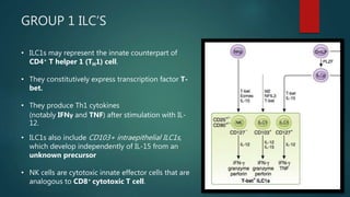
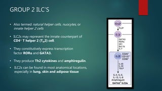
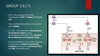
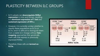
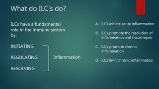
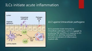
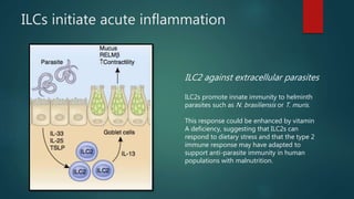
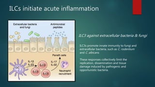
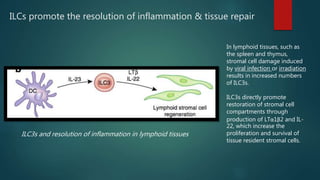
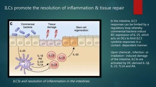
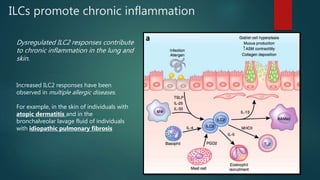
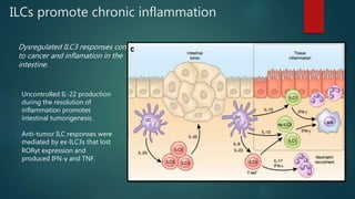
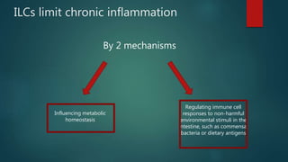
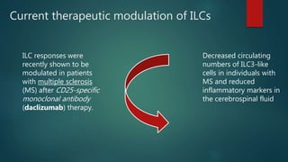
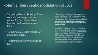
INNATE LYMPHOID
CELLS
ABDUL MAHIR FATHIMA SAKEENA
What and who are ILC’s?
They are the cells acting at the intersection of immunity.
Just like Innate Immune cells:
• They respond to infection quickly.
• They do not express Antigen-specific
receptors.
• They do not undergo clonal selection
and expansion.
Just like T-cells:
• They have similar functions and
phenotypes.
Where do we find them?
 They are enriched at barrier surfaces of the mammalian body,
such as:
Skin, lung & Intestine
• They are also found in adipose tissue & MALT
How do we classify ILC’s?
• Based on common expression of:
A. Surface markers
B. Transcription factors
C. Production of specific cytokines
• The emerging family of ILC’s are compose of three subsets termed:
GROUP 1, 2 or 3.
Development and
Heterogeneity of ILCs
GROUP 1 ILC’S
• ILC1s may represent the innate counterpart of
CD4+ T helper 1 (TH1) cell.
• They constitutively express transcription factor T-
bet.
• They produce Th1 cytokines
(notably IFNγ and TNF) after stimulation with IL-
12.
• ILC1s also include CD103+ intraepithelial ILC1s,
which develop independently of IL-15 from an
unknown precursor
• NK cells are cytotoxic innate effector cells that are
analogous to CD8+ cytotoxic T cell.
GROUP 2 ILC’S
• Also termed natural helper cells, nuocytes, or
innate helper 2 cells
• ILC2s may represent the innate counterpart of
CD4+ T helper 2 (TH2) cell.
• They constitutively express transcription
factor RORα and GATA3.
• They produce Th2 cytokines and amphiregulin.
• ILC2s can be found in most anatomical locations,
especially in lung, skin and adipose tissue
GROUP 3 ILC’S
• ILC3s may represent the innate
counterpart of CD4+ T helper 17 (TH17)
cell.
• They constitutively express transcription
factor RORγt.
• LTi cells - are essential for development
of lymphoid organs during
embryogenesis and after birth regulate
the architecture of lymphoid tissue.
• A subset of adult ILC3s are heterogeneous
in expression of natural cytotoxicity
receptors and require commensal flora
and Ahr for development.
PLASTICITY BETWEEN ILC GROUPS
• ILC3 subsets can downregulate RORγt
expression in mice and human, resulting
in a dominant expression of T-bet and
sustained expression of IFN-γ.
• However, it is currently unclear whether to
classify these populations as ILC1s or
ILC3s, and it is difficult to distinguish them
from a stable ILC1 lineage without fate-
mapping approaches that genetically
mark cells that express or previously
expressed RORγt.
• Therefore, these cells are termed ex-
ILC3s.
Role of ILC’s in inflammatory
processes
What do ILC’s do?
ILCs have a fundamental
role in the immune system
by:
INITIATING
REGULATING
RESOLVING
Inflammation
A. ILCs initiate acute inflammation
B. ILCs promote the resolution of
inflammation and tissue repair
C. ILCs promote chronic
inflammation
D. ILCs limit chronic inflammation
ILCs initiate acute inflammation
ILC1 against Intracellular pathogens
ILC1s promote innate immunity to
intracellular pathogens, such as T. gondii, by
producing TNF and IFN-γ in response to DC-
derived IL-12, and they subsequently
promote recruitment of inflammatory
myeloid cells.
ILCs initiate acute inflammation
ILC2 against extracellular parasites
ILC2s promote innate immunity to helminth
parasites such as N. brasiliensis or T. muris.
This response could be enhanced by vitamin
A deficiency, suggesting that ILC2s can
respond to dietary stress and that the type 2
immune response may have adapted to
support anti-parasite immunity in human
populations with malnutrition.
ILCs initiate acute inflammation
ILC3 against extracellular bacteria & fungi
ILC3s promote innate immunity to fungi and
extracellular bacteria, such as C. rodentium
and C. albicans.
These responses collectively limit the
replication, dissemination and tissue
damage induced by pathogenic and
opportunistic bacteria.
ILCs promote the resolution of inflammation & tissue repair
ILC2s and resolution of inflammation in the lung.
After viral infection in the
lung, airway epithelial cells
are damaged and, in
conjunction with resident
myeloid cell populations,
produce IL-33. ILC2s respond
to IL-33 and produce
amphiregulin, which
promotes repair of the airway
epithelium.
ILCs promote the resolution of inflammation & tissue repair
ILC3s and resolution of inflammation in lymphoid tissues
In lymphoid tissues, such as
the spleen and thymus,
stromal cell damage induced
by viral infection or irradiation
results in increased numbers
of ILC3s.
ILC3s directly promote
restoration of stromal cell
compartments through
production of LTα1β2 and IL-
22, which increase the
proliferation and survival of
tissue resident stromal cells.
ILCs promote the resolution of inflammation & tissue repair
ILC3s and resolution of inflammation in the intestines.
In the intestine, ILC3
responses can be limited by a
regulatory loop whereby
commensal bacteria induce
IEC expression of IL-25, which
acts on DCs to limit ILC3
cytokine responses in a
contact- dependent manner.
Upon chemical-, infection- or
irradiation- induced damage
of the intestine, ILC3s are
activated by DC-derived IL-1β,
IL-23, TL1A and RA.
ILCs promote chronic inflammation
Dysregulated ILC2 responses contribute
to chronic inflammation in the lung and
skin.
Increased ILC2 responses have been
observed in multiple allergic diseases.
For example, in the skin of individuals with
atopic dermatitis and in the
bronchalveolar lavage fluid of individuals
with idiopathic pulmonary fibrosis
ILCs promote chronic inflammation
Dysregulated ILC3 responses contribute
to cancer and inflamation in the
intestine.
Uncontrolled IL-22 production
during the resolution of
inflammation promotes
intestinal tumorigenesis.
Anti-tumor ILC responses were
mediated by ex-ILC3s that lost
RORγt expression and
produced IFN-γ and TNF.
ILCs limit chronic inflammation
By 2 mechanisms
Influencing metabolic
homeostasis
Regulating immune cell
responses to non-harmful
environmental stimuli in the
intestine, such as commensal
bacteria or dietary antigens
ILC2s and metabolic homeostasis.
Tissue recruitment of eosinophils in the
intestine is regulated by nutrient intake and
central circadian rhythms that induce
vasoactive intestinal peptide (VIP) expression
by IECs.
Differentiation of AAMacs or direct
stimulation of adipocytes leads to beiging, a
process which induces thermogenesis,
protects against insulin resistance and
regulates metabolic homeostasis.
In obese humans, frequencies of ILC2s are
reduced in the adipose tissues as compared
to non-obese controls, and this reduction in
ILC2s is associated with a reciprocal increase
in chronic low-grade systemic inflammation.
ILC3s in chronic systemic & intestinal inflammation.
Production of IL-22 restricts the
anatomical localization or replication of
specific species of commensal bacteria
in mice.
This occurs via fucosylation or
glycosylation of IECs, thus providing a
sugar food source for beneficial
microbiota, which in turn limits the
growth of pathogens or opportunistic
pathogens; AND IgA production.
Impairment of this pathway results in
low-grade systemic and intestinal
inflammation
Reduced frequencies of ILC3s is
associated with IBD or with HIV infection
Current therapeutic modulation of ILCs
ILC responses were
recently shown to be
modulated in patients
with multiple sclerosis
(MS) after CD25-specific
monoclonal antibody
(daclizumab) therapy.
Decreased circulating
numbers of ILC3-like
cells in individuals with
MS and reduced
inflammatory markers in
the cerebrospinal fluid
Potential therapeutic modulation of ILCs
 Targeting the cytokine–cytokine
receptor pathways that are
critical for the differentiation,
function and maintenance of
ILCs
 Targeting molecules critical for
migration of ILCs
 Targeting effector molecules of
ILCs
The design of novel therapeutics
may be necessary in order to find
strategies that selectively modulate
protective versus pathologic ILC
responses.
Example: targeting the IL-17 pathway
has demonstrated efficacy in psoriasis
and rheumatoid arthritis, however, in
IBD patients, blockade of IL-17 had
limited efficacy and in some cases
resulted in enhanced disease and
susceptibility to fungal infections
Outstanding Q’s and Future directions
 How to better define this family of ILC’s?
 How do they specifically interact with other innate,
adaptive
and non-hematopoietic cells?
 A universal consensus on gating strategies on
human and mouse
ILCs should be considered.
 How to define the potential plasticity of ILC
populations?
 To identify novel functions and regulatory
pathways influencing ILC responses.
End-references
Innate lymphoid cells in the initiation, regulation and resolution of inflammation
Gregory F Sonnenberg & David Artis
Nature Medicine
https://en.wikipedia.org/wiki/Innate_lymphoid_cell

Innate lymphoid cells

  • 1.
  • 2.
    What and whoare ILC’s? They are the cells acting at the intersection of immunity. Just like Innate Immune cells: • They respond to infection quickly. • They do not express Antigen-specific receptors. • They do not undergo clonal selection and expansion. Just like T-cells: • They have similar functions and phenotypes.
  • 3.
    Where do wefind them?  They are enriched at barrier surfaces of the mammalian body, such as: Skin, lung & Intestine • They are also found in adipose tissue & MALT
  • 4.
    How do weclassify ILC’s? • Based on common expression of: A. Surface markers B. Transcription factors C. Production of specific cytokines • The emerging family of ILC’s are compose of three subsets termed: GROUP 1, 2 or 3.
  • 5.
  • 7.
    GROUP 1 ILC’S •ILC1s may represent the innate counterpart of CD4+ T helper 1 (TH1) cell. • They constitutively express transcription factor T- bet. • They produce Th1 cytokines (notably IFNγ and TNF) after stimulation with IL- 12. • ILC1s also include CD103+ intraepithelial ILC1s, which develop independently of IL-15 from an unknown precursor • NK cells are cytotoxic innate effector cells that are analogous to CD8+ cytotoxic T cell.
  • 8.
    GROUP 2 ILC’S •Also termed natural helper cells, nuocytes, or innate helper 2 cells • ILC2s may represent the innate counterpart of CD4+ T helper 2 (TH2) cell. • They constitutively express transcription factor RORα and GATA3. • They produce Th2 cytokines and amphiregulin. • ILC2s can be found in most anatomical locations, especially in lung, skin and adipose tissue
  • 9.
    GROUP 3 ILC’S •ILC3s may represent the innate counterpart of CD4+ T helper 17 (TH17) cell. • They constitutively express transcription factor RORγt. • LTi cells - are essential for development of lymphoid organs during embryogenesis and after birth regulate the architecture of lymphoid tissue. • A subset of adult ILC3s are heterogeneous in expression of natural cytotoxicity receptors and require commensal flora and Ahr for development.
  • 10.
    PLASTICITY BETWEEN ILCGROUPS • ILC3 subsets can downregulate RORγt expression in mice and human, resulting in a dominant expression of T-bet and sustained expression of IFN-γ. • However, it is currently unclear whether to classify these populations as ILC1s or ILC3s, and it is difficult to distinguish them from a stable ILC1 lineage without fate- mapping approaches that genetically mark cells that express or previously expressed RORγt. • Therefore, these cells are termed ex- ILC3s.
  • 11.
    Role of ILC’sin inflammatory processes
  • 12.
    What do ILC’sdo? ILCs have a fundamental role in the immune system by: INITIATING REGULATING RESOLVING Inflammation A. ILCs initiate acute inflammation B. ILCs promote the resolution of inflammation and tissue repair C. ILCs promote chronic inflammation D. ILCs limit chronic inflammation
  • 13.
    ILCs initiate acuteinflammation ILC1 against Intracellular pathogens ILC1s promote innate immunity to intracellular pathogens, such as T. gondii, by producing TNF and IFN-γ in response to DC- derived IL-12, and they subsequently promote recruitment of inflammatory myeloid cells.
  • 14.
    ILCs initiate acuteinflammation ILC2 against extracellular parasites ILC2s promote innate immunity to helminth parasites such as N. brasiliensis or T. muris. This response could be enhanced by vitamin A deficiency, suggesting that ILC2s can respond to dietary stress and that the type 2 immune response may have adapted to support anti-parasite immunity in human populations with malnutrition.
  • 15.
    ILCs initiate acuteinflammation ILC3 against extracellular bacteria & fungi ILC3s promote innate immunity to fungi and extracellular bacteria, such as C. rodentium and C. albicans. These responses collectively limit the replication, dissemination and tissue damage induced by pathogenic and opportunistic bacteria.
  • 16.
    ILCs promote theresolution of inflammation & tissue repair ILC2s and resolution of inflammation in the lung. After viral infection in the lung, airway epithelial cells are damaged and, in conjunction with resident myeloid cell populations, produce IL-33. ILC2s respond to IL-33 and produce amphiregulin, which promotes repair of the airway epithelium.
  • 17.
    ILCs promote theresolution of inflammation & tissue repair ILC3s and resolution of inflammation in lymphoid tissues In lymphoid tissues, such as the spleen and thymus, stromal cell damage induced by viral infection or irradiation results in increased numbers of ILC3s. ILC3s directly promote restoration of stromal cell compartments through production of LTα1β2 and IL- 22, which increase the proliferation and survival of tissue resident stromal cells.
  • 18.
    ILCs promote theresolution of inflammation & tissue repair ILC3s and resolution of inflammation in the intestines. In the intestine, ILC3 responses can be limited by a regulatory loop whereby commensal bacteria induce IEC expression of IL-25, which acts on DCs to limit ILC3 cytokine responses in a contact- dependent manner. Upon chemical-, infection- or irradiation- induced damage of the intestine, ILC3s are activated by DC-derived IL-1β, IL-23, TL1A and RA.
  • 19.
    ILCs promote chronicinflammation Dysregulated ILC2 responses contribute to chronic inflammation in the lung and skin. Increased ILC2 responses have been observed in multiple allergic diseases. For example, in the skin of individuals with atopic dermatitis and in the bronchalveolar lavage fluid of individuals with idiopathic pulmonary fibrosis
  • 20.
    ILCs promote chronicinflammation Dysregulated ILC3 responses contribute to cancer and inflamation in the intestine. Uncontrolled IL-22 production during the resolution of inflammation promotes intestinal tumorigenesis. Anti-tumor ILC responses were mediated by ex-ILC3s that lost RORγt expression and produced IFN-γ and TNF.
  • 21.
    ILCs limit chronicinflammation By 2 mechanisms Influencing metabolic homeostasis Regulating immune cell responses to non-harmful environmental stimuli in the intestine, such as commensal bacteria or dietary antigens
  • 22.
    ILC2s and metabolichomeostasis. Tissue recruitment of eosinophils in the intestine is regulated by nutrient intake and central circadian rhythms that induce vasoactive intestinal peptide (VIP) expression by IECs. Differentiation of AAMacs or direct stimulation of adipocytes leads to beiging, a process which induces thermogenesis, protects against insulin resistance and regulates metabolic homeostasis. In obese humans, frequencies of ILC2s are reduced in the adipose tissues as compared to non-obese controls, and this reduction in ILC2s is associated with a reciprocal increase in chronic low-grade systemic inflammation.
  • 23.
    ILC3s in chronicsystemic & intestinal inflammation. Production of IL-22 restricts the anatomical localization or replication of specific species of commensal bacteria in mice. This occurs via fucosylation or glycosylation of IECs, thus providing a sugar food source for beneficial microbiota, which in turn limits the growth of pathogens or opportunistic pathogens; AND IgA production. Impairment of this pathway results in low-grade systemic and intestinal inflammation Reduced frequencies of ILC3s is associated with IBD or with HIV infection
  • 24.
    Current therapeutic modulationof ILCs ILC responses were recently shown to be modulated in patients with multiple sclerosis (MS) after CD25-specific monoclonal antibody (daclizumab) therapy. Decreased circulating numbers of ILC3-like cells in individuals with MS and reduced inflammatory markers in the cerebrospinal fluid
  • 25.
    Potential therapeutic modulationof ILCs  Targeting the cytokine–cytokine receptor pathways that are critical for the differentiation, function and maintenance of ILCs  Targeting molecules critical for migration of ILCs  Targeting effector molecules of ILCs The design of novel therapeutics may be necessary in order to find strategies that selectively modulate protective versus pathologic ILC responses. Example: targeting the IL-17 pathway has demonstrated efficacy in psoriasis and rheumatoid arthritis, however, in IBD patients, blockade of IL-17 had limited efficacy and in some cases resulted in enhanced disease and susceptibility to fungal infections
  • 26.
    Outstanding Q’s andFuture directions  How to better define this family of ILC’s?  How do they specifically interact with other innate, adaptive and non-hematopoietic cells?  A universal consensus on gating strategies on human and mouse ILCs should be considered.  How to define the potential plasticity of ILC populations?  To identify novel functions and regulatory pathways influencing ILC responses.
  • 27.
    End-references Innate lymphoid cellsin the initiation, regulation and resolution of inflammation Gregory F Sonnenberg & David Artis Nature Medicine https://en.wikipedia.org/wiki/Innate_lymphoid_cell

Editor's Notes

  • #3 For years, scientists divided the immune system into two arms: innate and adaptive. The cell types involved in the two arms differ in specificity and in how quickly they respond to infections. More recently, immunologists discovered a family of immune cells termed “innate lymphoid cells,” which straddle these two arms.
  • #7 ILCs initially develop in the fetal liver and later in the adult bone marrow from CLP’s. inhibitor of DNA binding 2 (Id2), nuclear factor interleukin-3 regulated (NFIL3) thymocyte selection-associated high mobility group box (Tox) promyelocytic leukemia zinc finger (PLZF) Lymphoid tissue inducer cells (LTi)