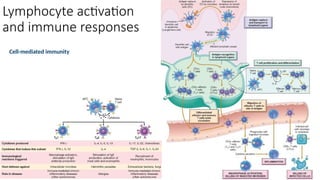
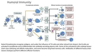
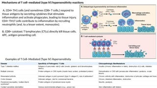
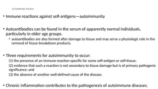
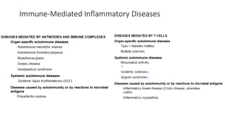
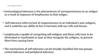
Immune Response
Normal immune response
• Defense against infectious pathogens
• The mechanisms of protection against infections fall into two broad categories.
• Innate immunity (also called natural, or native, immunity) refers to defense mechanisms
that are present even before infection and that have evolved to specifically recognize
microbes and protect individuals against infections.
• Innate immunity is the first line of defense
• Adaptive immunity (also called acquired, or specific, immunity) consists of mechanisms
that are stimulated by (“adapt to”) microbes and are capable of recognizing microbial and
nonmicrobial substances.
Innate immunity
• The major components of innate immunity are epithelial barriers that block
entry of microbes, phagocytic cells (mainly neutrophils and macrophages),
dendritic cells, natural killer (NK) cells, and several plasma proteins, including
the proteins of the complement system.
• The two cellular reactions of innate immunity are:
• inflammation, the process in which phagocytic leukocytes are recruited and activated to
kill microbes, and
• anti-viral defense, mediated by dendritic cells and NK cells.
• The leukocytes and epithelial cells recognize components of microbes
(pathogen-associated molecular patterns) that are often essential for the
infectivity of the pathogens
Innate immunity
• Leukocytes also recognize molecules released by injured and necrotic cells
called danger-associated molecular patterns.
• The cells recognize the microbes and the molecules through receptors called
pattern recognition receptors (eg toll-like receptors (TLR)).
• Upon recognition of microbes, the TLRs and other sensors activate of
transcription factors, eg NF-κB (nuclear factor κB) which turns on the
production of cytokines and proteins that stimulate the microbicidal activities of
various cells (phagocytes).
Innate immunity
• Epithelia of the skin and gastrointestinal and respiratory tracts provide mechanical
barriers to the entry of microbes from the external environment.
• The epithelial cells also produce anti-microbial molecules such as defensins, and
lymphocytes located in the epithelia combat microbes at these sites.
• Monocytes (macrophages) and neutrophils are phagocytes in the blood that can
rapidly be recruited to any site of infection.
• Dendritic cells produce type I interferons, anti-viral cytokines that inhibit viral
infection and replication
• Natural killer cells protect against many viruses and intracellular bacteria
Adaptive immunity
• Consists of lymphocytes and their products, including antibodies.
• The receptors of lymphocytes are much more diverse than those of the innate
immune system, but lymphocytes are not inherently specific for microbes but are
capable of recognizing a vast array of foreign substances called antigens.
• There are two types of adaptive immunity:
• humoral immunity, which protects against extracellular microbes and their toxins, and
• cell-mediated (or cellular) immunity, which is responsible for defense against
intracellular microbes.
• Humoral immunity is mediated by B (bone marrow–derived) lymphocytes and their
secreted products, antibodies (also called immunoglobulins, Ig), and cellular
immunity is mediated by T (thymus-derived) lymphocytes.
Components of the immune system
Cells
• Lymphocytes
The principal
classes of
lymphocytes
and their
functions in
adaptive immunity
Dendritic cells
• There are two types of cells with dendritic morphology that are functionally quite
different: interdigitating dendritic cells (dendritic cells)
• APCs for initiating primary T-cell responses against protein antigens.
• under epithelia, the common site of entry of microbes and foreign antigens, and in the
interstitia of all tissues
• Immature dendritic cells within the epidermis are called Langerhans cells.
• follicular dendritic cell - present in the germinal centers of lymphoid follicles in
the spleen and lymph nodes.
• bears Fc receptors for IgG and receptors for C3b and can trap antigens bound to antibodies
or complement proteins.
• APCs to B cells, select B cells that have the highest affinity for the antigen, thus improving
the quality of the antibody produced
Macrophages
• phagocytosed microbes and protein antigens process the antigens and present
peptide fragments to T cells. Thus, macrophages function as APCs in T-cell
activation.
• effector cells in certain forms of cell-mediated immunity, by eliminating
intracellular microbes. In this type of response.
• participate in the effector phase of humoral immunity; efficiently phagocytose
and destroy microbes that are opsonized (coated) by IgG or C3b.
Natural Killer Cells
• NK cells are approximately 10% to 15% of peripheral blood lymphocytes.
• They do not express TCRs or Ig.
• larger than small lymphocytes, and they contain abundant azurophilic granules (large
granular lymphocytes).
• Have the ability to kill a variety of infected and tumor cells, without prior exposure to
or activation by these microbes or tumors (using CD16 and CD56).
• CD16 on NK cells enables them to lyse IgG-coated target cells by a phenomenon is
known as antibody-dependent cell-mediated cytotoxicity (ADCC).
Activating and inhibitory receptors of
natural killer (NK) cells. A - Healthy cells
express self–class I MHC molecules, which
are recognized by inhibitory receptors, thus
ensuring that NK cells do not attack normal
cells. B - In infected and stressed cells, class
I MHC expression is reduced so that the
inhibitory receptors are not engaged, and
ligands for activating receptors are
expressed.
The result is that NK cells are activated and
the infected cells are killed.
Tissues of the Immune System
• They consist of 1. Lymphoid
organs
the generative (also called
primary, or central) lymphoid
organs: T and B lymphocytes
mature and become competent
to respond to antigens, and
the peripheral (or secondary)
lymphoid organs, in which
adaptive immune responses to
microbes are initiated.
Morphology of a lymph
node. A, The histology of a
lymph node, with an outer
cortex containing follicles
and an inner medulla. B,
The segregation of B cells
and T cells in different
regions of the lymph node,
illustrated schematically. C,
The location of B cells
(stained green, using the
immunofluorescence
technique) and T cells
(stained red) in a lymph
node.
• The cutaneous and mucosal lymphoid systems are located under the epithelia
of the skin and the gastrointestinal and respiratory tracts, respectively.
• Pharyngeal tonsils and Peyer's patches are mucosal lymphoid tissues in the
intestine
• In lymph nodes the B cells are concentrated in discrete structures, called
follicles (which contain the follicular dendritic cells that are involved in the
activation of B cells), located around the periphery, or cortex, of each node.
• Activated B cells are found in the central region of the follicle called a germinal
center.
• The T lymphocytes are concentrated in the paracortex, adjacent to the follicles.
2. The spleen - an abdominal organ that serves the same role in immune
responses to blood-borne antigens as that of lymph nodes in responses to
lymph-borne antigens.
• Blood entering the spleen flows through a network of sinusoids.
• Blood-borne antigens are trapped by dendritic cells and macrophages in
the spleen.
Lymphocyte Recirculation
• Lymphocytes
constantly recirculate
between tissues and
particular sites
• Naive lymphocytes
traverse the peripheral
lymphoid organs where
immune responses are
initiated, and effector
lymphocytes migrate to
sites of infection and
inflammation
Major Histocompatibility Complex (MHC) Molecules
• MHC molecules (Human Leukocyte Antigen (HLA)) are fundamental to the
recognition of antigens by T cells and are linked to many autoimmune diseases,
Inflammation, and inherited errors of metabolism.
• They display peptide fragments of proteins for recognition by antigen-specific T
cells.
• Based on their structure, cellular distribution, and function, MHC gene products
are classified into three groups: Class I, II, and MHC locus (encode some
complement components and the cytokines; tumor necrosis factor (TNF) and
lymphotoxin and other protein in the immune system
Antigen processing and display by major
histocompatibility complex (MHC)
molecules. A, Class I MHC pathway,
peptides are produced from proteins in the
cytosol and transported to the endoplasmic
reticulum (ER), where they bind to class I
MHC molecules. The peptide-MHC
complexes are transported to the cell
surface and displayed for recognition by
CD8+ T cells. B, Class II MHC pathway,
proteins are ingested into vesicles and
degraded into peptides, which bind to class
II MHC molecules being transported in the
same vesicles. The class II–peptide
complexes are expressed on the cell
surface and recognized by CD4+ T cells.
Lymphocyte activation
and immune responses
Cell-mediated immunity
Humoral Immunity
Naive B lymphocytes recognize antigens, and under the influence of TH cells and other stimuli (not shown), the B cells are
activated to proliferate and to differentiate into antibody-secreting plasma cells. Some of the activated B cells undergo heavy-
chain class switching and affinity maturation, and some become long-lived memory cells. Antibodies of different heavy-chain
classes (isotypes) perform different effector functions
Hypersensitivity and autoimmune disorders
• Hypersensitivity - an excessive response to an antigen.
• There are several important general features of hypersensitivity disorders.
• Exogenous and endogenous antigens induce hypersensitivity resulting in
trivial discomforts, such as itching of the skin, to potentially fatal diseases,
such as bronchial asthma and anaphylaxis.
• Autoimmune diseases - Immune responses against self, or autologous
antigens.
Hypersensitivity
• Hypersensitivity diseases can be classified on the basis of the immunologic mechanism that
mediates the disease.
• The main types of hypersensitivity reactions are the following:
Immediate hypersensitivity (type I hypersensitivity) - the immune response is mediated by TH2
cells, IgE antibodies, and mast cells
results in the release of mediators that act on vessels and smooth muscle and of pro-inflammatory
cytokines that recruit inflammatory cells.
Susceptibility to immediate hypersensitivity reactions is genetically determined
Antibody-mediated disorders (type II hypersensitivity) - IgG and IgM antibodies participate directly
in injury to cells by promoting their phagocytosis or lysis and in injury to tissues by inducing
inflammation.
The antibodies may also interfere with cellular functions and cause disease without tissue injury.
Hypersensitivity
Immune complex–mediated disorders (type III hypersensitivity) - IgG and IgM
antibodies bind antigens usually in the circulation, and the antigen-antibody
complexes deposit in tissues and induce inflammation.
The leukocytes are recruited (neutrophils and monocytes) and cause tissue
damage by the release of lysosomal enzymes and generation of free radicals.
Cell-mediated immune disorders (type IV hypersensitivity) - sensitized T
lymphocytes (TH1 and TH17 cells and CTLs) cause cellular and tissue injury.
Mechanisms of Immunologically Mediated Hypersensitivity Reactions
Systemic anaphylaxis is characterized by vascular shock, widespread edema, and difficulty in breathing
Immediate hypersensitivity reactions
are initiated by the introduction of
an allergen, which stimulates TH2
responses and IgE production in
genetically susceptible individuals.
IgE binds to Fc receptors (FcεRI) on
mast cells, and subsequent exposure
to the allergen activates the mast
cells to secrete the mediators that
are responsible for the pathologic
manifestations of immediate
hypersensitivity
PAF, platelet-activating factor; TNF, tumor necrosis factor.
Summary of the Action of Mast Cell Mediators in Immediate (Type I)
Hypersensitivity
Mechanism of Antibody-Mediated
(Type II) Hypersensitivity
• Antibodies that react with
antigens present on cell
surfaces or in the extracellular
matrix.
• The antigenic determinants
may be intrinsic to the cell
membrane or matrix, or they
may take the form of an
exogenous antigen, such as a
drug metabolite, that is
adsorbed on a cell surface or
matrix.
Immune Complex–Mediated (Type III) Hypersensitivity
• Antigen-antibody complexes produce tissue damage mainly by eliciting
inflammation at the sites of deposition.
• The reaction is initiated when antigen combines with antibody within the
circulation (circulating immune complexes), and these are deposited typically in
vessel walls.
• The complexes are formed at extravascular sites where antigen may have been
“planted” previously (called in situ immune complexes).
• The antigens may be exogenous, such as a foreign protein that is injected or
produced by an infectious microbe, or endogenous, if the individual produces
antibody against self-components (autoimmunity).
Immune Complex–Mediated (Type III) Hypersensitivity
• Immune complex disorders can be systemic, ie immune complexes are formed
in the circulation and are deposited in many organs, or localized to particular
organs, such as the kidney (glomerulonephritis), joints (arthritis), or the small
blood vessels of the skin if the complexes are deposited or formed in these
tissues.
Examples of Immune Complex–Mediated Diseases
T Cell-Mediated (Type IV) Hypersensitivity
• The cell-mediated type of hypersensitivity is initiated by antigen-activated
(sensitized) T lymphocytes, including CD4+ and CD8+ T cells
• CD4+ T cell–mediated hypersensitivity induced by environmental and self-
antigens can be a cause of chronic inflammatory disease.
• Many autoimmune diseases are now known to be caused by inflammatory
reactions driven by CD4+ T cells.
• CD8+ cells may also be involved T cell–mediated autoimmune diseases.
especially after viral infections.
Mechanisms of T cell–mediated (type IV) hypersensitivity reactions
A, CD4+ TH1 cells (and sometimes CD8+ T cells,) respond to
tissue antigens by secreting cytokines that stimulate
inflammation and activate phagocytes, leading to tissue injury.
CD4+ TH17 cells contribute to inflammation by recruiting
neutrophils (and, to a lesser extent, monocytes).
B, CD8+ cytotoxic T lymphocytes (CTLs) directly kill tissue cells.
APC, antigen-presenting cell.
Examples of T Cell–Mediated (Type IV) Hypersensitivity
AUTOIMMUNE DISEASES
• Immune reactions against self-antigens—autoimmunity
• Autoantibodies can be found in the serum of apparently normal individuals,
particularly in older age groups.
• autoantibodies are also formed after damage to tissue and may serve a physiologic role in the
removal of tissue breakdown products.
• Three requirements for autoimmunity to occur:
(1) the presence of an immune reaction specific for some self-antigen or self-tissue;
(2) evidence that such a reaction is not secondary to tissue damage but is of primary pathogenic
significance; and
(3) the absence of another well-defined cause of the disease.
• Chronic inflammation contributes to the pathogenesis of autoimmune diseases.
Immune-Mediated Inflammatory Diseases
Immunological Tolerance
• Immunological tolerance is the phenomenon of unresponsiveness to an antigen
as a result of exposure of lymphocytes to that antigen.
• Self-tolerance refers to lack of responsiveness to an individual's own antigens,
and it underlies our ability to live in harmony with our cells and tissues.
• Lymphocytes capable of recognizing self-antigens and these cells have to be
eliminated or inactivated as soon as they recognize the antigens, to prevent
them from causing harm.
• The mechanisms of self-tolerance can be broadly classified into two groups:
central tolerance and peripheral tolerance
Central Tolerance.
• Immature self-reactive T- and B-lymphocyte clones that recognize self-antigens
during their maturation in the central (or generative) lymphoid organs (the
thymus for T cells and the bone marrow for B cells) are killed or rendered
harmless
• The mechanisms of central tolerance in T and B cells show some similarities and
differences.
• In developing T cells, random somatic gene rearrangements generate diverse
TCRs. Such antigen-independent TCR generation produces many lymphocytes
that express high-affinity receptors for self-antigens.
• When immature lymphocytes encounter the antigens in the thymus, the cells
die by apoptosis, called negative selection or deletion
Central Tolerance.
• A protein called AIRE (autoimmune regulator) stimulates expression of some
“peripheral tissue-restricted” self-antigens in the thymus and is thus critical
for deletion of immature T cells specific for these antigens
• Mutations in the AIRE gene are the cause of an autoimmune
polyendocrinopathy
• Some CD4+ T-cell lineage, see self antigens in the thymus but do not die but
develop into regulatory T cells.
Central Tolerance.
• B cells strongly recognize self-antigens in the bone marrow, many of them reactivate
the machinery of antigen receptor gene rearrangement and begin to express new
antigen receptors, not specific for self-antigens, called receptor editing.
• If receptor editing does not occur, the self-reactive cells undergo apoptosis, thus
purging potentially dangerous lymphocytes from the mature pool.
• Not all self-antigens may be present in the thymus, and hence T cells bearing
receptors for such autoantigens escape into the periphery.
• similar to “slippage” in the B-cell system.
Peripheral Tolerance
• Several mechanisms silence potentially autoreactive T and B cells in peripheral
tissues; these are best defined for T cells.
• These mechanisms include the following:
• Anergy: prolonged or irreversible functional inactivation of lymphocytes, induced
by encounter with antigens under certain conditions.
T cells requires two signals for activation: first,recognition of peptide antigen in
association with self-MHC molecules on the surface of APCs and a set of
costimulatory signals (“second signals”) from APCs.
These second signals are provided by certain T cell–associated molecules, such as
CD28, that bind to their ligands (the costimulators B7-1 and B7-2) on APCs.
 If the antigen is presented by cells that do not bear the costimulators a negative
signal is delivered, and the cell becomes anergic.
Two mechanisms of T-cell anergy have been demonstrated in various
experimental systems.
First, the cells lose their ability to trigger biochemical signals from the TCR complex, in
part because of activation of ubiquitin ligases and proteolytic degradation of receptor-
associated signaling proteins.
Second, T cells that recognize self-antigens receive an inhibitory signal from receptors that
are structurally homologous to CD28 but serve the opposite functions.

6.Immune System notes fir nursingstudents.pptx

  • 1.
  • 2.
    Normal immune response •Defense against infectious pathogens • The mechanisms of protection against infections fall into two broad categories. • Innate immunity (also called natural, or native, immunity) refers to defense mechanisms that are present even before infection and that have evolved to specifically recognize microbes and protect individuals against infections. • Innate immunity is the first line of defense • Adaptive immunity (also called acquired, or specific, immunity) consists of mechanisms that are stimulated by (“adapt to”) microbes and are capable of recognizing microbial and nonmicrobial substances.
  • 3.
    Innate immunity • Themajor components of innate immunity are epithelial barriers that block entry of microbes, phagocytic cells (mainly neutrophils and macrophages), dendritic cells, natural killer (NK) cells, and several plasma proteins, including the proteins of the complement system. • The two cellular reactions of innate immunity are: • inflammation, the process in which phagocytic leukocytes are recruited and activated to kill microbes, and • anti-viral defense, mediated by dendritic cells and NK cells. • The leukocytes and epithelial cells recognize components of microbes (pathogen-associated molecular patterns) that are often essential for the infectivity of the pathogens
  • 4.
    Innate immunity • Leukocytesalso recognize molecules released by injured and necrotic cells called danger-associated molecular patterns. • The cells recognize the microbes and the molecules through receptors called pattern recognition receptors (eg toll-like receptors (TLR)). • Upon recognition of microbes, the TLRs and other sensors activate of transcription factors, eg NF-κB (nuclear factor κB) which turns on the production of cytokines and proteins that stimulate the microbicidal activities of various cells (phagocytes).
  • 5.
    Innate immunity • Epitheliaof the skin and gastrointestinal and respiratory tracts provide mechanical barriers to the entry of microbes from the external environment. • The epithelial cells also produce anti-microbial molecules such as defensins, and lymphocytes located in the epithelia combat microbes at these sites. • Monocytes (macrophages) and neutrophils are phagocytes in the blood that can rapidly be recruited to any site of infection. • Dendritic cells produce type I interferons, anti-viral cytokines that inhibit viral infection and replication • Natural killer cells protect against many viruses and intracellular bacteria
  • 6.
    Adaptive immunity • Consistsof lymphocytes and their products, including antibodies. • The receptors of lymphocytes are much more diverse than those of the innate immune system, but lymphocytes are not inherently specific for microbes but are capable of recognizing a vast array of foreign substances called antigens. • There are two types of adaptive immunity: • humoral immunity, which protects against extracellular microbes and their toxins, and • cell-mediated (or cellular) immunity, which is responsible for defense against intracellular microbes. • Humoral immunity is mediated by B (bone marrow–derived) lymphocytes and their secreted products, antibodies (also called immunoglobulins, Ig), and cellular immunity is mediated by T (thymus-derived) lymphocytes.
  • 7.
    Components of theimmune system Cells • Lymphocytes The principal classes of lymphocytes and their functions in adaptive immunity
  • 8.
    Dendritic cells • Thereare two types of cells with dendritic morphology that are functionally quite different: interdigitating dendritic cells (dendritic cells) • APCs for initiating primary T-cell responses against protein antigens. • under epithelia, the common site of entry of microbes and foreign antigens, and in the interstitia of all tissues • Immature dendritic cells within the epidermis are called Langerhans cells. • follicular dendritic cell - present in the germinal centers of lymphoid follicles in the spleen and lymph nodes. • bears Fc receptors for IgG and receptors for C3b and can trap antigens bound to antibodies or complement proteins. • APCs to B cells, select B cells that have the highest affinity for the antigen, thus improving the quality of the antibody produced
  • 9.
    Macrophages • phagocytosed microbesand protein antigens process the antigens and present peptide fragments to T cells. Thus, macrophages function as APCs in T-cell activation. • effector cells in certain forms of cell-mediated immunity, by eliminating intracellular microbes. In this type of response. • participate in the effector phase of humoral immunity; efficiently phagocytose and destroy microbes that are opsonized (coated) by IgG or C3b.
  • 10.
    Natural Killer Cells •NK cells are approximately 10% to 15% of peripheral blood lymphocytes. • They do not express TCRs or Ig. • larger than small lymphocytes, and they contain abundant azurophilic granules (large granular lymphocytes). • Have the ability to kill a variety of infected and tumor cells, without prior exposure to or activation by these microbes or tumors (using CD16 and CD56). • CD16 on NK cells enables them to lyse IgG-coated target cells by a phenomenon is known as antibody-dependent cell-mediated cytotoxicity (ADCC).
  • 11.
    Activating and inhibitoryreceptors of natural killer (NK) cells. A - Healthy cells express self–class I MHC molecules, which are recognized by inhibitory receptors, thus ensuring that NK cells do not attack normal cells. B - In infected and stressed cells, class I MHC expression is reduced so that the inhibitory receptors are not engaged, and ligands for activating receptors are expressed. The result is that NK cells are activated and the infected cells are killed.
  • 12.
    Tissues of theImmune System • They consist of 1. Lymphoid organs the generative (also called primary, or central) lymphoid organs: T and B lymphocytes mature and become competent to respond to antigens, and the peripheral (or secondary) lymphoid organs, in which adaptive immune responses to microbes are initiated. Morphology of a lymph node. A, The histology of a lymph node, with an outer cortex containing follicles and an inner medulla. B, The segregation of B cells and T cells in different regions of the lymph node, illustrated schematically. C, The location of B cells (stained green, using the immunofluorescence technique) and T cells (stained red) in a lymph node.
  • 13.
    • The cutaneousand mucosal lymphoid systems are located under the epithelia of the skin and the gastrointestinal and respiratory tracts, respectively. • Pharyngeal tonsils and Peyer's patches are mucosal lymphoid tissues in the intestine • In lymph nodes the B cells are concentrated in discrete structures, called follicles (which contain the follicular dendritic cells that are involved in the activation of B cells), located around the periphery, or cortex, of each node. • Activated B cells are found in the central region of the follicle called a germinal center. • The T lymphocytes are concentrated in the paracortex, adjacent to the follicles.
  • 14.
    2. The spleen- an abdominal organ that serves the same role in immune responses to blood-borne antigens as that of lymph nodes in responses to lymph-borne antigens. • Blood entering the spleen flows through a network of sinusoids. • Blood-borne antigens are trapped by dendritic cells and macrophages in the spleen.
  • 15.
    Lymphocyte Recirculation • Lymphocytes constantlyrecirculate between tissues and particular sites • Naive lymphocytes traverse the peripheral lymphoid organs where immune responses are initiated, and effector lymphocytes migrate to sites of infection and inflammation
  • 16.
    Major Histocompatibility Complex(MHC) Molecules • MHC molecules (Human Leukocyte Antigen (HLA)) are fundamental to the recognition of antigens by T cells and are linked to many autoimmune diseases, Inflammation, and inherited errors of metabolism. • They display peptide fragments of proteins for recognition by antigen-specific T cells. • Based on their structure, cellular distribution, and function, MHC gene products are classified into three groups: Class I, II, and MHC locus (encode some complement components and the cytokines; tumor necrosis factor (TNF) and lymphotoxin and other protein in the immune system
  • 17.
    Antigen processing anddisplay by major histocompatibility complex (MHC) molecules. A, Class I MHC pathway, peptides are produced from proteins in the cytosol and transported to the endoplasmic reticulum (ER), where they bind to class I MHC molecules. The peptide-MHC complexes are transported to the cell surface and displayed for recognition by CD8+ T cells. B, Class II MHC pathway, proteins are ingested into vesicles and degraded into peptides, which bind to class II MHC molecules being transported in the same vesicles. The class II–peptide complexes are expressed on the cell surface and recognized by CD4+ T cells.
  • 18.
    Lymphocyte activation and immuneresponses Cell-mediated immunity
  • 19.
    Humoral Immunity Naive Blymphocytes recognize antigens, and under the influence of TH cells and other stimuli (not shown), the B cells are activated to proliferate and to differentiate into antibody-secreting plasma cells. Some of the activated B cells undergo heavy- chain class switching and affinity maturation, and some become long-lived memory cells. Antibodies of different heavy-chain classes (isotypes) perform different effector functions
  • 20.
    Hypersensitivity and autoimmunedisorders • Hypersensitivity - an excessive response to an antigen. • There are several important general features of hypersensitivity disorders. • Exogenous and endogenous antigens induce hypersensitivity resulting in trivial discomforts, such as itching of the skin, to potentially fatal diseases, such as bronchial asthma and anaphylaxis. • Autoimmune diseases - Immune responses against self, or autologous antigens.
  • 21.
    Hypersensitivity • Hypersensitivity diseasescan be classified on the basis of the immunologic mechanism that mediates the disease. • The main types of hypersensitivity reactions are the following: Immediate hypersensitivity (type I hypersensitivity) - the immune response is mediated by TH2 cells, IgE antibodies, and mast cells results in the release of mediators that act on vessels and smooth muscle and of pro-inflammatory cytokines that recruit inflammatory cells. Susceptibility to immediate hypersensitivity reactions is genetically determined Antibody-mediated disorders (type II hypersensitivity) - IgG and IgM antibodies participate directly in injury to cells by promoting their phagocytosis or lysis and in injury to tissues by inducing inflammation. The antibodies may also interfere with cellular functions and cause disease without tissue injury.
  • 22.
    Hypersensitivity Immune complex–mediated disorders(type III hypersensitivity) - IgG and IgM antibodies bind antigens usually in the circulation, and the antigen-antibody complexes deposit in tissues and induce inflammation. The leukocytes are recruited (neutrophils and monocytes) and cause tissue damage by the release of lysosomal enzymes and generation of free radicals. Cell-mediated immune disorders (type IV hypersensitivity) - sensitized T lymphocytes (TH1 and TH17 cells and CTLs) cause cellular and tissue injury.
  • 23.
    Mechanisms of ImmunologicallyMediated Hypersensitivity Reactions Systemic anaphylaxis is characterized by vascular shock, widespread edema, and difficulty in breathing
  • 24.
    Immediate hypersensitivity reactions areinitiated by the introduction of an allergen, which stimulates TH2 responses and IgE production in genetically susceptible individuals. IgE binds to Fc receptors (FcεRI) on mast cells, and subsequent exposure to the allergen activates the mast cells to secrete the mediators that are responsible for the pathologic manifestations of immediate hypersensitivity
  • 25.
    PAF, platelet-activating factor;TNF, tumor necrosis factor. Summary of the Action of Mast Cell Mediators in Immediate (Type I) Hypersensitivity
  • 26.
    Mechanism of Antibody-Mediated (TypeII) Hypersensitivity • Antibodies that react with antigens present on cell surfaces or in the extracellular matrix. • The antigenic determinants may be intrinsic to the cell membrane or matrix, or they may take the form of an exogenous antigen, such as a drug metabolite, that is adsorbed on a cell surface or matrix.
  • 27.
    Immune Complex–Mediated (TypeIII) Hypersensitivity • Antigen-antibody complexes produce tissue damage mainly by eliciting inflammation at the sites of deposition. • The reaction is initiated when antigen combines with antibody within the circulation (circulating immune complexes), and these are deposited typically in vessel walls. • The complexes are formed at extravascular sites where antigen may have been “planted” previously (called in situ immune complexes). • The antigens may be exogenous, such as a foreign protein that is injected or produced by an infectious microbe, or endogenous, if the individual produces antibody against self-components (autoimmunity).
  • 28.
    Immune Complex–Mediated (TypeIII) Hypersensitivity • Immune complex disorders can be systemic, ie immune complexes are formed in the circulation and are deposited in many organs, or localized to particular organs, such as the kidney (glomerulonephritis), joints (arthritis), or the small blood vessels of the skin if the complexes are deposited or formed in these tissues. Examples of Immune Complex–Mediated Diseases
  • 29.
    T Cell-Mediated (TypeIV) Hypersensitivity • The cell-mediated type of hypersensitivity is initiated by antigen-activated (sensitized) T lymphocytes, including CD4+ and CD8+ T cells • CD4+ T cell–mediated hypersensitivity induced by environmental and self- antigens can be a cause of chronic inflammatory disease. • Many autoimmune diseases are now known to be caused by inflammatory reactions driven by CD4+ T cells. • CD8+ cells may also be involved T cell–mediated autoimmune diseases. especially after viral infections.
  • 30.
    Mechanisms of Tcell–mediated (type IV) hypersensitivity reactions A, CD4+ TH1 cells (and sometimes CD8+ T cells,) respond to tissue antigens by secreting cytokines that stimulate inflammation and activate phagocytes, leading to tissue injury. CD4+ TH17 cells contribute to inflammation by recruiting neutrophils (and, to a lesser extent, monocytes). B, CD8+ cytotoxic T lymphocytes (CTLs) directly kill tissue cells. APC, antigen-presenting cell. Examples of T Cell–Mediated (Type IV) Hypersensitivity
  • 31.
    AUTOIMMUNE DISEASES • Immunereactions against self-antigens—autoimmunity • Autoantibodies can be found in the serum of apparently normal individuals, particularly in older age groups. • autoantibodies are also formed after damage to tissue and may serve a physiologic role in the removal of tissue breakdown products. • Three requirements for autoimmunity to occur: (1) the presence of an immune reaction specific for some self-antigen or self-tissue; (2) evidence that such a reaction is not secondary to tissue damage but is of primary pathogenic significance; and (3) the absence of another well-defined cause of the disease. • Chronic inflammation contributes to the pathogenesis of autoimmune diseases.
  • 32.
  • 33.
    Immunological Tolerance • Immunologicaltolerance is the phenomenon of unresponsiveness to an antigen as a result of exposure of lymphocytes to that antigen. • Self-tolerance refers to lack of responsiveness to an individual's own antigens, and it underlies our ability to live in harmony with our cells and tissues. • Lymphocytes capable of recognizing self-antigens and these cells have to be eliminated or inactivated as soon as they recognize the antigens, to prevent them from causing harm. • The mechanisms of self-tolerance can be broadly classified into two groups: central tolerance and peripheral tolerance
  • 34.
    Central Tolerance. • Immatureself-reactive T- and B-lymphocyte clones that recognize self-antigens during their maturation in the central (or generative) lymphoid organs (the thymus for T cells and the bone marrow for B cells) are killed or rendered harmless • The mechanisms of central tolerance in T and B cells show some similarities and differences. • In developing T cells, random somatic gene rearrangements generate diverse TCRs. Such antigen-independent TCR generation produces many lymphocytes that express high-affinity receptors for self-antigens. • When immature lymphocytes encounter the antigens in the thymus, the cells die by apoptosis, called negative selection or deletion
  • 35.
    Central Tolerance. • Aprotein called AIRE (autoimmune regulator) stimulates expression of some “peripheral tissue-restricted” self-antigens in the thymus and is thus critical for deletion of immature T cells specific for these antigens • Mutations in the AIRE gene are the cause of an autoimmune polyendocrinopathy • Some CD4+ T-cell lineage, see self antigens in the thymus but do not die but develop into regulatory T cells.
  • 36.
    Central Tolerance. • Bcells strongly recognize self-antigens in the bone marrow, many of them reactivate the machinery of antigen receptor gene rearrangement and begin to express new antigen receptors, not specific for self-antigens, called receptor editing. • If receptor editing does not occur, the self-reactive cells undergo apoptosis, thus purging potentially dangerous lymphocytes from the mature pool. • Not all self-antigens may be present in the thymus, and hence T cells bearing receptors for such autoantigens escape into the periphery. • similar to “slippage” in the B-cell system.
  • 37.
    Peripheral Tolerance • Severalmechanisms silence potentially autoreactive T and B cells in peripheral tissues; these are best defined for T cells. • These mechanisms include the following: • Anergy: prolonged or irreversible functional inactivation of lymphocytes, induced by encounter with antigens under certain conditions. T cells requires two signals for activation: first,recognition of peptide antigen in association with self-MHC molecules on the surface of APCs and a set of costimulatory signals (“second signals”) from APCs. These second signals are provided by certain T cell–associated molecules, such as CD28, that bind to their ligands (the costimulators B7-1 and B7-2) on APCs.
  • 38.
     If theantigen is presented by cells that do not bear the costimulators a negative signal is delivered, and the cell becomes anergic. Two mechanisms of T-cell anergy have been demonstrated in various experimental systems. First, the cells lose their ability to trigger biochemical signals from the TCR complex, in part because of activation of ubiquitin ligases and proteolytic degradation of receptor- associated signaling proteins. Second, T cells that recognize self-antigens receive an inhibitory signal from receptors that are structurally homologous to CD28 but serve the opposite functions.

Editor's Notes

  • #15 Naive T lymphocytes home to lymph nodes as a result of L-selectin and integrin binding to their ligands on high endothelial venules (HEVs). Chemokines expressed in lymph nodes (called CCL19 and CCL21) bind to receptors (CCR7) on naive T cells, enhancing integrin-dependent adhesion and inducing migration of the cells through the HEV wall. Activated T lymphocytes, including effector and memory cells, home to sites of infection in peripheral tissues, and this migration is mediated by E-selectin and P-selectin, integrins, and chemokines secreted at inflammatory sites (e.g., CXCL10) that are recognized by chemokine receptors (e.g., CXCR3) that are expressed on activated T cells. APC, antigen-presenting cell; ICAM-1, intercellular adhesion molecule 1; VCAM-1, vascular cell adhesion molecule 1.
  • #18 Cell-mediated immunity. Dendritic cells (DCs) capture microbial antigens from epithelia and tissues and transport the antigens to lymph nodes. During this process, the DCs mature, and express high levels of MHC molecules and costimulators. Naive T cells recognize MHC-associated peptide antigens displayed on DCs. The T cells are activated to proliferate and to differentiate into effector and memory cells, which migrate to sites of infection and serve various functions in cell-mediated immunity. CD4+ effector T cells of the TH1 subset recognize the antigens of microbes ingested by phagocytes, and activate the phagocytes to kill the microbes. CD4+ T cells also induce inflammation. CD8+ cytotoxic T lymphocytes (CTLs) kill infected cells harboring microbes in the cytoplasm. Not shown are TH2 cells, which are especially important in defense against helminthic infections. Some activated T cells differentiate into long-lived memory cells. APC, antigen-presenting cell.
  • #21 This classification is of value in distinguishing the manner in which the immune response causes tissue injury and disease, and the accompanying pathologic and clinical manifestations. However, it is now increasingly recognized that multiple mechanisms may be operative in any one hypersensitivity disease.
  • #26 A, Opsonization of cells by antibodies and complement components and ingestion by phagocytes. B, Inflammation induced by antibody binding to Fc receptors of leukocytes and by complement breakdown products. C, Anti-receptor antibodies disturb the normal function of receptors. In these examples, antibodies to the acetylcholine (ACh) receptor impair neuromuscular transmission in myasthenia gravis, and antibodies against the thyroid-stimulating hormone (TSH) receptor activate thyroid cells in Graves disease
  • #38 Two of these inhibitory receptors are CTLA-4, which (like CD28) also binds to B7 molecules, and PD-1, which binds to two ligands that are expressed on a wide variety of cells How T cells choose to use CD28 to recognize B7 molecules and be activated, or CTLA-4 to recognize the same B7 molecules and become anergic, is an intriguing question to which there are no clear answers. Nevertheless, the importance of these inhibitory mechanisms has been established by the finding that mice in which the gene encoding CTLA-4 or PD-1 is knocked out develop autoimmune diseases. Furthermore, polymorphisms in the CTLA4 gene are associated with some autoimmune endocrine diseases in humans. Interestingly, some tumors and viruses may have evolved to use the same pathways of immune regulation to evade immune attack.