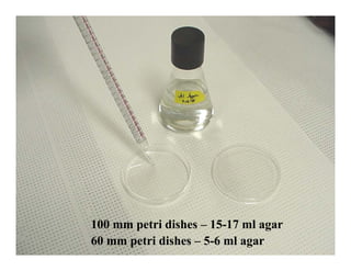
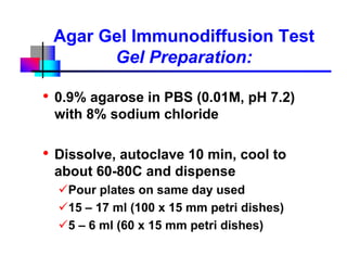
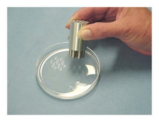
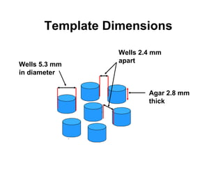
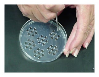
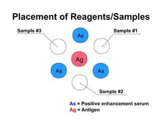
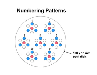
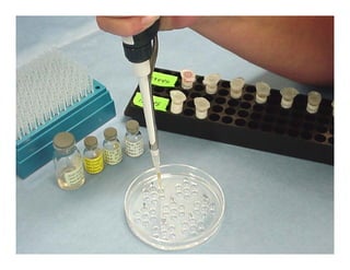
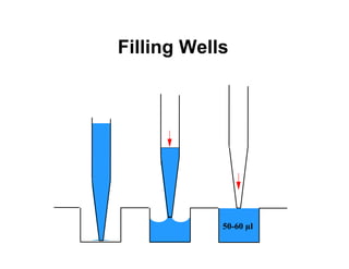
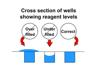
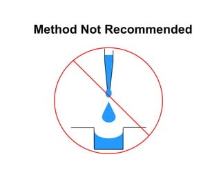
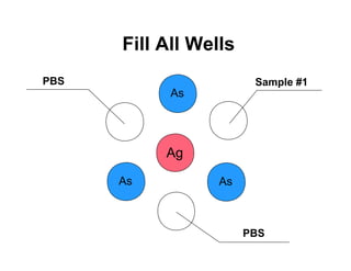
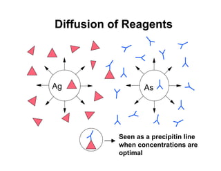
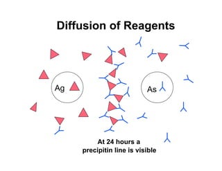
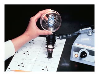
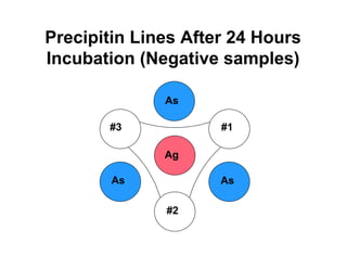
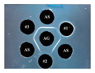
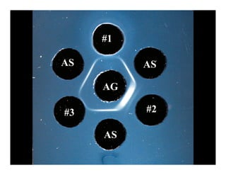
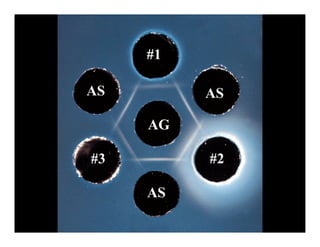
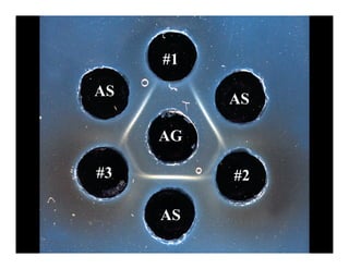
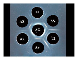
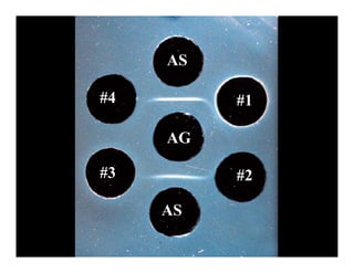
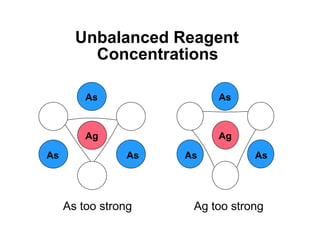
Agar Gel Immunodiffusion
        (AGID) Test
Principles and Techniques
           Dennis A. Senne
   dennis.a.senne@aphis.usda.gov

             USDA, APHIS, VS,
 National Veterinary Services Laboratories,
              Ames, IA 50010
Agar Gel Immunodiffusion Test
            Definition:
• The passive diffusion of soluble
 antigens and/or antibodies
 toward each other leading to
 their precipitation in a gel matrix
Synonyms:
  • Ouchterlony test (1949)
  • Double immunodiffusion test
  • Agar gel precipitin (AGP)     USDA-APHIS
Agar Gel Immunodiffusion
       Test (AGID)
Advantages:
  • Group-specific test (detects
    antibodies to all subtypes of flu-A,
    determine antigenic relationships)
  • Easy, requires few reagents/equip
Disadvantages:
  •   Semi quantitative
  •   Moderate sensitivity
  •   Subjective interpretation
  •   Requires 24 hr                  USDA-APHIS
100 mm petri dishes – 15-17 ml agar
60 mm petri dishes – 5-6 ml agar
Agar Gel Immunodiffusion Test
          Gel Preparation:

•   0.9% agarose in PBS (0.01M, pH 7.2)
    with 8% sodium chloride

•   Dissolve, autoclave 10 min, cool to
    about 60-80C and dispense
      Pour plates on same day used
      15 – 17 ml (100 x 15 mm petri dishes)
      5 – 6 ml (60 x 15 mm petri dishes)
                                              USDA-APHIS
Template Dimensions

                  Wells 2.4 mm
                  apart
Wells 5.3 mm
in diameter


                          Agar 2.8 mm
                          thick
Source of Template
         Cutters
Veterinary Diagnostic Technology, Inc.
4890 Van Gordon St., Suite 101
Wheat Ridge, Co. 80033
Phone: (303) 467-2741
Fax: (303) 467-2799

                                 USDA-APHIS
VACUUM ASSEMBLY
 Single hole stopper
         Side-arm flask
              Glass or plasic tubing


                               VACUUM PUMP
                          IN

                       OUT


                  Vacuum hose          Vacuum set at
                                       12 to 20 psi
                          12 to 14 gauge cannula
Placement of Reagents/Samples
 Sample #3                         Sample #1
                     As


                    Ag
             As              As


                             Sample #2

                  As = Positive enhancement serum
                  Ag = Antigen
Numbering Patterns
      1       2


  3       4       5


      6       7
                      100 x 15 mm
                      petri dish
Agar Gel Immunodiffusion Test
       Reagent Sources (AIV):
•   NVSL, Ames, IA:
     Domestic AIV surveillance – antigen and
     enhancement serum are available, free of
     charge
     Export testing, international – available for
     sale

•   Charles River (SPAFAS), Norwich, CT:
     AIV antigen and enhancement serum
     available for sale
                                            USDA-APHIS
Filling Wells


            50-60 µl
Cross section of wells
showing reagent levels

  Over     Under
                    Correct
  filled   filled
Hold Tip Vertical When
     Filling Wells
Method Not Recommended
Fill All Wells
PBS                     Sample #1
            As


           Ag
      As          As


                       PBS
Diffusion of Reagents


Ag                 As


           Seen as a precipitin line
           when concentrations are
           optimal
Diffusion of Reagents


Ag                         As


          At 24 hours a
     precipitin line is visible
Precipitin Lines After 24 Hours
Incubation (Negative samples)

              As

       #3            #1

              Ag

        As           As

              #2
Examples of Positive Reactions
               Negative


+3        +1                               +4


     +2                         +5


               May be seen as light haze
Examples of Nonspecific Lines

                              Negative
Negative


                                   Line of partial
                                   identity


 Line of non identity   Negative
Incomplete Line Formation


                  +                  +


                         -


Before 24 hours   At 24 - 48 hours
Potential Positive Sample
Additional Problems
     “Halos”


    +
    +        -


         +
         +
AS

#3        #1

     AG

AS        AS

     #2
#1

AS        AS

     AG

#3        #2

     AS
#1

AS        AS

     AG

#3        #2

     AS
#1
AS        AS

     AG

#3        #2

     AS
#1

AS        AS

     AG

#3        #2

     AS
AS

#4        #1

     AG

#3        #2

     AS
Unbalanced Reagent
         Concentrations

         As               As


         Ag               Ag

As              As   As          As


     As too strong    Ag too strong
Cutting Errors


 May miss the weak
 positives

Inmunodiagnostico AGID

  • 1.
    Agar Gel Immunodiffusion (AGID) Test Principles and Techniques Dennis A. Senne dennis.a.senne@aphis.usda.gov USDA, APHIS, VS, National Veterinary Services Laboratories, Ames, IA 50010
  • 2.
    Agar Gel ImmunodiffusionTest Definition: • The passive diffusion of soluble antigens and/or antibodies toward each other leading to their precipitation in a gel matrix Synonyms: • Ouchterlony test (1949) • Double immunodiffusion test • Agar gel precipitin (AGP) USDA-APHIS
  • 3.
    Agar Gel Immunodiffusion Test (AGID) Advantages: • Group-specific test (detects antibodies to all subtypes of flu-A, determine antigenic relationships) • Easy, requires few reagents/equip Disadvantages: • Semi quantitative • Moderate sensitivity • Subjective interpretation • Requires 24 hr USDA-APHIS
  • 4.
    100 mm petridishes – 15-17 ml agar 60 mm petri dishes – 5-6 ml agar
  • 5.
    Agar Gel ImmunodiffusionTest Gel Preparation: • 0.9% agarose in PBS (0.01M, pH 7.2) with 8% sodium chloride • Dissolve, autoclave 10 min, cool to about 60-80C and dispense Pour plates on same day used 15 – 17 ml (100 x 15 mm petri dishes) 5 – 6 ml (60 x 15 mm petri dishes) USDA-APHIS
  • 7.
    Template Dimensions Wells 2.4 mm apart Wells 5.3 mm in diameter Agar 2.8 mm thick
  • 8.
    Source of Template Cutters Veterinary Diagnostic Technology, Inc. 4890 Van Gordon St., Suite 101 Wheat Ridge, Co. 80033 Phone: (303) 467-2741 Fax: (303) 467-2799 USDA-APHIS
  • 10.
    VACUUM ASSEMBLY Singlehole stopper Side-arm flask Glass or plasic tubing VACUUM PUMP IN OUT Vacuum hose Vacuum set at 12 to 20 psi 12 to 14 gauge cannula
  • 11.
    Placement of Reagents/Samples Sample #3 Sample #1 As Ag As As Sample #2 As = Positive enhancement serum Ag = Antigen
  • 12.
    Numbering Patterns 1 2 3 4 5 6 7 100 x 15 mm petri dish
  • 14.
    Agar Gel ImmunodiffusionTest Reagent Sources (AIV): • NVSL, Ames, IA: Domestic AIV surveillance – antigen and enhancement serum are available, free of charge Export testing, international – available for sale • Charles River (SPAFAS), Norwich, CT: AIV antigen and enhancement serum available for sale USDA-APHIS
  • 15.
    Filling Wells 50-60 µl
  • 16.
    Cross section ofwells showing reagent levels Over Under Correct filled filled
  • 17.
    Hold Tip VerticalWhen Filling Wells
  • 18.
  • 19.
    Fill All Wells PBS Sample #1 As Ag As As PBS
  • 20.
    Diffusion of Reagents Ag As Seen as a precipitin line when concentrations are optimal
  • 21.
    Diffusion of Reagents Ag As At 24 hours a precipitin line is visible
  • 23.
    Precipitin Lines After24 Hours Incubation (Negative samples) As #3 #1 Ag As As #2
  • 24.
    Examples of PositiveReactions Negative +3 +1 +4 +2 +5 May be seen as light haze
  • 25.
    Examples of NonspecificLines Negative Negative Line of partial identity Line of non identity Negative
  • 26.
    Incomplete Line Formation + + - Before 24 hours At 24 - 48 hours
  • 27.
  • 28.
    Additional Problems “Halos” + + - + +
  • 29.
    AS #3 #1 AG AS AS #2
  • 30.
    #1 AS AS AG #3 #2 AS
  • 31.
    #1 AS AS AG #3 #2 AS
  • 32.
    #1 AS AS AG #3 #2 AS
  • 33.
    #1 AS AS AG #3 #2 AS
  • 34.
    AS #4 #1 AG #3 #2 AS
  • 35.
    Unbalanced Reagent Concentrations As As Ag Ag As As As As As too strong Ag too strong
  • 36.
    Cutting Errors Maymiss the weak positives