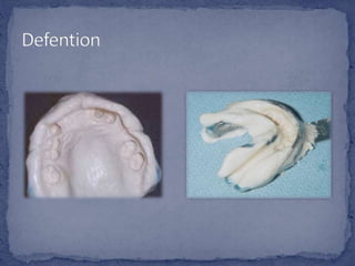
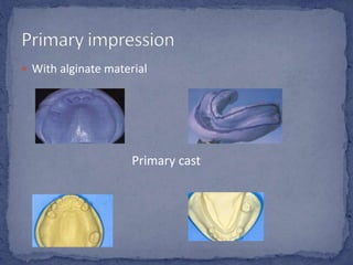
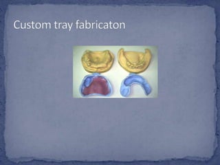
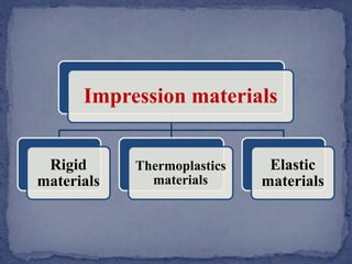
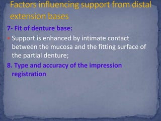
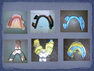
By Noha Taha
 Definition of impression
 Types of impression
 Impression materials
 Impression methods
 Factors influencing support of distal extension base
Primary
impression
Secondary
impression
 Selection of stock tray
 With alginate material
Primary cast
Impression materials
Rigid
materials
Thermoplastics
materials
Elastic
materials
 It record tooth and tissue details
accurately but it cannot be removed from
the mouth without fracture.
 Plaster of paris
 It have been used in dentistry for over 200 years but now
elastic materials completely replaced it.
 Metallic oxide past
 Not used as primary impression materials
 Used for extension base edentulous ridge areas for RPD
Cannot record minute details
accurately because they under go
permanent distortion during removal
from the tooth and tissue undercuts.
• Used mostly for border molding
of custom impression tray.
Modeling plastic
• They have the ability to record
border details accurately.
Impression
waxes and
Natural Resins
 Remain in an elastic state after they set and
removed from the mouth.
 Used for making impression for RPD, when
tissue undercuts and surface detail must be
record with accuracy.
• It is accurate for making master
cast for RPD ,
• It is mainly used for duplication
of cast
Reversible
hydrocolloid
(agar-agar)
• Are used for Making study
cast and Master cast
Irreversible
hydrocolloid
(alginate)
• Should not be used when
several undercuts are present.
Mercaptan
rubber –base
impression
materials
• Provide good surface details
and make them useful as
border molding materials
Polyether
impression
materials
• More accurate and easier to use
than other elastic impression
material
Silicone
impression
material
1- Anatomic ridge form:
 for tooth suppoted R.P.D. (Kenedy‘s class
III, short span class IV)
 so the edentulous ridges don´t
contribute to the support of the R.P.D.
 Single, pressure-free imp. records the
teeth and soft tissues in their
anatomic form .
Impression Techniques
2-Physiologic or functional ridge form:
 for tooth- tissue supported R.P.D.
(Kenedy‘s class I,II,long span class IV)
 When the occlusal forces fall on tooth-
tissue supported R.P.D., the ridge
contribute to support as well as teeth
 This imp. recordteeth in their anatomic
form and the ridge in its functional form
under pressure.
Impression Techniques
The objectives of technique is :
 to provide maximum support for the
removable partial denture bases. This
allows for:
1. maintenance of occlusal contact
between both natural and artificial
dentition
2. minimum movement of the base,
which would create leverage on the
abutment teeth.
1- Quality of soft tissues covering edentulous ridge
2- Type of bone making up denture bearing area
3- Design of partial denture
4- Amount of tissue coverage of denture base:
5- Amount of occlusal forces
6- Anatomy of denture bearing area:
7- Fit of denture base:
8. Type and accuracy of the impression registration:
1- Quality of soft tissues covering
edentulous ridge
 It should be firm, dense fibrous C.T. of even thickness
 slightly compressible and firmly attached to the bone
Factors influencing support from
distal extension bases
2- Type of bone making up denture bearing
area:
The ideal ridge would consist of:
 Cortical bone that covers dense
Cancellous bone with broad rounded
crest and high vertical slops.
 Cortical bone can resist vertical forces better than
cancellous bone.
Factors influencing support from distal extension
bases (factors influencing the amount of tissue
displacement
3- Design of partial denture:
 The use of indirect retainer will control
rotational movement of distal extension RPD.
Factors influencing support
from distal extension bases
4- Amount of tissue coverage of denture
base:
The broader the coverage of the
edentulous ridge, the greater the
distribution of the load & the smaller the
force per unit area
Factors influencing support
from distal extension bases
5- Amount of occlusal forces:
1- Number of artificial teeth.
2-Width of the occlusal table.
3- type of the opposing dentition
4-powerfull musculature of the patient
It influences the amount of support required to
stabilize the denture base..
Factors influencing support
from distal extension bases
6- Anatomy of denture bearing area:
 To distribute the forces of mastication to
the ridge most efficiently, the majority of
force must be directed to the primary
stress bearing areas, that are capable of
withstanding that force.
Factors influencing support
from distal extension bases
7- Fit of denture base:
 Support is enhanced by intimate contact
between the mucosa and the fitting surface of
the partial denture;
8. Type and accuracy of the impression
registration
 which records the ridge portion of the cast in
its physiologic or functioning form by placing
an occlusal load on the impression tray as the
impression is being made.
3-Selective tissue placement
impression technique.
At the imp. stage:
Mclean´s and Hindel´s methods = dual imp. Technique =
pseudo-functional imp. or
Impressions with custom trays.
At the framework stage:
Altered cast method either by functional imp.method
At the finished denture stage:
Functional relining method using fluid wax or zinc oxide
euginol or rubber base relining method.
Impression for distal
extension R.P.D.
1- At the imp. stage:
 McLean‘s technique (closed mouth)
 The technique consists of making an impression of the
edentulous ridge in border-moulded denture base tray
which is provided with occlusion rims.
 Impression paste is used to record ridge areas under
biting stresses
 After setting of ZnO eugenol it is removed, tested,
reinserted; overall alginate impression is made with the
ZnO imp.seated in the mouth.
Imp. for Dis. Ex. R.P.D.
1- At the imp. stage:
 Hindle‘s technique (opened mouth)
 the same idea of McLean‘s
technique but instead of the
occlusion rims, use finger pressure
through 2 circular openings in the
posterior region of the
hydrocolloid imp. Tray.
Imp. for Dis. Ex. R.P.D.
1- At the imp. stage:
 Disadvantages
 If the clasp action is sufficient to maintain the
denture base in its intended position, This may
result in compromised blood flow with adverse
soft tissue reaction and bone resorption.
 If clasp action is not sufficient to maintain that
functional relationship of the denture base to the
soft tissue, this will result in floating denture with
premature contact and patient dissatisfaction.
Imp. for Dis. Ex. R.P.D.
Steps:
1- after the RPD frame work is constucted on anatomic
imp.cast.it should be evaluated
for any metal projections and sharp
edges.
2-check the RPD metal frame
work in the patient’s mouth
3-the impression tray is made
using chemically activated resin, a
the frame work with the
attached impression tray is
placed in the patient’s mouth
and correct peripheral extension
4-border molding the impression
tray using low fusing modeling
plastic < green or grey sticks >
5-the final impression is made by
using zinc-oxide euginol paste
with the mouth opened and
tripod pressure is applied on
occlusal rests and indirect retainer
6-after the impression material is
set, the tray is removed and
checked for any discrepancies
7. The metal framework with the
attached imp. is positioned on
the master cast with all
occlusal rests properly seated
in their prepared recesses.
8. The entire assembly is boxed
and poured in a different
colored stone.
Altered cast method
3- At the finished denture stage:
Functional relining method:
 The finished denture is relined by applying
for example ZnO eugenol imp. paste to the
acrylic fitting surface of the distal extension
saddle
 the impression is made with the denture
being seated by pressure on the occlusal
rests and indirect retainers only.
 No pressure is applied to the occlusal surface
of the artificial teeth
Imp. for Dis. Ex. R.P.D.
fdocuments.in_impression-materials-for-partial-denture.pptx

fdocuments.in_impression-materials-for-partial-denture.pptx

  • 1.
  • 2.
     Definition ofimpression  Types of impression  Impression materials  Impression methods  Factors influencing support of distal extension base
  • 4.
  • 5.
     Selection ofstock tray
  • 8.
     With alginatematerial Primary cast
  • 10.
  • 11.
     It recordtooth and tissue details accurately but it cannot be removed from the mouth without fracture.
  • 12.
     Plaster ofparis  It have been used in dentistry for over 200 years but now elastic materials completely replaced it.  Metallic oxide past  Not used as primary impression materials  Used for extension base edentulous ridge areas for RPD
  • 13.
    Cannot record minutedetails accurately because they under go permanent distortion during removal from the tooth and tissue undercuts.
  • 14.
    • Used mostlyfor border molding of custom impression tray. Modeling plastic • They have the ability to record border details accurately. Impression waxes and Natural Resins
  • 15.
     Remain inan elastic state after they set and removed from the mouth.  Used for making impression for RPD, when tissue undercuts and surface detail must be record with accuracy.
  • 16.
    • It isaccurate for making master cast for RPD , • It is mainly used for duplication of cast Reversible hydrocolloid (agar-agar) • Are used for Making study cast and Master cast Irreversible hydrocolloid (alginate)
  • 17.
    • Should notbe used when several undercuts are present. Mercaptan rubber –base impression materials • Provide good surface details and make them useful as border molding materials Polyether impression materials • More accurate and easier to use than other elastic impression material Silicone impression material
  • 18.
    1- Anatomic ridgeform:  for tooth suppoted R.P.D. (Kenedy‘s class III, short span class IV)  so the edentulous ridges don´t contribute to the support of the R.P.D.  Single, pressure-free imp. records the teeth and soft tissues in their anatomic form . Impression Techniques
  • 19.
    2-Physiologic or functionalridge form:  for tooth- tissue supported R.P.D. (Kenedy‘s class I,II,long span class IV)  When the occlusal forces fall on tooth- tissue supported R.P.D., the ridge contribute to support as well as teeth  This imp. recordteeth in their anatomic form and the ridge in its functional form under pressure. Impression Techniques
  • 20.
    The objectives oftechnique is :  to provide maximum support for the removable partial denture bases. This allows for: 1. maintenance of occlusal contact between both natural and artificial dentition 2. minimum movement of the base, which would create leverage on the abutment teeth.
  • 21.
    1- Quality ofsoft tissues covering edentulous ridge 2- Type of bone making up denture bearing area 3- Design of partial denture 4- Amount of tissue coverage of denture base: 5- Amount of occlusal forces 6- Anatomy of denture bearing area: 7- Fit of denture base: 8. Type and accuracy of the impression registration:
  • 22.
    1- Quality ofsoft tissues covering edentulous ridge  It should be firm, dense fibrous C.T. of even thickness  slightly compressible and firmly attached to the bone Factors influencing support from distal extension bases
  • 23.
    2- Type ofbone making up denture bearing area: The ideal ridge would consist of:  Cortical bone that covers dense Cancellous bone with broad rounded crest and high vertical slops.  Cortical bone can resist vertical forces better than cancellous bone. Factors influencing support from distal extension bases (factors influencing the amount of tissue displacement
  • 24.
    3- Design ofpartial denture:  The use of indirect retainer will control rotational movement of distal extension RPD. Factors influencing support from distal extension bases
  • 25.
    4- Amount oftissue coverage of denture base: The broader the coverage of the edentulous ridge, the greater the distribution of the load & the smaller the force per unit area Factors influencing support from distal extension bases
  • 26.
    5- Amount ofocclusal forces: 1- Number of artificial teeth. 2-Width of the occlusal table. 3- type of the opposing dentition 4-powerfull musculature of the patient It influences the amount of support required to stabilize the denture base.. Factors influencing support from distal extension bases
  • 27.
    6- Anatomy ofdenture bearing area:  To distribute the forces of mastication to the ridge most efficiently, the majority of force must be directed to the primary stress bearing areas, that are capable of withstanding that force. Factors influencing support from distal extension bases
  • 28.
    7- Fit ofdenture base:  Support is enhanced by intimate contact between the mucosa and the fitting surface of the partial denture; 8. Type and accuracy of the impression registration
  • 29.
     which recordsthe ridge portion of the cast in its physiologic or functioning form by placing an occlusal load on the impression tray as the impression is being made. 3-Selective tissue placement impression technique.
  • 30.
    At the imp.stage: Mclean´s and Hindel´s methods = dual imp. Technique = pseudo-functional imp. or Impressions with custom trays. At the framework stage: Altered cast method either by functional imp.method At the finished denture stage: Functional relining method using fluid wax or zinc oxide euginol or rubber base relining method. Impression for distal extension R.P.D.
  • 31.
    1- At theimp. stage:  McLean‘s technique (closed mouth)  The technique consists of making an impression of the edentulous ridge in border-moulded denture base tray which is provided with occlusion rims.  Impression paste is used to record ridge areas under biting stresses  After setting of ZnO eugenol it is removed, tested, reinserted; overall alginate impression is made with the ZnO imp.seated in the mouth. Imp. for Dis. Ex. R.P.D.
  • 32.
    1- At theimp. stage:  Hindle‘s technique (opened mouth)  the same idea of McLean‘s technique but instead of the occlusion rims, use finger pressure through 2 circular openings in the posterior region of the hydrocolloid imp. Tray. Imp. for Dis. Ex. R.P.D.
  • 34.
    1- At theimp. stage:  Disadvantages  If the clasp action is sufficient to maintain the denture base in its intended position, This may result in compromised blood flow with adverse soft tissue reaction and bone resorption.  If clasp action is not sufficient to maintain that functional relationship of the denture base to the soft tissue, this will result in floating denture with premature contact and patient dissatisfaction. Imp. for Dis. Ex. R.P.D.
  • 35.
    Steps: 1- after theRPD frame work is constucted on anatomic imp.cast.it should be evaluated for any metal projections and sharp edges. 2-check the RPD metal frame work in the patient’s mouth
  • 36.
    3-the impression trayis made using chemically activated resin, a the frame work with the attached impression tray is placed in the patient’s mouth and correct peripheral extension 4-border molding the impression tray using low fusing modeling plastic < green or grey sticks >
  • 37.
    5-the final impressionis made by using zinc-oxide euginol paste with the mouth opened and tripod pressure is applied on occlusal rests and indirect retainer 6-after the impression material is set, the tray is removed and checked for any discrepancies
  • 38.
    7. The metalframework with the attached imp. is positioned on the master cast with all occlusal rests properly seated in their prepared recesses. 8. The entire assembly is boxed and poured in a different colored stone. Altered cast method
  • 39.
    3- At thefinished denture stage: Functional relining method:  The finished denture is relined by applying for example ZnO eugenol imp. paste to the acrylic fitting surface of the distal extension saddle  the impression is made with the denture being seated by pressure on the occlusal rests and indirect retainers only.  No pressure is applied to the occlusal surface of the artificial teeth Imp. for Dis. Ex. R.P.D.