SlideShare a Scribd company logo
1 of 5
Download to read offline
Figure:
Immunohistochemical
analysis of paraffin
embedded Human liver
tissue. 1: Cleaved-Notch
2 (D1733) Polyclonal
Antibody was diluted at
1:200 (4 degree
Celsius,overnight). 2:
Sodium citrate pH 6.0
was used for antibody
retrieval (>98 degree
Celsius,20min). 3:
Secondary antibody was
diluted at 1:200 (room
temperature, 30min).
Negative control was
used by secondary
antibody only.
Report Number 90068-a Host Rabbit
Application IHC-P Clonality Polyclonal
Model Number STJ90068 Clone ID NA
Antibody Name Anti-Cleaved-Notch 2 (D1733) antibody
Testing Species HUMAN Testing Tissue LIVER
ANTIBODY VALIDATION REPORT
a. (A small amount of distilled water was added into the incubation
box to prevent evaporation of antibody).
52. Secondary antibody incubation
a. Slides were washed 3 times, with PBS on a shaker for 5min.
Shortly after the slides were dried the corresponding secondary
antibody solution was added (HRP labelled), covering the
tissues, and incubated at room temperature for 30min.
b.
53. DAB staining
a. Slides were washed 3 times, with PBS on a shaker for 5min.
b. Shortly after, the slides were dried and fresh DAB staining buffer
was added inside the circles. The staining time was adjusted
under a microscope. Yellow-brown colour represented a positive
result. Slides were washed with water to stop the staining.
c.
54. Haematoxylin staining
a. Haematoxylin was used to counter-staining for 1min, and then
the slides were washed with water. 1% Hydrochloric acid and
alcohol was added for several seconds and then washed with
water. Ammonia was used to reveal blue colour, and then
flushed with water.
b.
55. Desolation and Clearing
i. Slides were incubated sequentially into: 75% alcohol 5min, 85%
alcohol 5min, Anhydrous ethanol - 5min, Anhydrous ethanol -
5min & Xylene - 5min. Shortly after slides were dried and neutral
gum was used to seal the slides.
ii.
56. Visualization
a. Results were validated with microscope, and the slides were
scanned.
Paraffin-Embedded
Immunohistochemistry Protocol
46.
47. Tissue processing
a. Slides were incubated sequentially into Xylene; 15min –
Xylene, 15min - Anhydrous ethanol, 5min - Anhydrous
ethanol, 5min - 85% alcohol, 5min - 75% alcohol & 5min –
wash in distilled water.
b.
48. Antigen retrieval
a. Tissue slides were incubated with citric acid (PH6.0) antigen
retrieval buffer and microwaved for antigen retrieval (heated
until boiled and then stopped heating) for 8min. Slides were
then heated with medium power for 7min. During this
process slides were kept from drying out. After cooling down
at room temperature, slides were washed with PBS on
shaker for 5min, repeated for 3 times.
b.
49. Inhibition of endogenous peroxidase
a. Slides were placed in 3% Hydrogen peroxide solution, and
incubated for 10 min at room temperature without light
exposure. Slides were then washed 3 times with PBS on a
shaker for 5mins.
b.
50. BSA Blocking
a. Shortly after slides were dried, a PAP pen was used to draw
circles around the tissue sections (and to prevent draining of
the antibody solution). Inside the circles, BSA was used to
cover the tissue evenly, blocking for 30min.
b.
51. Primary antibody incubation
After blocking solution was removed a 1:200 solution of
primary antibody/PBS was added on the slide, and incubated
overnight at 4°C.
St John's Laboratory Ltd.
www.stjohnslabs.com
Figure:
Immunohistochemical
analysis of paraffin
embedded Rat lung
tissue. 1: Cleaved-Notch
2 (D1733) Polyclonal
Antibody was diluted at
1:200 (4 degree
Celsius,overnight). 2:
Sodium citrate pH 6.0
was used for antibody
retrieval (>98 degree
Celsius,20min). 3:
Secondary antibody was
diluted at 1:200 (room
temperature, 30min).
Negative control was
used by secondary
antibody only.
Report Number 90068-b Host Rabbit
Application IHC-P Clonality Polyclonal
Model Number STJ90068 Clone ID NA
Antibody Name Anti-Cleaved-Notch 2 (D1733) antibody
Testing Species RAT Testing Tissue LUNG
ANTIBODY VALIDATION REPORT
a. (A small amount of distilled water was added into the incubation
box to prevent evaporation of antibody).
41. Secondary antibody incubation
a. Slides were washed 3 times, with PBS on a shaker for 5min.
Shortly after the slides were dried the corresponding secondary
antibody solution was added (HRP labelled), covering the
tissues, and incubated at room temperature for 30min.
b.
42. DAB staining
a. Slides were washed 3 times, with PBS on a shaker for 5min.
b. Shortly after, the slides were dried and fresh DAB staining buffer
was added inside the circles. The staining time was adjusted
under a microscope. Yellow-brown colour represented a positive
result. Slides were washed with water to stop the staining.
c.
43. Haematoxylin staining
a. Haematoxylin was used to counter-staining for 1min, and then
the slides were washed with water. 1% Hydrochloric acid and
alcohol was added for several seconds and then washed with
water. Ammonia was used to reveal blue colour, and then
flushed with water.
b.
44. Desolation and Clearing
i. Slides were incubated sequentially into: 75% alcohol 5min, 85%
alcohol 5min, Anhydrous ethanol - 5min, Anhydrous ethanol -
5min & Xylene - 5min. Shortly after slides were dried and neutral
gum was used to seal the slides.
ii.
45. Visualization
a. Results were validated with microscope, and the slides were
scanned.
Paraffin-Embedded
Immunohistochemistry Protocol
35.
36. Tissue processing
a. Slides were incubated sequentially into Xylene; 15min –
Xylene, 15min - Anhydrous ethanol, 5min - Anhydrous
ethanol, 5min - 85% alcohol, 5min - 75% alcohol & 5min –
wash in distilled water.
b.
37. Antigen retrieval
a. Tissue slides were incubated with citric acid (PH6.0) antigen
retrieval buffer and microwaved for antigen retrieval (heated
until boiled and then stopped heating) for 8min. Slides were
then heated with medium power for 7min. During this
process slides were kept from drying out. After cooling down
at room temperature, slides were washed with PBS on
shaker for 5min, repeated for 3 times.
b.
38. Inhibition of endogenous peroxidase
a. Slides were placed in 3% Hydrogen peroxide solution, and
incubated for 10 min at room temperature without light
exposure. Slides were then washed 3 times with PBS on a
shaker for 5mins.
b.
39. BSA Blocking
a. Shortly after slides were dried, a PAP pen was used to draw
circles around the tissue sections (and to prevent draining of
the antibody solution). Inside the circles, BSA was used to
cover the tissue evenly, blocking for 30min.
b.
40. Primary antibody incubation
After blocking solution was removed a 1:200 solution of
primary antibody/PBS was added on the slide, and incubated
overnight at 4°C.
St John's Laboratory Ltd.
www.stjohnslabs.com
Figure:
Immunohistochemical
analysis of paraffin
embedded Rat spleen
tissue. 1: Cleaved-Notch
2 (D1733) Polyclonal
Antibody was diluted at
1:200 (4 degree
Celsius,overnight). 2:
Sodium citrate pH 6.0
was used for antibody
retrieval (>98 degree
Celsius,20min). 3:
Secondary antibody was
diluted at 1:200 (room
temperature, 30min).
Negative control was
used by secondary
antibody only.
Report Number 90068-c Host Rabbit
Application IHC-P Clonality Polyclonal
Model Number STJ90068 Clone ID NA
Antibody Name Anti-Cleaved-Notch 2 (D1733) antibody
Testing Species RAT Testing Tissue SPLEEN
ANTIBODY VALIDATION REPORT
a. (A small amount of distilled water was added into the incubation
box to prevent evaporation of antibody).
30. Secondary antibody incubation
a. Slides were washed 3 times, with PBS on a shaker for 5min.
Shortly after the slides were dried the corresponding secondary
antibody solution was added (HRP labelled), covering the
tissues, and incubated at room temperature for 30min.
b.
31. DAB staining
a. Slides were washed 3 times, with PBS on a shaker for 5min.
b. Shortly after, the slides were dried and fresh DAB staining buffer
was added inside the circles. The staining time was adjusted
under a microscope. Yellow-brown colour represented a positive
result. Slides were washed with water to stop the staining.
c.
32. Haematoxylin staining
a. Haematoxylin was used to counter-staining for 1min, and then
the slides were washed with water. 1% Hydrochloric acid and
alcohol was added for several seconds and then washed with
water. Ammonia was used to reveal blue colour, and then
flushed with water.
b.
33. Desolation and Clearing
i. Slides were incubated sequentially into: 75% alcohol 5min, 85%
alcohol 5min, Anhydrous ethanol - 5min, Anhydrous ethanol -
5min & Xylene - 5min. Shortly after slides were dried and neutral
gum was used to seal the slides.
ii.
34. Visualization
a. Results were validated with microscope, and the slides were
scanned.
Paraffin-Embedded
Immunohistochemistry Protocol
24.
25. Tissue processing
a. Slides were incubated sequentially into Xylene; 15min –
Xylene, 15min - Anhydrous ethanol, 5min - Anhydrous
ethanol, 5min - 85% alcohol, 5min - 75% alcohol & 5min –
wash in distilled water.
b.
26. Antigen retrieval
a. Tissue slides were incubated with citric acid (PH6.0) antigen
retrieval buffer and microwaved for antigen retrieval (heated
until boiled and then stopped heating) for 8min. Slides were
then heated with medium power for 7min. During this
process slides were kept from drying out. After cooling down
at room temperature, slides were washed with PBS on
shaker for 5min, repeated for 3 times.
b.
27. Inhibition of endogenous peroxidase
a. Slides were placed in 3% Hydrogen peroxide solution, and
incubated for 10 min at room temperature without light
exposure. Slides were then washed 3 times with PBS on a
shaker for 5mins.
b.
28. BSA Blocking
a. Shortly after slides were dried, a PAP pen was used to draw
circles around the tissue sections (and to prevent draining of
the antibody solution). Inside the circles, BSA was used to
cover the tissue evenly, blocking for 30min.
b.
29. Primary antibody incubation
After blocking solution was removed a 1:200 solution of
primary antibody/PBS was added on the slide, and incubated
overnight at 4°C.
St John's Laboratory Ltd.
www.stjohnslabs.com
Figure:
Immunohistochemical
analysis of paraffin
embedded Mouse lung
tissue. 1: Cleaved-Notch
2 (D1733) Polyclonal
Antibody was diluted at
1:200 (4 degree
Celsius,overnight). 2:
Sodium citrate pH 6.0
was used for antibody
retrieval (>98 degree
Celsius,20min). 3:
Secondary antibody was
diluted at 1:200 (room
temperature, 30min).
Negative control was
used by secondary
antibody only.
Report Number 90068-d Host Rabbit
Application IHC-P Clonality Polyclonal
Model Number STJ90068 Clone ID NA
Antibody Name Anti-Cleaved-Notch 2 (D1733) antibody
Testing Species MOUSE Testing Tissue LUNG
ANTIBODY VALIDATION REPORT
a. (A small amount of distilled water was added into the incubation
box to prevent evaporation of antibody).
19. Secondary antibody incubation
a. Slides were washed 3 times, with PBS on a shaker for 5min.
Shortly after the slides were dried the corresponding secondary
antibody solution was added (HRP labelled), covering the
tissues, and incubated at room temperature for 30min.
b.
20. DAB staining
a. Slides were washed 3 times, with PBS on a shaker for 5min.
b. Shortly after, the slides were dried and fresh DAB staining buffer
was added inside the circles. The staining time was adjusted
under a microscope. Yellow-brown colour represented a positive
result. Slides were washed with water to stop the staining.
c.
21. Haematoxylin staining
a. Haematoxylin was used to counter-staining for 1min, and then
the slides were washed with water. 1% Hydrochloric acid and
alcohol was added for several seconds and then washed with
water. Ammonia was used to reveal blue colour, and then
flushed with water.
b.
22. Desolation and Clearing
i. Slides were incubated sequentially into: 75% alcohol 5min, 85%
alcohol 5min, Anhydrous ethanol - 5min, Anhydrous ethanol -
5min & Xylene - 5min. Shortly after slides were dried and neutral
gum was used to seal the slides.
ii.
23. Visualization
a. Results were validated with microscope, and the slides were
scanned.
Paraffin-Embedded
Immunohistochemistry Protocol
13.
14. Tissue processing
a. Slides were incubated sequentially into Xylene; 15min –
Xylene, 15min - Anhydrous ethanol, 5min - Anhydrous
ethanol, 5min - 85% alcohol, 5min - 75% alcohol & 5min –
wash in distilled water.
b.
15. Antigen retrieval
a. Tissue slides were incubated with citric acid (PH6.0) antigen
retrieval buffer and microwaved for antigen retrieval (heated
until boiled and then stopped heating) for 8min. Slides were
then heated with medium power for 7min. During this
process slides were kept from drying out. After cooling down
at room temperature, slides were washed with PBS on
shaker for 5min, repeated for 3 times.
b.
16. Inhibition of endogenous peroxidase
a. Slides were placed in 3% Hydrogen peroxide solution, and
incubated for 10 min at room temperature without light
exposure. Slides were then washed 3 times with PBS on a
shaker for 5mins.
b.
17. BSA Blocking
a. Shortly after slides were dried, a PAP pen was used to draw
circles around the tissue sections (and to prevent draining of
the antibody solution). Inside the circles, BSA was used to
cover the tissue evenly, blocking for 30min.
b.
18. Primary antibody incubation
After blocking solution was removed a 1:200 solution of
primary antibody/PBS was added on the slide, and incubated
overnight at 4°C.
St John's Laboratory Ltd.
www.stjohnslabs.com
Figure:
Immunohistochemical
analysis of paraffin
embedded Mouse
kidney tissue. 1:
Cleaved-Notch 2
(D1733) Polyclonal
Antibody was diluted at
1:200 (4 degree
Celsius,overnight). 2:
Sodium citrate pH 6.0
was used for antibody
retrieval (>98 degree
Celsius,20min). 3:
Secondary antibody was
diluted at 1:200 (room
temperature, 30min).
Negative control was
used by secondary
antibody only.
Report Number 90068-e Host Rabbit
Application IHC-P Clonality Polyclonal
Model Number STJ90068 Clone ID NA
Antibody Name Anti-Cleaved-Notch 2 (D1733) antibody
Testing Species MOUSE Testing Tissue KIDNEY
ANTIBODY VALIDATION REPORT
a. (A small amount of distilled water was added into the incubation
box to prevent evaporation of antibody).
8. Secondary antibody incubation
a. Slides were washed 3 times, with PBS on a shaker for 5min.
Shortly after the slides were dried the corresponding secondary
antibody solution was added (HRP labelled), covering the
tissues, and incubated at room temperature for 30min.
b.
9. DAB staining
a. Slides were washed 3 times, with PBS on a shaker for 5min.
b. Shortly after, the slides were dried and fresh DAB staining buffer
was added inside the circles. The staining time was adjusted
under a microscope. Yellow-brown colour represented a positive
result. Slides were washed with water to stop the staining.
c.
10. Haematoxylin staining
a. Haematoxylin was used to counter-staining for 1min, and then
the slides were washed with water. 1% Hydrochloric acid and
alcohol was added for several seconds and then washed with
water. Ammonia was used to reveal blue colour, and then
flushed with water.
b.
11. Desolation and Clearing
i. Slides were incubated sequentially into: 75% alcohol 5min, 85%
alcohol 5min, Anhydrous ethanol - 5min, Anhydrous ethanol -
5min & Xylene - 5min. Shortly after slides were dried and neutral
gum was used to seal the slides.
ii.
12. Visualization
a. Results were validated with microscope, and the slides were
scanned.
Paraffin-Embedded
Immunohistochemistry Protocol
2.
3. Tissue processing
a. Slides were incubated sequentially into Xylene; 15min –
Xylene, 15min - Anhydrous ethanol, 5min - Anhydrous
ethanol, 5min - 85% alcohol, 5min - 75% alcohol & 5min –
wash in distilled water.
b.
4. Antigen retrieval
a. Tissue slides were incubated with citric acid (PH6.0) antigen
retrieval buffer and microwaved for antigen retrieval (heated
until boiled and then stopped heating) for 8min. Slides were
then heated with medium power for 7min. During this
process slides were kept from drying out. After cooling down
at room temperature, slides were washed with PBS on
shaker for 5min, repeated for 3 times.
b.
5. Inhibition of endogenous peroxidase
a. Slides were placed in 3% Hydrogen peroxide solution, and
incubated for 10 min at room temperature without light
exposure. Slides were then washed 3 times with PBS on a
shaker for 5mins.
b.
6. BSA Blocking
a. Shortly after slides were dried, a PAP pen was used to draw
circles around the tissue sections (and to prevent draining of
the antibody solution). Inside the circles, BSA was used to
cover the tissue evenly, blocking for 30min.
b.
7. Primary antibody incubation
After blocking solution was removed a 1:200 solution of
primary antibody/PBS was added on the slide, and incubated
overnight at 4°C.
St John's Laboratory Ltd.
www.stjohnslabs.com

More Related Content

What's hot

Immunohistochemistry Antibody Validation Report for Anti-GAPDH Antibody (STJ9...
Immunohistochemistry Antibody Validation Report for Anti-GAPDH Antibody (STJ9...Immunohistochemistry Antibody Validation Report for Anti-GAPDH Antibody (STJ9...
Immunohistochemistry Antibody Validation Report for Anti-GAPDH Antibody (STJ9...
St John's Laboratory Ltd
 
Immunohistochemistry Antibody Validation Report for Anti-Phospho-ERK 1/2 (Y22...
Immunohistochemistry Antibody Validation Report for Anti-Phospho-ERK 1/2 (Y22...Immunohistochemistry Antibody Validation Report for Anti-Phospho-ERK 1/2 (Y22...
Immunohistochemistry Antibody Validation Report for Anti-Phospho-ERK 1/2 (Y22...
St John's Laboratory Ltd
 

What's hot (20)

Immunofluorescence Antibody Validation Report for Anti-ERK 1/2 Antibody (STJ9...
Immunofluorescence Antibody Validation Report for Anti-ERK 1/2 Antibody (STJ9...Immunofluorescence Antibody Validation Report for Anti-ERK 1/2 Antibody (STJ9...
Immunofluorescence Antibody Validation Report for Anti-ERK 1/2 Antibody (STJ9...
 
Immunofluorescence Antibody Validation Report for Anti-Amyloid-β Antibody (ST...
Immunofluorescence Antibody Validation Report for Anti-Amyloid-β Antibody (ST...Immunofluorescence Antibody Validation Report for Anti-Amyloid-β Antibody (ST...
Immunofluorescence Antibody Validation Report for Anti-Amyloid-β Antibody (ST...
 
Immunohistochemistry Antibody Validation Report for Anti-ERK 1/2 Antibody (ST...
Immunohistochemistry Antibody Validation Report for Anti-ERK 1/2 Antibody (ST...Immunohistochemistry Antibody Validation Report for Anti-ERK 1/2 Antibody (ST...
Immunohistochemistry Antibody Validation Report for Anti-ERK 1/2 Antibody (ST...
 
Immunofluorescence Antibody Validation Report for Anti-β-Tubulin Antibody (ST...
Immunofluorescence Antibody Validation Report for Anti-β-Tubulin Antibody (ST...Immunofluorescence Antibody Validation Report for Anti-β-Tubulin Antibody (ST...
Immunofluorescence Antibody Validation Report for Anti-β-Tubulin Antibody (ST...
 
Immunohistochemistry Antibody Validation Report for Anti-Amyloid-β Antibody (...
Immunohistochemistry Antibody Validation Report for Anti-Amyloid-β Antibody (...Immunohistochemistry Antibody Validation Report for Anti-Amyloid-β Antibody (...
Immunohistochemistry Antibody Validation Report for Anti-Amyloid-β Antibody (...
 
Immunofluorescence Antibody Validation Report for Anti-GAPDH Antibody (STJ96931)
Immunofluorescence Antibody Validation Report for Anti-GAPDH Antibody (STJ96931)Immunofluorescence Antibody Validation Report for Anti-GAPDH Antibody (STJ96931)
Immunofluorescence Antibody Validation Report for Anti-GAPDH Antibody (STJ96931)
 
Immunohistochemistry Antibody Validation Report for Anti-GAPDH Antibody (STJ9...
Immunohistochemistry Antibody Validation Report for Anti-GAPDH Antibody (STJ9...Immunohistochemistry Antibody Validation Report for Anti-GAPDH Antibody (STJ9...
Immunohistochemistry Antibody Validation Report for Anti-GAPDH Antibody (STJ9...
 
Immunofluorescence Antibody Validation Report for Anti-Phospho-ERK 1/2 (Y222/...
Immunofluorescence Antibody Validation Report for Anti-Phospho-ERK 1/2 (Y222/...Immunofluorescence Antibody Validation Report for Anti-Phospho-ERK 1/2 (Y222/...
Immunofluorescence Antibody Validation Report for Anti-Phospho-ERK 1/2 (Y222/...
 
Immunohistochemistry Antibody Validation Report for Anti-Phospho-ERK 1/2 (Y22...
Immunohistochemistry Antibody Validation Report for Anti-Phospho-ERK 1/2 (Y22...Immunohistochemistry Antibody Validation Report for Anti-Phospho-ERK 1/2 (Y22...
Immunohistochemistry Antibody Validation Report for Anti-Phospho-ERK 1/2 (Y22...
 
Immunohistochemistry Antibody Validation Report for Anti-HSP70 Antibody (STJ9...
Immunohistochemistry Antibody Validation Report for Anti-HSP70 Antibody (STJ9...Immunohistochemistry Antibody Validation Report for Anti-HSP70 Antibody (STJ9...
Immunohistochemistry Antibody Validation Report for Anti-HSP70 Antibody (STJ9...
 
Immunofluorescence Antibody Validation Report for Anti-Cleaved-PARP-1 (D214) ...
Immunofluorescence Antibody Validation Report for Anti-Cleaved-PARP-1 (D214) ...Immunofluorescence Antibody Validation Report for Anti-Cleaved-PARP-1 (D214) ...
Immunofluorescence Antibody Validation Report for Anti-Cleaved-PARP-1 (D214) ...
 
Immunofluorescence Antibody Validation Report for Anti-HSP70 Antibody (STJ96944)
Immunofluorescence Antibody Validation Report for Anti-HSP70 Antibody (STJ96944)Immunofluorescence Antibody Validation Report for Anti-HSP70 Antibody (STJ96944)
Immunofluorescence Antibody Validation Report for Anti-HSP70 Antibody (STJ96944)
 
Immunohistochemistry Antibody Validation Report for Anti-Cleaved-PARP-1 (D214...
Immunohistochemistry Antibody Validation Report for Anti-Cleaved-PARP-1 (D214...Immunohistochemistry Antibody Validation Report for Anti-Cleaved-PARP-1 (D214...
Immunohistochemistry Antibody Validation Report for Anti-Cleaved-PARP-1 (D214...
 
Immunofluorescence Antibody Validation Report for Anti-p21 Antibody (STJ94862)
Immunofluorescence Antibody Validation Report for Anti-p21 Antibody (STJ94862)Immunofluorescence Antibody Validation Report for Anti-p21 Antibody (STJ94862)
Immunofluorescence Antibody Validation Report for Anti-p21 Antibody (STJ94862)
 
Immunohistochemistry Antibody Validation Report for Anti-HER2 Antibody (STJ96...
Immunohistochemistry Antibody Validation Report for Anti-HER2 Antibody (STJ96...Immunohistochemistry Antibody Validation Report for Anti-HER2 Antibody (STJ96...
Immunohistochemistry Antibody Validation Report for Anti-HER2 Antibody (STJ96...
 
Immunohistochemistry Antibody Validation Report for Anti-Active Caspase-3 Ant...
Immunohistochemistry Antibody Validation Report for Anti-Active Caspase-3 Ant...Immunohistochemistry Antibody Validation Report for Anti-Active Caspase-3 Ant...
Immunohistochemistry Antibody Validation Report for Anti-Active Caspase-3 Ant...
 
Immunofluorescence Antibody Validation Report for Anti-Lamin B1 Antibody (STJ...
Immunofluorescence Antibody Validation Report for Anti-Lamin B1 Antibody (STJ...Immunofluorescence Antibody Validation Report for Anti-Lamin B1 Antibody (STJ...
Immunofluorescence Antibody Validation Report for Anti-Lamin B1 Antibody (STJ...
 
Immunofluorescence Antibody Validation Report for Anti-Lamin B1 Antibody (STJ...
Immunofluorescence Antibody Validation Report for Anti-Lamin B1 Antibody (STJ...Immunofluorescence Antibody Validation Report for Anti-Lamin B1 Antibody (STJ...
Immunofluorescence Antibody Validation Report for Anti-Lamin B1 Antibody (STJ...
 
Immunohistochemistry Antibody Validation Report for Anti-p21 Antibody (STJ94862)
Immunohistochemistry Antibody Validation Report for Anti-p21 Antibody (STJ94862)Immunohistochemistry Antibody Validation Report for Anti-p21 Antibody (STJ94862)
Immunohistochemistry Antibody Validation Report for Anti-p21 Antibody (STJ94862)
 
Immunofluorescence Antibody Validation Report for Anti-Active Caspase-3 Antib...
Immunofluorescence Antibody Validation Report for Anti-Active Caspase-3 Antib...Immunofluorescence Antibody Validation Report for Anti-Active Caspase-3 Antib...
Immunofluorescence Antibody Validation Report for Anti-Active Caspase-3 Antib...
 

Viewers also liked

Viewers also liked (20)

Immunofluorescence Antibody Validation Report for Anti-Cleaved-Caspase-3 p12 ...
Immunofluorescence Antibody Validation Report for Anti-Cleaved-Caspase-3 p12 ...Immunofluorescence Antibody Validation Report for Anti-Cleaved-Caspase-3 p12 ...
Immunofluorescence Antibody Validation Report for Anti-Cleaved-Caspase-3 p12 ...
 
Immunohistochemistry Antibody Validation Report for Anti-Phospho-Chk2 (T68) A...
Immunohistochemistry Antibody Validation Report for Anti-Phospho-Chk2 (T68) A...Immunohistochemistry Antibody Validation Report for Anti-Phospho-Chk2 (T68) A...
Immunohistochemistry Antibody Validation Report for Anti-Phospho-Chk2 (T68) A...
 
Immunohistochemistry Antibody Validation Report for Anti-Phospho-AP-1 (S63) A...
Immunohistochemistry Antibody Validation Report for Anti-Phospho-AP-1 (S63) A...Immunohistochemistry Antibody Validation Report for Anti-Phospho-AP-1 (S63) A...
Immunohistochemistry Antibody Validation Report for Anti-Phospho-AP-1 (S63) A...
 
Immunofluorescence Antibody Validation Report for Anti-Phospho-Chk2 (T68) Ant...
Immunofluorescence Antibody Validation Report for Anti-Phospho-Chk2 (T68) Ant...Immunofluorescence Antibody Validation Report for Anti-Phospho-Chk2 (T68) Ant...
Immunofluorescence Antibody Validation Report for Anti-Phospho-Chk2 (T68) Ant...
 
Immunofluorescence Antibody Validation Report for Anti-Caspase-7 Antibody (ST...
Immunofluorescence Antibody Validation Report for Anti-Caspase-7 Antibody (ST...Immunofluorescence Antibody Validation Report for Anti-Caspase-7 Antibody (ST...
Immunofluorescence Antibody Validation Report for Anti-Caspase-7 Antibody (ST...
 
Immunofluorescence Antibody Validation Report for Anti-Histone H3 (Acetyl K9)...
Immunofluorescence Antibody Validation Report for Anti-Histone H3 (Acetyl K9)...Immunofluorescence Antibody Validation Report for Anti-Histone H3 (Acetyl K9)...
Immunofluorescence Antibody Validation Report for Anti-Histone H3 (Acetyl K9)...
 
Immunohistochemistry Antibody Validation Report for Anti-Phospho-ERK 1/2 (Y20...
Immunohistochemistry Antibody Validation Report for Anti-Phospho-ERK 1/2 (Y20...Immunohistochemistry Antibody Validation Report for Anti-Phospho-ERK 1/2 (Y20...
Immunohistochemistry Antibody Validation Report for Anti-Phospho-ERK 1/2 (Y20...
 
Immunohistochemistry Antibody Validation Report for Anti-Caspase-7 Antibody (...
Immunohistochemistry Antibody Validation Report for Anti-Caspase-7 Antibody (...Immunohistochemistry Antibody Validation Report for Anti-Caspase-7 Antibody (...
Immunohistochemistry Antibody Validation Report for Anti-Caspase-7 Antibody (...
 
Immunohistochemistry Antibody Validation Report for Anti-Cleaved-Caspase-3 p1...
Immunohistochemistry Antibody Validation Report for Anti-Cleaved-Caspase-3 p1...Immunohistochemistry Antibody Validation Report for Anti-Cleaved-Caspase-3 p1...
Immunohistochemistry Antibody Validation Report for Anti-Cleaved-Caspase-3 p1...
 
Immunohistochemistry Antibody Validation Report for Anti-Cleaved-Caspase-3 p1...
Immunohistochemistry Antibody Validation Report for Anti-Cleaved-Caspase-3 p1...Immunohistochemistry Antibody Validation Report for Anti-Cleaved-Caspase-3 p1...
Immunohistochemistry Antibody Validation Report for Anti-Cleaved-Caspase-3 p1...
 
Immunohistochemistry Antibody Validation Report for Anti-Histone H3 (Acetyl K...
Immunohistochemistry Antibody Validation Report for Anti-Histone H3 (Acetyl K...Immunohistochemistry Antibody Validation Report for Anti-Histone H3 (Acetyl K...
Immunohistochemistry Antibody Validation Report for Anti-Histone H3 (Acetyl K...
 
Immunohistochemistry Antibody Validation Report for Anti-Phospho-Catenin-β (S...
Immunohistochemistry Antibody Validation Report for Anti-Phospho-Catenin-β (S...Immunohistochemistry Antibody Validation Report for Anti-Phospho-Catenin-β (S...
Immunohistochemistry Antibody Validation Report for Anti-Phospho-Catenin-β (S...
 
Immunofluorescence Antibody Validation Report for Anti-Histone H4 (Acetyl K16...
Immunofluorescence Antibody Validation Report for Anti-Histone H4 (Acetyl K16...Immunofluorescence Antibody Validation Report for Anti-Histone H4 (Acetyl K16...
Immunofluorescence Antibody Validation Report for Anti-Histone H4 (Acetyl K16...
 
Immunofluorescence Antibody Validation Report for Anti-Cleaved-Caspase-3 p17 ...
Immunofluorescence Antibody Validation Report for Anti-Cleaved-Caspase-3 p17 ...Immunofluorescence Antibody Validation Report for Anti-Cleaved-Caspase-3 p17 ...
Immunofluorescence Antibody Validation Report for Anti-Cleaved-Caspase-3 p17 ...
 
Immunohistochemistry Antibody Validation Report for Anti-Histone H4 (Acetyl K...
Immunohistochemistry Antibody Validation Report for Anti-Histone H4 (Acetyl K...Immunohistochemistry Antibody Validation Report for Anti-Histone H4 (Acetyl K...
Immunohistochemistry Antibody Validation Report for Anti-Histone H4 (Acetyl K...
 
Immunohistochemistry Antibody Validation Report for Anti-Cleaved-Caspase-9 (D...
Immunohistochemistry Antibody Validation Report for Anti-Cleaved-Caspase-9 (D...Immunohistochemistry Antibody Validation Report for Anti-Cleaved-Caspase-9 (D...
Immunohistochemistry Antibody Validation Report for Anti-Cleaved-Caspase-9 (D...
 
Immunofluorescence Antibody Validation Report for Anti-Cleaved-Caspase-9 (D35...
Immunofluorescence Antibody Validation Report for Anti-Cleaved-Caspase-9 (D35...Immunofluorescence Antibody Validation Report for Anti-Cleaved-Caspase-9 (D35...
Immunofluorescence Antibody Validation Report for Anti-Cleaved-Caspase-9 (D35...
 
Immunohistochemistry Antibody Validation Report for Anti-Cleaved-Caspase-1 (D...
Immunohistochemistry Antibody Validation Report for Anti-Cleaved-Caspase-1 (D...Immunohistochemistry Antibody Validation Report for Anti-Cleaved-Caspase-1 (D...
Immunohistochemistry Antibody Validation Report for Anti-Cleaved-Caspase-1 (D...
 
Immunofluorescence Antibody Validation Report for Anti-Cleaved-Caspase-1 (D21...
Immunofluorescence Antibody Validation Report for Anti-Cleaved-Caspase-1 (D21...Immunofluorescence Antibody Validation Report for Anti-Cleaved-Caspase-1 (D21...
Immunofluorescence Antibody Validation Report for Anti-Cleaved-Caspase-1 (D21...
 
Immunohistochemistry Antibody Validation Report for Anti-Cleaved-Caspase-9 p3...
Immunohistochemistry Antibody Validation Report for Anti-Cleaved-Caspase-9 p3...Immunohistochemistry Antibody Validation Report for Anti-Cleaved-Caspase-9 p3...
Immunohistochemistry Antibody Validation Report for Anti-Cleaved-Caspase-9 p3...
 

Similar to Immunohistochemistry Antibody Validation Report for Anti-Cleaved-Notch 2 (D1733) Antibody (STJ90068)

Immunohistochemistry Antibody Validation Report for Anti-Caspase-8 Antibody (...
Immunohistochemistry Antibody Validation Report for Anti-Caspase-8 Antibody (...Immunohistochemistry Antibody Validation Report for Anti-Caspase-8 Antibody (...
Immunohistochemistry Antibody Validation Report for Anti-Caspase-8 Antibody (...
St John's Laboratory Ltd
 

Similar to Immunohistochemistry Antibody Validation Report for Anti-Cleaved-Notch 2 (D1733) Antibody (STJ90068) (20)

Immunohistochemistry Antibody Validation Report for Anti-Dnmt3b Antibody (STJ...
Immunohistochemistry Antibody Validation Report for Anti-Dnmt3b Antibody (STJ...Immunohistochemistry Antibody Validation Report for Anti-Dnmt3b Antibody (STJ...
Immunohistochemistry Antibody Validation Report for Anti-Dnmt3b Antibody (STJ...
 
Immunohistochemistry Antibody Validation Report for Anti-Survivin Antibody (S...
Immunohistochemistry Antibody Validation Report for Anti-Survivin Antibody (S...Immunohistochemistry Antibody Validation Report for Anti-Survivin Antibody (S...
Immunohistochemistry Antibody Validation Report for Anti-Survivin Antibody (S...
 
Immunohistochemistry Antibody Validation Report for Anti-JNK1/2/3 Antibody (S...
Immunohistochemistry Antibody Validation Report for Anti-JNK1/2/3 Antibody (S...Immunohistochemistry Antibody Validation Report for Anti-JNK1/2/3 Antibody (S...
Immunohistochemistry Antibody Validation Report for Anti-JNK1/2/3 Antibody (S...
 
Immunohistochemistry Antibody Validation Report for Anti-MyD88 Antibody (STJ9...
Immunohistochemistry Antibody Validation Report for Anti-MyD88 Antibody (STJ9...Immunohistochemistry Antibody Validation Report for Anti-MyD88 Antibody (STJ9...
Immunohistochemistry Antibody Validation Report for Anti-MyD88 Antibody (STJ9...
 
Immunohistochemistry Antibody Validation Report for Anti-N-cadherin Antibody ...
Immunohistochemistry Antibody Validation Report for Anti-N-cadherin Antibody ...Immunohistochemistry Antibody Validation Report for Anti-N-cadherin Antibody ...
Immunohistochemistry Antibody Validation Report for Anti-N-cadherin Antibody ...
 
Immunohistochemistry Antibody Validation Report for Anti-Cytokeratin 18 Antib...
Immunohistochemistry Antibody Validation Report for Anti-Cytokeratin 18 Antib...Immunohistochemistry Antibody Validation Report for Anti-Cytokeratin 18 Antib...
Immunohistochemistry Antibody Validation Report for Anti-Cytokeratin 18 Antib...
 
Immunohistochemistry Antibody Validation Report for Anti-OPG Antibody (STJ94831)
Immunohistochemistry Antibody Validation Report for Anti-OPG Antibody (STJ94831)Immunohistochemistry Antibody Validation Report for Anti-OPG Antibody (STJ94831)
Immunohistochemistry Antibody Validation Report for Anti-OPG Antibody (STJ94831)
 
Immunohistochemistry Antibody Validation Report for Anti-Akt Antibody (STJ91550)
Immunohistochemistry Antibody Validation Report for Anti-Akt Antibody (STJ91550)Immunohistochemistry Antibody Validation Report for Anti-Akt Antibody (STJ91550)
Immunohistochemistry Antibody Validation Report for Anti-Akt Antibody (STJ91550)
 
Immunohistochemistry Antibody Validation Report for Anti-NBR1 Antibody (STJ98...
Immunohistochemistry Antibody Validation Report for Anti-NBR1 Antibody (STJ98...Immunohistochemistry Antibody Validation Report for Anti-NBR1 Antibody (STJ98...
Immunohistochemistry Antibody Validation Report for Anti-NBR1 Antibody (STJ98...
 
Immunohistochemistry Antibody Validation Report for Anti-COL2A1 Antibody (STJ...
Immunohistochemistry Antibody Validation Report for Anti-COL2A1 Antibody (STJ...Immunohistochemistry Antibody Validation Report for Anti-COL2A1 Antibody (STJ...
Immunohistochemistry Antibody Validation Report for Anti-COL2A1 Antibody (STJ...
 
Immunohistochemistry Antibody Validation Report for Anti-p38 Antibody (STJ94878)
Immunohistochemistry Antibody Validation Report for Anti-p38 Antibody (STJ94878)Immunohistochemistry Antibody Validation Report for Anti-p38 Antibody (STJ94878)
Immunohistochemistry Antibody Validation Report for Anti-p38 Antibody (STJ94878)
 
Immunohistochemistry Antibody Validation Report for Anti-Glut1 Antibody (STJ9...
Immunohistochemistry Antibody Validation Report for Anti-Glut1 Antibody (STJ9...Immunohistochemistry Antibody Validation Report for Anti-Glut1 Antibody (STJ9...
Immunohistochemistry Antibody Validation Report for Anti-Glut1 Antibody (STJ9...
 
Immunohistochemistry Antibody Validation Report for Anti-Akt1 Antibody (STJ91...
Immunohistochemistry Antibody Validation Report for Anti-Akt1 Antibody (STJ91...Immunohistochemistry Antibody Validation Report for Anti-Akt1 Antibody (STJ91...
Immunohistochemistry Antibody Validation Report for Anti-Akt1 Antibody (STJ91...
 
Immunohistochemistry Antibody Validation Report for Anti-Nrf2 Antibody (STJ96...
Immunohistochemistry Antibody Validation Report for Anti-Nrf2 Antibody (STJ96...Immunohistochemistry Antibody Validation Report for Anti-Nrf2 Antibody (STJ96...
Immunohistochemistry Antibody Validation Report for Anti-Nrf2 Antibody (STJ96...
 
Immunohistochemistry Antibody Validation Report for Anti-AR Antibody (STJ97233)
Immunohistochemistry Antibody Validation Report for Anti-AR Antibody (STJ97233)Immunohistochemistry Antibody Validation Report for Anti-AR Antibody (STJ97233)
Immunohistochemistry Antibody Validation Report for Anti-AR Antibody (STJ97233)
 
Immunohistochemistry Antibody Validation Report for Anti-CYCS Antibody (STJ98...
Immunohistochemistry Antibody Validation Report for Anti-CYCS Antibody (STJ98...Immunohistochemistry Antibody Validation Report for Anti-CYCS Antibody (STJ98...
Immunohistochemistry Antibody Validation Report for Anti-CYCS Antibody (STJ98...
 
Immunohistochemistry Antibody Validation Report for Anti-Synapsin I Antibody ...
Immunohistochemistry Antibody Validation Report for Anti-Synapsin I Antibody ...Immunohistochemistry Antibody Validation Report for Anti-Synapsin I Antibody ...
Immunohistochemistry Antibody Validation Report for Anti-Synapsin I Antibody ...
 
Immunohistochemistry Antibody Validation Report for Anti-Caspase-8 Antibody (...
Immunohistochemistry Antibody Validation Report for Anti-Caspase-8 Antibody (...Immunohistochemistry Antibody Validation Report for Anti-Caspase-8 Antibody (...
Immunohistochemistry Antibody Validation Report for Anti-Caspase-8 Antibody (...
 
Immunohistochemistry Antibody Validation Report for Anti-PERK Antibody (STJ95...
Immunohistochemistry Antibody Validation Report for Anti-PERK Antibody (STJ95...Immunohistochemistry Antibody Validation Report for Anti-PERK Antibody (STJ95...
Immunohistochemistry Antibody Validation Report for Anti-PERK Antibody (STJ95...
 
Immunohistochemistry Antibody Validation Report for Anti-Desmin Antibody (STJ...
Immunohistochemistry Antibody Validation Report for Anti-Desmin Antibody (STJ...Immunohistochemistry Antibody Validation Report for Anti-Desmin Antibody (STJ...
Immunohistochemistry Antibody Validation Report for Anti-Desmin Antibody (STJ...
 

More from St John's Laboratory Ltd

Western Blot Customer Review Anti Glucocorticoid Receptor antibody (STJ97101)
Western Blot Customer Review Anti Glucocorticoid Receptor antibody (STJ97101)Western Blot Customer Review Anti Glucocorticoid Receptor antibody (STJ97101)
Western Blot Customer Review Anti Glucocorticoid Receptor antibody (STJ97101)
St John's Laboratory Ltd
 

More from St John's Laboratory Ltd (20)

Immunohistochemistry Customer Review Anti-Smo antibody (STJ95710)
Immunohistochemistry Customer Review Anti-Smo antibody (STJ95710)Immunohistochemistry Customer Review Anti-Smo antibody (STJ95710)
Immunohistochemistry Customer Review Anti-Smo antibody (STJ95710)
 
Western Blot Customer Review Anti Glucocorticoid Receptor antibody (STJ97101)
Western Blot Customer Review Anti Glucocorticoid Receptor antibody (STJ97101)Western Blot Customer Review Anti Glucocorticoid Receptor antibody (STJ97101)
Western Blot Customer Review Anti Glucocorticoid Receptor antibody (STJ97101)
 
Western Blot Customer Review Anti-Phospho-Cofilin (S3) Antibody (STJ90230)
Western Blot Customer Review Anti-Phospho-Cofilin (S3) Antibody (STJ90230)Western Blot Customer Review Anti-Phospho-Cofilin (S3) Antibody (STJ90230)
Western Blot Customer Review Anti-Phospho-Cofilin (S3) Antibody (STJ90230)
 
Scientist of the Month - May Byron Baron
Scientist of the Month - May Byron BaronScientist of the Month - May Byron Baron
Scientist of the Month - May Byron Baron
 
Western Blot Customer Review Anti-WAVE2 Antibody (STJ96263)
Western Blot Customer Review Anti-WAVE2 Antibody (STJ96263)Western Blot Customer Review Anti-WAVE2 Antibody (STJ96263)
Western Blot Customer Review Anti-WAVE2 Antibody (STJ96263)
 
Western Blot Customer Review Anti-Endophilin I Antibody (STJ92924)
Western Blot Customer Review Anti-Endophilin I Antibody (STJ92924)Western Blot Customer Review Anti-Endophilin I Antibody (STJ92924)
Western Blot Customer Review Anti-Endophilin I Antibody (STJ92924)
 
Western Blot Customer Review Anti-β-tubulin Antibody (STJ97037)
Western Blot Customer Review Anti-β-tubulin Antibody (STJ97037)Western Blot Customer Review Anti-β-tubulin Antibody (STJ97037)
Western Blot Customer Review Anti-β-tubulin Antibody (STJ97037)
 
Immunohistochemistry Antibody Validation Report for Anti-Gamma Tubulin Antibo...
Immunohistochemistry Antibody Validation Report for Anti-Gamma Tubulin Antibo...Immunohistochemistry Antibody Validation Report for Anti-Gamma Tubulin Antibo...
Immunohistochemistry Antibody Validation Report for Anti-Gamma Tubulin Antibo...
 
Immunohistochemistry Antibody Validation Report for Anti-Epsilon Tubulin Anti...
Immunohistochemistry Antibody Validation Report for Anti-Epsilon Tubulin Anti...Immunohistochemistry Antibody Validation Report for Anti-Epsilon Tubulin Anti...
Immunohistochemistry Antibody Validation Report for Anti-Epsilon Tubulin Anti...
 
Immunohistochemistry Antibody Validation Report for Anti-Luciferase Antibody ...
Immunohistochemistry Antibody Validation Report for Anti-Luciferase Antibody ...Immunohistochemistry Antibody Validation Report for Anti-Luciferase Antibody ...
Immunohistochemistry Antibody Validation Report for Anti-Luciferase Antibody ...
 
Immunohistochemistry Antibody Validation Report for Anti-LC3A Antibody (STJ97...
Immunohistochemistry Antibody Validation Report for Anti-LC3A Antibody (STJ97...Immunohistochemistry Antibody Validation Report for Anti-LC3A Antibody (STJ97...
Immunohistochemistry Antibody Validation Report for Anti-LC3A Antibody (STJ97...
 
Immunohistochemistry Antibody Validation Report for Anti-CHOP Antibody (STJ97...
Immunohistochemistry Antibody Validation Report for Anti-CHOP Antibody (STJ97...Immunohistochemistry Antibody Validation Report for Anti-CHOP Antibody (STJ97...
Immunohistochemistry Antibody Validation Report for Anti-CHOP Antibody (STJ97...
 
Immunohistochemistry Antibody Validation Report for Anti-phospho-MLKL (S358) ...
Immunohistochemistry Antibody Validation Report for Anti-phospho-MLKL (S358) ...Immunohistochemistry Antibody Validation Report for Anti-phospho-MLKL (S358) ...
Immunohistochemistry Antibody Validation Report for Anti-phospho-MLKL (S358) ...
 
Immunohistochemistry Antibody Validation Report for Anti-ERK1 Antibody (STJ97...
Immunohistochemistry Antibody Validation Report for Anti-ERK1 Antibody (STJ97...Immunohistochemistry Antibody Validation Report for Anti-ERK1 Antibody (STJ97...
Immunohistochemistry Antibody Validation Report for Anti-ERK1 Antibody (STJ97...
 
Immunohistochemistry Antibody Validation Report for Anti-PDGFRα Antibody (STJ...
Immunohistochemistry Antibody Validation Report for Anti-PDGFRα Antibody (STJ...Immunohistochemistry Antibody Validation Report for Anti-PDGFRα Antibody (STJ...
Immunohistochemistry Antibody Validation Report for Anti-PDGFRα Antibody (STJ...
 
Immunohistochemistry Antibody Validation Report for Anti-Caspase-1 Antibody (...
Immunohistochemistry Antibody Validation Report for Anti-Caspase-1 Antibody (...Immunohistochemistry Antibody Validation Report for Anti-Caspase-1 Antibody (...
Immunohistochemistry Antibody Validation Report for Anti-Caspase-1 Antibody (...
 
Immunohistochemistry Antibody Validation Report for Anti-Bcl-2 Antibody (STJ9...
Immunohistochemistry Antibody Validation Report for Anti-Bcl-2 Antibody (STJ9...Immunohistochemistry Antibody Validation Report for Anti-Bcl-2 Antibody (STJ9...
Immunohistochemistry Antibody Validation Report for Anti-Bcl-2 Antibody (STJ9...
 
Immunohistochemistry Antibody Validation Report for Anti-α-SMA Antibody (STJ9...
Immunohistochemistry Antibody Validation Report for Anti-α-SMA Antibody (STJ9...Immunohistochemistry Antibody Validation Report for Anti-α-SMA Antibody (STJ9...
Immunohistochemistry Antibody Validation Report for Anti-α-SMA Antibody (STJ9...
 
Immunohistochemistry Antibody Validation Report for Anti-E2F-1 Antibody (STJ9...
Immunohistochemistry Antibody Validation Report for Anti-E2F-1 Antibody (STJ9...Immunohistochemistry Antibody Validation Report for Anti-E2F-1 Antibody (STJ9...
Immunohistochemistry Antibody Validation Report for Anti-E2F-1 Antibody (STJ9...
 
Immunohistochemistry Antibody Validation Report for Anti-Annexin I Antibody (...
Immunohistochemistry Antibody Validation Report for Anti-Annexin I Antibody (...Immunohistochemistry Antibody Validation Report for Anti-Annexin I Antibody (...
Immunohistochemistry Antibody Validation Report for Anti-Annexin I Antibody (...
 

Recently uploaded

POGONATUM : morphology, anatomy, reproduction etc.
POGONATUM : morphology, anatomy, reproduction etc.POGONATUM : morphology, anatomy, reproduction etc.
POGONATUM : morphology, anatomy, reproduction etc.
Silpa
 
(May 9, 2024) Enhanced Ultrafast Vector Flow Imaging (VFI) Using Multi-Angle ...
(May 9, 2024) Enhanced Ultrafast Vector Flow Imaging (VFI) Using Multi-Angle ...(May 9, 2024) Enhanced Ultrafast Vector Flow Imaging (VFI) Using Multi-Angle ...
(May 9, 2024) Enhanced Ultrafast Vector Flow Imaging (VFI) Using Multi-Angle ...
Scintica Instrumentation
 
Human genetics..........................pptx
Human genetics..........................pptxHuman genetics..........................pptx
Human genetics..........................pptx
Silpa
 
biology HL practice questions IB BIOLOGY
biology HL practice questions IB BIOLOGYbiology HL practice questions IB BIOLOGY
biology HL practice questions IB BIOLOGY
1301aanya
 
Digital Dentistry.Digital Dentistryvv.pptx
Digital Dentistry.Digital Dentistryvv.pptxDigital Dentistry.Digital Dentistryvv.pptx
Digital Dentistry.Digital Dentistryvv.pptx
MohamedFarag457087
 
Biogenic Sulfur Gases as Biosignatures on Temperate Sub-Neptune Waterworlds
Biogenic Sulfur Gases as Biosignatures on Temperate Sub-Neptune WaterworldsBiogenic Sulfur Gases as Biosignatures on Temperate Sub-Neptune Waterworlds
Biogenic Sulfur Gases as Biosignatures on Temperate Sub-Neptune Waterworlds
Sérgio Sacani
 

Recently uploaded (20)

POGONATUM : morphology, anatomy, reproduction etc.
POGONATUM : morphology, anatomy, reproduction etc.POGONATUM : morphology, anatomy, reproduction etc.
POGONATUM : morphology, anatomy, reproduction etc.
 
PATNA CALL GIRLS 8617370543 LOW PRICE ESCORT SERVICE
PATNA CALL GIRLS 8617370543 LOW PRICE ESCORT SERVICEPATNA CALL GIRLS 8617370543 LOW PRICE ESCORT SERVICE
PATNA CALL GIRLS 8617370543 LOW PRICE ESCORT SERVICE
 
(May 9, 2024) Enhanced Ultrafast Vector Flow Imaging (VFI) Using Multi-Angle ...
(May 9, 2024) Enhanced Ultrafast Vector Flow Imaging (VFI) Using Multi-Angle ...(May 9, 2024) Enhanced Ultrafast Vector Flow Imaging (VFI) Using Multi-Angle ...
(May 9, 2024) Enhanced Ultrafast Vector Flow Imaging (VFI) Using Multi-Angle ...
 
GBSN - Microbiology (Unit 2)
GBSN - Microbiology (Unit 2)GBSN - Microbiology (Unit 2)
GBSN - Microbiology (Unit 2)
 
Use of mutants in understanding seedling development.pptx
Use of mutants in understanding seedling development.pptxUse of mutants in understanding seedling development.pptx
Use of mutants in understanding seedling development.pptx
 
pumpkin fruit fly, water melon fruit fly, cucumber fruit fly
pumpkin fruit fly, water melon fruit fly, cucumber fruit flypumpkin fruit fly, water melon fruit fly, cucumber fruit fly
pumpkin fruit fly, water melon fruit fly, cucumber fruit fly
 
Selaginella: features, morphology ,anatomy and reproduction.
Selaginella: features, morphology ,anatomy and reproduction.Selaginella: features, morphology ,anatomy and reproduction.
Selaginella: features, morphology ,anatomy and reproduction.
 
Proteomics: types, protein profiling steps etc.
Proteomics: types, protein profiling steps etc.Proteomics: types, protein profiling steps etc.
Proteomics: types, protein profiling steps etc.
 
Molecular markers- RFLP, RAPD, AFLP, SNP etc.
Molecular markers- RFLP, RAPD, AFLP, SNP etc.Molecular markers- RFLP, RAPD, AFLP, SNP etc.
Molecular markers- RFLP, RAPD, AFLP, SNP etc.
 
Factory Acceptance Test( FAT).pptx .
Factory Acceptance Test( FAT).pptx       .Factory Acceptance Test( FAT).pptx       .
Factory Acceptance Test( FAT).pptx .
 
Human genetics..........................pptx
Human genetics..........................pptxHuman genetics..........................pptx
Human genetics..........................pptx
 
biology HL practice questions IB BIOLOGY
biology HL practice questions IB BIOLOGYbiology HL practice questions IB BIOLOGY
biology HL practice questions IB BIOLOGY
 
Call Girls Ahmedabad +917728919243 call me Independent Escort Service
Call Girls Ahmedabad +917728919243 call me Independent Escort ServiceCall Girls Ahmedabad +917728919243 call me Independent Escort Service
Call Girls Ahmedabad +917728919243 call me Independent Escort Service
 
Digital Dentistry.Digital Dentistryvv.pptx
Digital Dentistry.Digital Dentistryvv.pptxDigital Dentistry.Digital Dentistryvv.pptx
Digital Dentistry.Digital Dentistryvv.pptx
 
Chemistry 5th semester paper 1st Notes.pdf
Chemistry 5th semester paper 1st Notes.pdfChemistry 5th semester paper 1st Notes.pdf
Chemistry 5th semester paper 1st Notes.pdf
 
Biogenic Sulfur Gases as Biosignatures on Temperate Sub-Neptune Waterworlds
Biogenic Sulfur Gases as Biosignatures on Temperate Sub-Neptune WaterworldsBiogenic Sulfur Gases as Biosignatures on Temperate Sub-Neptune Waterworlds
Biogenic Sulfur Gases as Biosignatures on Temperate Sub-Neptune Waterworlds
 
Bhiwandi Bhiwandi ❤CALL GIRL 7870993772 ❤CALL GIRLS ESCORT SERVICE In Bhiwan...
Bhiwandi Bhiwandi ❤CALL GIRL 7870993772 ❤CALL GIRLS  ESCORT SERVICE In Bhiwan...Bhiwandi Bhiwandi ❤CALL GIRL 7870993772 ❤CALL GIRLS  ESCORT SERVICE In Bhiwan...
Bhiwandi Bhiwandi ❤CALL GIRL 7870993772 ❤CALL GIRLS ESCORT SERVICE In Bhiwan...
 
Grade 7 - Lesson 1 - Microscope and Its Functions
Grade 7 - Lesson 1 - Microscope and Its FunctionsGrade 7 - Lesson 1 - Microscope and Its Functions
Grade 7 - Lesson 1 - Microscope and Its Functions
 
GBSN - Biochemistry (Unit 1)
GBSN - Biochemistry (Unit 1)GBSN - Biochemistry (Unit 1)
GBSN - Biochemistry (Unit 1)
 
Human & Veterinary Respiratory Physilogy_DR.E.Muralinath_Associate Professor....
Human & Veterinary Respiratory Physilogy_DR.E.Muralinath_Associate Professor....Human & Veterinary Respiratory Physilogy_DR.E.Muralinath_Associate Professor....
Human & Veterinary Respiratory Physilogy_DR.E.Muralinath_Associate Professor....
 

Immunohistochemistry Antibody Validation Report for Anti-Cleaved-Notch 2 (D1733) Antibody (STJ90068)

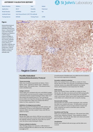
  • 1. Figure: Immunohistochemical analysis of paraffin embedded Human liver tissue. 1: Cleaved-Notch 2 (D1733) Polyclonal Antibody was diluted at 1:200 (4 degree Celsius,overnight). 2: Sodium citrate pH 6.0 was used for antibody retrieval (>98 degree Celsius,20min). 3: Secondary antibody was diluted at 1:200 (room temperature, 30min). Negative control was used by secondary antibody only. Report Number 90068-a Host Rabbit Application IHC-P Clonality Polyclonal Model Number STJ90068 Clone ID NA Antibody Name Anti-Cleaved-Notch 2 (D1733) antibody Testing Species HUMAN Testing Tissue LIVER ANTIBODY VALIDATION REPORT a. (A small amount of distilled water was added into the incubation box to prevent evaporation of antibody). 52. Secondary antibody incubation a. Slides were washed 3 times, with PBS on a shaker for 5min. Shortly after the slides were dried the corresponding secondary antibody solution was added (HRP labelled), covering the tissues, and incubated at room temperature for 30min. b. 53. DAB staining a. Slides were washed 3 times, with PBS on a shaker for 5min. b. Shortly after, the slides were dried and fresh DAB staining buffer was added inside the circles. The staining time was adjusted under a microscope. Yellow-brown colour represented a positive result. Slides were washed with water to stop the staining. c. 54. Haematoxylin staining a. Haematoxylin was used to counter-staining for 1min, and then the slides were washed with water. 1% Hydrochloric acid and alcohol was added for several seconds and then washed with water. Ammonia was used to reveal blue colour, and then flushed with water. b. 55. Desolation and Clearing i. Slides were incubated sequentially into: 75% alcohol 5min, 85% alcohol 5min, Anhydrous ethanol - 5min, Anhydrous ethanol - 5min & Xylene - 5min. Shortly after slides were dried and neutral gum was used to seal the slides. ii. 56. Visualization a. Results were validated with microscope, and the slides were scanned. Paraffin-Embedded Immunohistochemistry Protocol 46. 47. Tissue processing a. Slides were incubated sequentially into Xylene; 15min – Xylene, 15min - Anhydrous ethanol, 5min - Anhydrous ethanol, 5min - 85% alcohol, 5min - 75% alcohol & 5min – wash in distilled water. b. 48. Antigen retrieval a. Tissue slides were incubated with citric acid (PH6.0) antigen retrieval buffer and microwaved for antigen retrieval (heated until boiled and then stopped heating) for 8min. Slides were then heated with medium power for 7min. During this process slides were kept from drying out. After cooling down at room temperature, slides were washed with PBS on shaker for 5min, repeated for 3 times. b. 49. Inhibition of endogenous peroxidase a. Slides were placed in 3% Hydrogen peroxide solution, and incubated for 10 min at room temperature without light exposure. Slides were then washed 3 times with PBS on a shaker for 5mins. b. 50. BSA Blocking a. Shortly after slides were dried, a PAP pen was used to draw circles around the tissue sections (and to prevent draining of the antibody solution). Inside the circles, BSA was used to cover the tissue evenly, blocking for 30min. b. 51. Primary antibody incubation After blocking solution was removed a 1:200 solution of primary antibody/PBS was added on the slide, and incubated overnight at 4°C. St John's Laboratory Ltd. www.stjohnslabs.com
  • 2. Figure: Immunohistochemical analysis of paraffin embedded Rat lung tissue. 1: Cleaved-Notch 2 (D1733) Polyclonal Antibody was diluted at 1:200 (4 degree Celsius,overnight). 2: Sodium citrate pH 6.0 was used for antibody retrieval (>98 degree Celsius,20min). 3: Secondary antibody was diluted at 1:200 (room temperature, 30min). Negative control was used by secondary antibody only. Report Number 90068-b Host Rabbit Application IHC-P Clonality Polyclonal Model Number STJ90068 Clone ID NA Antibody Name Anti-Cleaved-Notch 2 (D1733) antibody Testing Species RAT Testing Tissue LUNG ANTIBODY VALIDATION REPORT a. (A small amount of distilled water was added into the incubation box to prevent evaporation of antibody). 41. Secondary antibody incubation a. Slides were washed 3 times, with PBS on a shaker for 5min. Shortly after the slides were dried the corresponding secondary antibody solution was added (HRP labelled), covering the tissues, and incubated at room temperature for 30min. b. 42. DAB staining a. Slides were washed 3 times, with PBS on a shaker for 5min. b. Shortly after, the slides were dried and fresh DAB staining buffer was added inside the circles. The staining time was adjusted under a microscope. Yellow-brown colour represented a positive result. Slides were washed with water to stop the staining. c. 43. Haematoxylin staining a. Haematoxylin was used to counter-staining for 1min, and then the slides were washed with water. 1% Hydrochloric acid and alcohol was added for several seconds and then washed with water. Ammonia was used to reveal blue colour, and then flushed with water. b. 44. Desolation and Clearing i. Slides were incubated sequentially into: 75% alcohol 5min, 85% alcohol 5min, Anhydrous ethanol - 5min, Anhydrous ethanol - 5min & Xylene - 5min. Shortly after slides were dried and neutral gum was used to seal the slides. ii. 45. Visualization a. Results were validated with microscope, and the slides were scanned. Paraffin-Embedded Immunohistochemistry Protocol 35. 36. Tissue processing a. Slides were incubated sequentially into Xylene; 15min – Xylene, 15min - Anhydrous ethanol, 5min - Anhydrous ethanol, 5min - 85% alcohol, 5min - 75% alcohol & 5min – wash in distilled water. b. 37. Antigen retrieval a. Tissue slides were incubated with citric acid (PH6.0) antigen retrieval buffer and microwaved for antigen retrieval (heated until boiled and then stopped heating) for 8min. Slides were then heated with medium power for 7min. During this process slides were kept from drying out. After cooling down at room temperature, slides were washed with PBS on shaker for 5min, repeated for 3 times. b. 38. Inhibition of endogenous peroxidase a. Slides were placed in 3% Hydrogen peroxide solution, and incubated for 10 min at room temperature without light exposure. Slides were then washed 3 times with PBS on a shaker for 5mins. b. 39. BSA Blocking a. Shortly after slides were dried, a PAP pen was used to draw circles around the tissue sections (and to prevent draining of the antibody solution). Inside the circles, BSA was used to cover the tissue evenly, blocking for 30min. b. 40. Primary antibody incubation After blocking solution was removed a 1:200 solution of primary antibody/PBS was added on the slide, and incubated overnight at 4°C. St John's Laboratory Ltd. www.stjohnslabs.com
  • 3. Figure: Immunohistochemical analysis of paraffin embedded Rat spleen tissue. 1: Cleaved-Notch 2 (D1733) Polyclonal Antibody was diluted at 1:200 (4 degree Celsius,overnight). 2: Sodium citrate pH 6.0 was used for antibody retrieval (>98 degree Celsius,20min). 3: Secondary antibody was diluted at 1:200 (room temperature, 30min). Negative control was used by secondary antibody only. Report Number 90068-c Host Rabbit Application IHC-P Clonality Polyclonal Model Number STJ90068 Clone ID NA Antibody Name Anti-Cleaved-Notch 2 (D1733) antibody Testing Species RAT Testing Tissue SPLEEN ANTIBODY VALIDATION REPORT a. (A small amount of distilled water was added into the incubation box to prevent evaporation of antibody). 30. Secondary antibody incubation a. Slides were washed 3 times, with PBS on a shaker for 5min. Shortly after the slides were dried the corresponding secondary antibody solution was added (HRP labelled), covering the tissues, and incubated at room temperature for 30min. b. 31. DAB staining a. Slides were washed 3 times, with PBS on a shaker for 5min. b. Shortly after, the slides were dried and fresh DAB staining buffer was added inside the circles. The staining time was adjusted under a microscope. Yellow-brown colour represented a positive result. Slides were washed with water to stop the staining. c. 32. Haematoxylin staining a. Haematoxylin was used to counter-staining for 1min, and then the slides were washed with water. 1% Hydrochloric acid and alcohol was added for several seconds and then washed with water. Ammonia was used to reveal blue colour, and then flushed with water. b. 33. Desolation and Clearing i. Slides were incubated sequentially into: 75% alcohol 5min, 85% alcohol 5min, Anhydrous ethanol - 5min, Anhydrous ethanol - 5min & Xylene - 5min. Shortly after slides were dried and neutral gum was used to seal the slides. ii. 34. Visualization a. Results were validated with microscope, and the slides were scanned. Paraffin-Embedded Immunohistochemistry Protocol 24. 25. Tissue processing a. Slides were incubated sequentially into Xylene; 15min – Xylene, 15min - Anhydrous ethanol, 5min - Anhydrous ethanol, 5min - 85% alcohol, 5min - 75% alcohol & 5min – wash in distilled water. b. 26. Antigen retrieval a. Tissue slides were incubated with citric acid (PH6.0) antigen retrieval buffer and microwaved for antigen retrieval (heated until boiled and then stopped heating) for 8min. Slides were then heated with medium power for 7min. During this process slides were kept from drying out. After cooling down at room temperature, slides were washed with PBS on shaker for 5min, repeated for 3 times. b. 27. Inhibition of endogenous peroxidase a. Slides were placed in 3% Hydrogen peroxide solution, and incubated for 10 min at room temperature without light exposure. Slides were then washed 3 times with PBS on a shaker for 5mins. b. 28. BSA Blocking a. Shortly after slides were dried, a PAP pen was used to draw circles around the tissue sections (and to prevent draining of the antibody solution). Inside the circles, BSA was used to cover the tissue evenly, blocking for 30min. b. 29. Primary antibody incubation After blocking solution was removed a 1:200 solution of primary antibody/PBS was added on the slide, and incubated overnight at 4°C. St John's Laboratory Ltd. www.stjohnslabs.com
  • 4. Figure: Immunohistochemical analysis of paraffin embedded Mouse lung tissue. 1: Cleaved-Notch 2 (D1733) Polyclonal Antibody was diluted at 1:200 (4 degree Celsius,overnight). 2: Sodium citrate pH 6.0 was used for antibody retrieval (>98 degree Celsius,20min). 3: Secondary antibody was diluted at 1:200 (room temperature, 30min). Negative control was used by secondary antibody only. Report Number 90068-d Host Rabbit Application IHC-P Clonality Polyclonal Model Number STJ90068 Clone ID NA Antibody Name Anti-Cleaved-Notch 2 (D1733) antibody Testing Species MOUSE Testing Tissue LUNG ANTIBODY VALIDATION REPORT a. (A small amount of distilled water was added into the incubation box to prevent evaporation of antibody). 19. Secondary antibody incubation a. Slides were washed 3 times, with PBS on a shaker for 5min. Shortly after the slides were dried the corresponding secondary antibody solution was added (HRP labelled), covering the tissues, and incubated at room temperature for 30min. b. 20. DAB staining a. Slides were washed 3 times, with PBS on a shaker for 5min. b. Shortly after, the slides were dried and fresh DAB staining buffer was added inside the circles. The staining time was adjusted under a microscope. Yellow-brown colour represented a positive result. Slides were washed with water to stop the staining. c. 21. Haematoxylin staining a. Haematoxylin was used to counter-staining for 1min, and then the slides were washed with water. 1% Hydrochloric acid and alcohol was added for several seconds and then washed with water. Ammonia was used to reveal blue colour, and then flushed with water. b. 22. Desolation and Clearing i. Slides were incubated sequentially into: 75% alcohol 5min, 85% alcohol 5min, Anhydrous ethanol - 5min, Anhydrous ethanol - 5min & Xylene - 5min. Shortly after slides were dried and neutral gum was used to seal the slides. ii. 23. Visualization a. Results were validated with microscope, and the slides were scanned. Paraffin-Embedded Immunohistochemistry Protocol 13. 14. Tissue processing a. Slides were incubated sequentially into Xylene; 15min – Xylene, 15min - Anhydrous ethanol, 5min - Anhydrous ethanol, 5min - 85% alcohol, 5min - 75% alcohol & 5min – wash in distilled water. b. 15. Antigen retrieval a. Tissue slides were incubated with citric acid (PH6.0) antigen retrieval buffer and microwaved for antigen retrieval (heated until boiled and then stopped heating) for 8min. Slides were then heated with medium power for 7min. During this process slides were kept from drying out. After cooling down at room temperature, slides were washed with PBS on shaker for 5min, repeated for 3 times. b. 16. Inhibition of endogenous peroxidase a. Slides were placed in 3% Hydrogen peroxide solution, and incubated for 10 min at room temperature without light exposure. Slides were then washed 3 times with PBS on a shaker for 5mins. b. 17. BSA Blocking a. Shortly after slides were dried, a PAP pen was used to draw circles around the tissue sections (and to prevent draining of the antibody solution). Inside the circles, BSA was used to cover the tissue evenly, blocking for 30min. b. 18. Primary antibody incubation After blocking solution was removed a 1:200 solution of primary antibody/PBS was added on the slide, and incubated overnight at 4°C. St John's Laboratory Ltd. www.stjohnslabs.com
  • 5. Figure: Immunohistochemical analysis of paraffin embedded Mouse kidney tissue. 1: Cleaved-Notch 2 (D1733) Polyclonal Antibody was diluted at 1:200 (4 degree Celsius,overnight). 2: Sodium citrate pH 6.0 was used for antibody retrieval (>98 degree Celsius,20min). 3: Secondary antibody was diluted at 1:200 (room temperature, 30min). Negative control was used by secondary antibody only. Report Number 90068-e Host Rabbit Application IHC-P Clonality Polyclonal Model Number STJ90068 Clone ID NA Antibody Name Anti-Cleaved-Notch 2 (D1733) antibody Testing Species MOUSE Testing Tissue KIDNEY ANTIBODY VALIDATION REPORT a. (A small amount of distilled water was added into the incubation box to prevent evaporation of antibody). 8. Secondary antibody incubation a. Slides were washed 3 times, with PBS on a shaker for 5min. Shortly after the slides were dried the corresponding secondary antibody solution was added (HRP labelled), covering the tissues, and incubated at room temperature for 30min. b. 9. DAB staining a. Slides were washed 3 times, with PBS on a shaker for 5min. b. Shortly after, the slides were dried and fresh DAB staining buffer was added inside the circles. The staining time was adjusted under a microscope. Yellow-brown colour represented a positive result. Slides were washed with water to stop the staining. c. 10. Haematoxylin staining a. Haematoxylin was used to counter-staining for 1min, and then the slides were washed with water. 1% Hydrochloric acid and alcohol was added for several seconds and then washed with water. Ammonia was used to reveal blue colour, and then flushed with water. b. 11. Desolation and Clearing i. Slides were incubated sequentially into: 75% alcohol 5min, 85% alcohol 5min, Anhydrous ethanol - 5min, Anhydrous ethanol - 5min & Xylene - 5min. Shortly after slides were dried and neutral gum was used to seal the slides. ii. 12. Visualization a. Results were validated with microscope, and the slides were scanned. Paraffin-Embedded Immunohistochemistry Protocol 2. 3. Tissue processing a. Slides were incubated sequentially into Xylene; 15min – Xylene, 15min - Anhydrous ethanol, 5min - Anhydrous ethanol, 5min - 85% alcohol, 5min - 75% alcohol & 5min – wash in distilled water. b. 4. Antigen retrieval a. Tissue slides were incubated with citric acid (PH6.0) antigen retrieval buffer and microwaved for antigen retrieval (heated until boiled and then stopped heating) for 8min. Slides were then heated with medium power for 7min. During this process slides were kept from drying out. After cooling down at room temperature, slides were washed with PBS on shaker for 5min, repeated for 3 times. b. 5. Inhibition of endogenous peroxidase a. Slides were placed in 3% Hydrogen peroxide solution, and incubated for 10 min at room temperature without light exposure. Slides were then washed 3 times with PBS on a shaker for 5mins. b. 6. BSA Blocking a. Shortly after slides were dried, a PAP pen was used to draw circles around the tissue sections (and to prevent draining of the antibody solution). Inside the circles, BSA was used to cover the tissue evenly, blocking for 30min. b. 7. Primary antibody incubation After blocking solution was removed a 1:200 solution of primary antibody/PBS was added on the slide, and incubated overnight at 4°C. St John's Laboratory Ltd. www.stjohnslabs.com