SlideShare a Scribd company logo
1 of 11
Download to read offline
Figure:
Immunohistochemical
analysis of paraffin
embedded Human liver
tissue. 1: E-cadherin
Polyclonal Antibody
was diluted at 1:200 (4
degree
Celsius,overnight). 2:
Sodium citrate pH 6.0
was used for antibody
retrieval (>98 degree
Celsius,20min). 3:
Secondary antibody was
diluted at 1:200 (room
temperature, 30min).
Negative control was
used by secondary
antibody only.
Report Number 92819-a Host Rabbit
Application IHC-P Clonality Polyclonal
Model Number STJ92819 Clone ID NA
Antibody Name Anti-E-cadherin antibody
Testing Species HUMAN Testing Tissue LIVER
ANTIBODY VALIDATION REPORT
b. (A small amount of distilled water was added into the incubation
box to prevent evaporation of antibody).
95. Secondary antibody incubation
a. Slides were washed 3 times, with PBS on a shaker for 5min.
Shortly after the slides were dried the corresponding secondary
antibody solution was added (HRP labelled), covering the
tissues, and incubated at room temperature for 30min.
b.
96. DAB staining
a. Slides were washed 3 times, with PBS on a shaker for 5min.
b. Shortly after, the slides were dried and fresh DAB staining buffer
was added inside the circles. The staining time was adjusted
under a microscope. Yellow-brown colour represented a positive
result. Slides were washed with water to stop the staining.
c.
97. Haematoxylin staining
a. Haematoxylin was used to counter-staining for 1min, and then
the slides were washed with water. 1% Hydrochloric acid and
alcohol was added for several seconds and then washed with
water. Ammonia was used to reveal blue colour, and then
flushed with water.
b.
98. Desolation and Clearing
i. Slides were incubated sequentially into: 75% alcohol 5min, 85%
alcohol 5min, Anhydrous ethanol - 5min, Anhydrous ethanol -
5min & Xylene - 5min. Shortly after slides were dried and neutral
gum was used to seal the slides.
ii.
99. Visualization
a. Results were validated with microscope, and the slides were
scanned.
Paraffin-Embedded
Immunohistochemistry Protocol
89.
90. Tissue processing
a. Slides were incubated sequentially into Xylene; 15min –
Xylene, 15min - Anhydrous ethanol, 5min - Anhydrous
ethanol, 5min - 85% alcohol, 5min - 75% alcohol & 5min –
wash in distilled water.
b.
91. Antigen retrieval
a. Tissue slides were incubated with citric acid (PH6.0) antigen
retrieval buffer and microwaved for antigen retrieval (heated
until boiled and then stopped heating) for 8min. Slides were
then heated with medium power for 7min. During this
process slides were kept from drying out. After cooling down
at room temperature, slides were washed with PBS on
shaker for 5min, repeated for 3 times.
b.
92. Inhibition of endogenous peroxidase
a. Slides were placed in 3% Hydrogen peroxide solution, and
incubated for 10 min at room temperature without light
exposure. Slides were then washed 3 times with PBS on a
shaker for 5mins.
b.
93. BSA Blocking
a. Shortly after slides were dried, a PAP pen was used to draw
circles around the tissue sections (and to prevent draining of
the antibody solution). Inside the circles, BSA was used to
cover the tissue evenly, blocking for 30min.
b.
94. Primary antibody incubation
After blocking solution was removed a 1:200 solution of
primary antibody/PBS was added on the slide, and incubated
overnight at 4°C.
St John's Laboratory Ltd.
www.stjohnslabs.com
Figure:
Immunohistochemical
analysis of paraffin
embedded Human liver
cancer tissue. 1: E-
cadherin Polyclonal
Antibody was diluted at
1:200 (4 degree
Celsius,overnight). 2:
Sodium citrate pH 6.0
was used for antibody
retrieval (>98 degree
Celsius,20min). 3:
Secondary antibody was
diluted at 1:200 (room
temperature, 30min).
Negative control was
used by secondary
antibody only.
Report Number 92819-b Host Rabbit
Application IHC-P Clonality Polyclonal
Model Number STJ92819 Clone ID NA
Antibody Name Anti-E-cadherin antibody
Testing Species HUMAN Testing Tissue LIVER CANCER
ANTIBODY VALIDATION REPORT
b. (A small amount of distilled water was added into the incubation
box to prevent evaporation of antibody).
84. Secondary antibody incubation
a. Slides were washed 3 times, with PBS on a shaker for 5min.
Shortly after the slides were dried the corresponding secondary
antibody solution was added (HRP labelled), covering the
tissues, and incubated at room temperature for 30min.
b.
85. DAB staining
a. Slides were washed 3 times, with PBS on a shaker for 5min.
b. Shortly after, the slides were dried and fresh DAB staining buffer
was added inside the circles. The staining time was adjusted
under a microscope. Yellow-brown colour represented a positive
result. Slides were washed with water to stop the staining.
c.
86. Haematoxylin staining
a. Haematoxylin was used to counter-staining for 1min, and then
the slides were washed with water. 1% Hydrochloric acid and
alcohol was added for several seconds and then washed with
water. Ammonia was used to reveal blue colour, and then
flushed with water.
b.
87. Desolation and Clearing
i. Slides were incubated sequentially into: 75% alcohol 5min, 85%
alcohol 5min, Anhydrous ethanol - 5min, Anhydrous ethanol -
5min & Xylene - 5min. Shortly after slides were dried and neutral
gum was used to seal the slides.
ii.
88. Visualization
a. Results were validated with microscope, and the slides were
scanned.
Paraffin-Embedded
Immunohistochemistry Protocol
78.
79. Tissue processing
a. Slides were incubated sequentially into Xylene; 15min –
Xylene, 15min - Anhydrous ethanol, 5min - Anhydrous
ethanol, 5min - 85% alcohol, 5min - 75% alcohol & 5min –
wash in distilled water.
b.
80. Antigen retrieval
a. Tissue slides were incubated with citric acid (PH6.0) antigen
retrieval buffer and microwaved for antigen retrieval (heated
until boiled and then stopped heating) for 8min. Slides were
then heated with medium power for 7min. During this
process slides were kept from drying out. After cooling down
at room temperature, slides were washed with PBS on
shaker for 5min, repeated for 3 times.
b.
81. Inhibition of endogenous peroxidase
a. Slides were placed in 3% Hydrogen peroxide solution, and
incubated for 10 min at room temperature without light
exposure. Slides were then washed 3 times with PBS on a
shaker for 5mins.
b.
82. BSA Blocking
a. Shortly after slides were dried, a PAP pen was used to draw
circles around the tissue sections (and to prevent draining of
the antibody solution). Inside the circles, BSA was used to
cover the tissue evenly, blocking for 30min.
b.
83. Primary antibody incubation
After blocking solution was removed a 1:200 solution of
primary antibody/PBS was added on the slide, and incubated
overnight at 4°C.
St John's Laboratory Ltd.
www.stjohnslabs.com
Figure:
Immunohistochemical
analysis of paraffin
embedded Human
stomach tissue. 1: E-
cadherin Polyclonal
Antibody was diluted at
1:200 (4 degree
Celsius,overnight). 2:
Sodium citrate pH 6.0
was used for antibody
retrieval (>98 degree
Celsius,20min). 3:
Secondary antibody was
diluted at 1:200 (room
temperature, 30min).
Negative control was
used by secondary
antibody only.
Report Number 92819-c Host Rabbit
Application IHC-P Clonality Polyclonal
Model Number STJ92819 Clone ID NA
Antibody Name Anti-E-cadherin antibody
Testing Species HUMAN Testing Tissue STOMACH
ANTIBODY VALIDATION REPORT
b. (A small amount of distilled water was added into the incubation
box to prevent evaporation of antibody).
73. Secondary antibody incubation
a. Slides were washed 3 times, with PBS on a shaker for 5min.
Shortly after the slides were dried the corresponding secondary
antibody solution was added (HRP labelled), covering the
tissues, and incubated at room temperature for 30min.
b.
74. DAB staining
a. Slides were washed 3 times, with PBS on a shaker for 5min.
b. Shortly after, the slides were dried and fresh DAB staining buffer
was added inside the circles. The staining time was adjusted
under a microscope. Yellow-brown colour represented a positive
result. Slides were washed with water to stop the staining.
c.
75. Haematoxylin staining
a. Haematoxylin was used to counter-staining for 1min, and then
the slides were washed with water. 1% Hydrochloric acid and
alcohol was added for several seconds and then washed with
water. Ammonia was used to reveal blue colour, and then
flushed with water.
b.
76. Desolation and Clearing
i. Slides were incubated sequentially into: 75% alcohol 5min, 85%
alcohol 5min, Anhydrous ethanol - 5min, Anhydrous ethanol -
5min & Xylene - 5min. Shortly after slides were dried and neutral
gum was used to seal the slides.
ii.
77. Visualization
a. Results were validated with microscope, and the slides were
scanned.
Paraffin-Embedded
Immunohistochemistry Protocol
67.
68. Tissue processing
a. Slides were incubated sequentially into Xylene; 15min –
Xylene, 15min - Anhydrous ethanol, 5min - Anhydrous
ethanol, 5min - 85% alcohol, 5min - 75% alcohol & 5min –
wash in distilled water.
b.
69. Antigen retrieval
a. Tissue slides were incubated with citric acid (PH6.0) antigen
retrieval buffer and microwaved for antigen retrieval (heated
until boiled and then stopped heating) for 8min. Slides were
then heated with medium power for 7min. During this
process slides were kept from drying out. After cooling down
at room temperature, slides were washed with PBS on
shaker for 5min, repeated for 3 times.
b.
70. Inhibition of endogenous peroxidase
a. Slides were placed in 3% Hydrogen peroxide solution, and
incubated for 10 min at room temperature without light
exposure. Slides were then washed 3 times with PBS on a
shaker for 5mins.
b.
71. BSA Blocking
a. Shortly after slides were dried, a PAP pen was used to draw
circles around the tissue sections (and to prevent draining of
the antibody solution). Inside the circles, BSA was used to
cover the tissue evenly, blocking for 30min.
b.
72. Primary antibody incubation
After blocking solution was removed a 1:200 solution of
primary antibody/PBS was added on the slide, and incubated
overnight at 4°C.
St John's Laboratory Ltd.
www.stjohnslabs.com
Figure:
Immunohistochemical
analysis of paraffin
embedded Rat heart
tissue. 1: E-cadherin
Polyclonal Antibody
was diluted at 1:200 (4
degree
Celsius,overnight). 2:
Sodium citrate pH 6.0
was used for antibody
retrieval (>98 degree
Celsius,20min). 3:
Secondary antibody was
diluted at 1:200 (room
temperature, 30min).
Negative control was
used by secondary
antibody only.
Report Number 92819-d Host Rabbit
Application IHC-P Clonality Polyclonal
Model Number STJ92819 Clone ID NA
Antibody Name Anti-E-cadherin antibody
Testing Species RAT Testing Tissue HEART
ANTIBODY VALIDATION REPORT
b. (A small amount of distilled water was added into the incubation
box to prevent evaporation of antibody).
62. Secondary antibody incubation
a. Slides were washed 3 times, with PBS on a shaker for 5min.
Shortly after the slides were dried the corresponding secondary
antibody solution was added (HRP labelled), covering the
tissues, and incubated at room temperature for 30min.
b.
63. DAB staining
a. Slides were washed 3 times, with PBS on a shaker for 5min.
b. Shortly after, the slides were dried and fresh DAB staining buffer
was added inside the circles. The staining time was adjusted
under a microscope. Yellow-brown colour represented a positive
result. Slides were washed with water to stop the staining.
c.
64. Haematoxylin staining
a. Haematoxylin was used to counter-staining for 1min, and then
the slides were washed with water. 1% Hydrochloric acid and
alcohol was added for several seconds and then washed with
water. Ammonia was used to reveal blue colour, and then
flushed with water.
b.
65. Desolation and Clearing
i. Slides were incubated sequentially into: 75% alcohol 5min, 85%
alcohol 5min, Anhydrous ethanol - 5min, Anhydrous ethanol -
5min & Xylene - 5min. Shortly after slides were dried and neutral
gum was used to seal the slides.
ii.
66. Visualization
a. Results were validated with microscope, and the slides were
scanned.
Paraffin-Embedded
Immunohistochemistry Protocol
56.
57. Tissue processing
a. Slides were incubated sequentially into Xylene; 15min –
Xylene, 15min - Anhydrous ethanol, 5min - Anhydrous
ethanol, 5min - 85% alcohol, 5min - 75% alcohol & 5min –
wash in distilled water.
b.
58. Antigen retrieval
a. Tissue slides were incubated with citric acid (PH6.0) antigen
retrieval buffer and microwaved for antigen retrieval (heated
until boiled and then stopped heating) for 8min. Slides were
then heated with medium power for 7min. During this
process slides were kept from drying out. After cooling down
at room temperature, slides were washed with PBS on
shaker for 5min, repeated for 3 times.
b.
59. Inhibition of endogenous peroxidase
a. Slides were placed in 3% Hydrogen peroxide solution, and
incubated for 10 min at room temperature without light
exposure. Slides were then washed 3 times with PBS on a
shaker for 5mins.
b.
60. BSA Blocking
a. Shortly after slides were dried, a PAP pen was used to draw
circles around the tissue sections (and to prevent draining of
the antibody solution). Inside the circles, BSA was used to
cover the tissue evenly, blocking for 30min.
b.
61. Primary antibody incubation
After blocking solution was removed a 1:200 solution of
primary antibody/PBS was added on the slide, and incubated
overnight at 4°C.
St John's Laboratory Ltd.
www.stjohnslabs.com
Figure:
Immunohistochemical
analysis of paraffin
embedded Rat lung
tissue. 1: E-cadherin
Polyclonal Antibody
was diluted at 1:200 (4
degree
Celsius,overnight). 2:
Sodium citrate pH 6.0
was used for antibody
retrieval (>98 degree
Celsius,20min). 3:
Secondary antibody was
diluted at 1:200 (room
temperature, 30min).
Negative control was
used by secondary
antibody only.
Report Number 92819-e Host Rabbit
Application IHC-P Clonality Polyclonal
Model Number STJ92819 Clone ID NA
Antibody Name Anti-E-cadherin antibody
Testing Species RAT Testing Tissue LUNG
ANTIBODY VALIDATION REPORT
b. (A small amount of distilled water was added into the incubation
box to prevent evaporation of antibody).
51. Secondary antibody incubation
a. Slides were washed 3 times, with PBS on a shaker for 5min.
Shortly after the slides were dried the corresponding secondary
antibody solution was added (HRP labelled), covering the
tissues, and incubated at room temperature for 30min.
b.
52. DAB staining
a. Slides were washed 3 times, with PBS on a shaker for 5min.
b. Shortly after, the slides were dried and fresh DAB staining buffer
was added inside the circles. The staining time was adjusted
under a microscope. Yellow-brown colour represented a positive
result. Slides were washed with water to stop the staining.
c.
53. Haematoxylin staining
a. Haematoxylin was used to counter-staining for 1min, and then
the slides were washed with water. 1% Hydrochloric acid and
alcohol was added for several seconds and then washed with
water. Ammonia was used to reveal blue colour, and then
flushed with water.
b.
54. Desolation and Clearing
i. Slides were incubated sequentially into: 75% alcohol 5min, 85%
alcohol 5min, Anhydrous ethanol - 5min, Anhydrous ethanol -
5min & Xylene - 5min. Shortly after slides were dried and neutral
gum was used to seal the slides.
ii.
55. Visualization
a. Results were validated with microscope, and the slides were
scanned.
Paraffin-Embedded
Immunohistochemistry Protocol
45.
46. Tissue processing
a. Slides were incubated sequentially into Xylene; 15min –
Xylene, 15min - Anhydrous ethanol, 5min - Anhydrous
ethanol, 5min - 85% alcohol, 5min - 75% alcohol & 5min –
wash in distilled water.
b.
47. Antigen retrieval
a. Tissue slides were incubated with citric acid (PH6.0) antigen
retrieval buffer and microwaved for antigen retrieval (heated
until boiled and then stopped heating) for 8min. Slides were
then heated with medium power for 7min. During this
process slides were kept from drying out. After cooling down
at room temperature, slides were washed with PBS on
shaker for 5min, repeated for 3 times.
b.
48. Inhibition of endogenous peroxidase
a. Slides were placed in 3% Hydrogen peroxide solution, and
incubated for 10 min at room temperature without light
exposure. Slides were then washed 3 times with PBS on a
shaker for 5mins.
b.
49. BSA Blocking
a. Shortly after slides were dried, a PAP pen was used to draw
circles around the tissue sections (and to prevent draining of
the antibody solution). Inside the circles, BSA was used to
cover the tissue evenly, blocking for 30min.
b.
50. Primary antibody incubation
After blocking solution was removed a 1:200 solution of
primary antibody/PBS was added on the slide, and incubated
overnight at 4°C.
St John's Laboratory Ltd.
www.stjohnslabs.com
Figure:
Immunohistochemical
analysis of paraffin
embedded Rat kidney
tissue. 1: E-cadherin
Polyclonal Antibody
was diluted at 1:200 (4
degree
Celsius,overnight). 2:
Sodium citrate pH 6.0
was used for antibody
retrieval (>98 degree
Celsius,20min). 3:
Secondary antibody was
diluted at 1:200 (room
temperature, 30min).
Negative control was
used by secondary
antibody only.
Report Number 92819-f Host Rabbit
Application IHC-P Clonality Polyclonal
Model Number STJ92819 Clone ID NA
Antibody Name Anti-E-cadherin antibody
Testing Species RAT Testing Tissue KIDNEY
ANTIBODY VALIDATION REPORT
b. (A small amount of distilled water was added into the incubation
box to prevent evaporation of antibody).
40. Secondary antibody incubation
a. Slides were washed 3 times, with PBS on a shaker for 5min.
Shortly after the slides were dried the corresponding secondary
antibody solution was added (HRP labelled), covering the
tissues, and incubated at room temperature for 30min.
b.
41. DAB staining
a. Slides were washed 3 times, with PBS on a shaker for 5min.
b. Shortly after, the slides were dried and fresh DAB staining buffer
was added inside the circles. The staining time was adjusted
under a microscope. Yellow-brown colour represented a positive
result. Slides were washed with water to stop the staining.
c.
42. Haematoxylin staining
a. Haematoxylin was used to counter-staining for 1min, and then
the slides were washed with water. 1% Hydrochloric acid and
alcohol was added for several seconds and then washed with
water. Ammonia was used to reveal blue colour, and then
flushed with water.
b.
43. Desolation and Clearing
i. Slides were incubated sequentially into: 75% alcohol 5min, 85%
alcohol 5min, Anhydrous ethanol - 5min, Anhydrous ethanol -
5min & Xylene - 5min. Shortly after slides were dried and neutral
gum was used to seal the slides.
ii.
44. Visualization
a. Results were validated with microscope, and the slides were
scanned.
Paraffin-Embedded
Immunohistochemistry Protocol
34.
35. Tissue processing
a. Slides were incubated sequentially into Xylene; 15min –
Xylene, 15min - Anhydrous ethanol, 5min - Anhydrous
ethanol, 5min - 85% alcohol, 5min - 75% alcohol & 5min –
wash in distilled water.
b.
36. Antigen retrieval
a. Tissue slides were incubated with citric acid (PH6.0) antigen
retrieval buffer and microwaved for antigen retrieval (heated
until boiled and then stopped heating) for 8min. Slides were
then heated with medium power for 7min. During this
process slides were kept from drying out. After cooling down
at room temperature, slides were washed with PBS on
shaker for 5min, repeated for 3 times.
b.
37. Inhibition of endogenous peroxidase
a. Slides were placed in 3% Hydrogen peroxide solution, and
incubated for 10 min at room temperature without light
exposure. Slides were then washed 3 times with PBS on a
shaker for 5mins.
b.
38. BSA Blocking
a. Shortly after slides were dried, a PAP pen was used to draw
circles around the tissue sections (and to prevent draining of
the antibody solution). Inside the circles, BSA was used to
cover the tissue evenly, blocking for 30min.
b.
39. Primary antibody incubation
After blocking solution was removed a 1:200 solution of
primary antibody/PBS was added on the slide, and incubated
overnight at 4°C.
St John's Laboratory Ltd.
www.stjohnslabs.com
Figure:
Immunohistochemical
analysis of paraffin
embedded Rat spleen
tissue. 1: E-cadherin
Polyclonal Antibody
was diluted at 1:200 (4
degree
Celsius,overnight). 2:
Sodium citrate pH 6.0
was used for antibody
retrieval (>98 degree
Celsius,20min). 3:
Secondary antibody was
diluted at 1:200 (room
temperature, 30min).
Negative control was
used by secondary
antibody only.
Report Number 92819-g Host Rabbit
Application IHC-P Clonality Polyclonal
Model Number STJ92819 Clone ID NA
Antibody Name Anti-E-cadherin antibody
Testing Species RAT Testing Tissue SPLEEN
ANTIBODY VALIDATION REPORT
b. (A small amount of distilled water was added into the incubation
box to prevent evaporation of antibody).
29. Secondary antibody incubation
a. Slides were washed 3 times, with PBS on a shaker for 5min.
Shortly after the slides were dried the corresponding secondary
antibody solution was added (HRP labelled), covering the
tissues, and incubated at room temperature for 30min.
b.
30. DAB staining
a. Slides were washed 3 times, with PBS on a shaker for 5min.
b. Shortly after, the slides were dried and fresh DAB staining buffer
was added inside the circles. The staining time was adjusted
under a microscope. Yellow-brown colour represented a positive
result. Slides were washed with water to stop the staining.
c.
31. Haematoxylin staining
a. Haematoxylin was used to counter-staining for 1min, and then
the slides were washed with water. 1% Hydrochloric acid and
alcohol was added for several seconds and then washed with
water. Ammonia was used to reveal blue colour, and then
flushed with water.
b.
32. Desolation and Clearing
i. Slides were incubated sequentially into: 75% alcohol 5min, 85%
alcohol 5min, Anhydrous ethanol - 5min, Anhydrous ethanol -
5min & Xylene - 5min. Shortly after slides were dried and neutral
gum was used to seal the slides.
ii.
33. Visualization
a. Results were validated with microscope, and the slides were
scanned.
Paraffin-Embedded
Immunohistochemistry Protocol
23.
24. Tissue processing
a. Slides were incubated sequentially into Xylene; 15min –
Xylene, 15min - Anhydrous ethanol, 5min - Anhydrous
ethanol, 5min - 85% alcohol, 5min - 75% alcohol & 5min –
wash in distilled water.
b.
25. Antigen retrieval
a. Tissue slides were incubated with citric acid (PH6.0) antigen
retrieval buffer and microwaved for antigen retrieval (heated
until boiled and then stopped heating) for 8min. Slides were
then heated with medium power for 7min. During this
process slides were kept from drying out. After cooling down
at room temperature, slides were washed with PBS on
shaker for 5min, repeated for 3 times.
b.
26. Inhibition of endogenous peroxidase
a. Slides were placed in 3% Hydrogen peroxide solution, and
incubated for 10 min at room temperature without light
exposure. Slides were then washed 3 times with PBS on a
shaker for 5mins.
b.
27. BSA Blocking
a. Shortly after slides were dried, a PAP pen was used to draw
circles around the tissue sections (and to prevent draining of
the antibody solution). Inside the circles, BSA was used to
cover the tissue evenly, blocking for 30min.
b.
28. Primary antibody incubation
After blocking solution was removed a 1:200 solution of
primary antibody/PBS was added on the slide, and incubated
overnight at 4°C.
St John's Laboratory Ltd.
www.stjohnslabs.com
Figure:
Immunohistochemical
analysis of paraffin
embedded Mouse testis
tissue. 1: E-cadherin
Polyclonal Antibody
was diluted at 1:200 (4
degree
Celsius,overnight). 2:
Sodium citrate pH 6.0
was used for antibody
retrieval (>98 degree
Celsius,20min). 3:
Secondary antibody was
diluted at 1:200 (room
temperature, 30min).
Negative control was
used by secondary
antibody only.
Report Number 92819-h Host Rabbit
Application IHC-P Clonality Polyclonal
Model Number STJ92819 Clone ID NA
Antibody Name Anti-E-cadherin antibody
Testing Species MOUSE Testing Tissue TESTIS
ANTIBODY VALIDATION REPORT
b. (A small amount of distilled water was added into the incubation
box to prevent evaporation of antibody).
18. Secondary antibody incubation
a. Slides were washed 3 times, with PBS on a shaker for 5min.
Shortly after the slides were dried the corresponding secondary
antibody solution was added (HRP labelled), covering the
tissues, and incubated at room temperature for 30min.
b.
19. DAB staining
a. Slides were washed 3 times, with PBS on a shaker for 5min.
b. Shortly after, the slides were dried and fresh DAB staining buffer
was added inside the circles. The staining time was adjusted
under a microscope. Yellow-brown colour represented a positive
result. Slides were washed with water to stop the staining.
c.
20. Haematoxylin staining
a. Haematoxylin was used to counter-staining for 1min, and then
the slides were washed with water. 1% Hydrochloric acid and
alcohol was added for several seconds and then washed with
water. Ammonia was used to reveal blue colour, and then
flushed with water.
b.
21. Desolation and Clearing
i. Slides were incubated sequentially into: 75% alcohol 5min, 85%
alcohol 5min, Anhydrous ethanol - 5min, Anhydrous ethanol -
5min & Xylene - 5min. Shortly after slides were dried and neutral
gum was used to seal the slides.
ii.
22. Visualization
a. Results were validated with microscope, and the slides were
scanned.
Paraffin-Embedded
Immunohistochemistry Protocol
12.
13. Tissue processing
a. Slides were incubated sequentially into Xylene; 15min –
Xylene, 15min - Anhydrous ethanol, 5min - Anhydrous
ethanol, 5min - 85% alcohol, 5min - 75% alcohol & 5min –
wash in distilled water.
b.
14. Antigen retrieval
a. Tissue slides were incubated with citric acid (PH6.0) antigen
retrieval buffer and microwaved for antigen retrieval (heated
until boiled and then stopped heating) for 8min. Slides were
then heated with medium power for 7min. During this
process slides were kept from drying out. After cooling down
at room temperature, slides were washed with PBS on
shaker for 5min, repeated for 3 times.
b.
15. Inhibition of endogenous peroxidase
a. Slides were placed in 3% Hydrogen peroxide solution, and
incubated for 10 min at room temperature without light
exposure. Slides were then washed 3 times with PBS on a
shaker for 5mins.
b.
16. BSA Blocking
a. Shortly after slides were dried, a PAP pen was used to draw
circles around the tissue sections (and to prevent draining of
the antibody solution). Inside the circles, BSA was used to
cover the tissue evenly, blocking for 30min.
b.
17. Primary antibody incubation
After blocking solution was removed a 1:200 solution of
primary antibody/PBS was added on the slide, and incubated
overnight at 4°C.
St John's Laboratory Ltd.
www.stjohnslabs.com
Figure:
Immunohistochemical
analysis of paraffin
embedded Mouse lung
tissue. 1: E-cadherin
Polyclonal Antibody
was diluted at 1:200 (4
degree
Celsius,overnight). 2:
Sodium citrate pH 6.0
was used for antibody
retrieval (>98 degree
Celsius,20min). 3:
Secondary antibody was
diluted at 1:200 (room
temperature, 30min).
Negative control was
used by secondary
antibody only.
Report Number 92819-i Host Rabbit
Application IHC-P Clonality Polyclonal
Model Number STJ92819 Clone ID NA
Antibody Name Anti-E-cadherin antibody
Testing Species MOUSE Testing Tissue LUNG
ANTIBODY VALIDATION REPORT
b. (A small amount of distilled water was added into the incubation
box to prevent evaporation of antibody).
7. Secondary antibody incubation
a. Slides were washed 3 times, with PBS on a shaker for 5min.
Shortly after the slides were dried the corresponding secondary
antibody solution was added (HRP labelled), covering the
tissues, and incubated at room temperature for 30min.
b.
8. DAB staining
a. Slides were washed 3 times, with PBS on a shaker for 5min.
b. Shortly after, the slides were dried and fresh DAB staining buffer
was added inside the circles. The staining time was adjusted
under a microscope. Yellow-brown colour represented a positive
result. Slides were washed with water to stop the staining.
c.
9. Haematoxylin staining
a. Haematoxylin was used to counter-staining for 1min, and then
the slides were washed with water. 1% Hydrochloric acid and
alcohol was added for several seconds and then washed with
water. Ammonia was used to reveal blue colour, and then
flushed with water.
b.
10. Desolation and Clearing
i. Slides were incubated sequentially into: 75% alcohol 5min, 85%
alcohol 5min, Anhydrous ethanol - 5min, Anhydrous ethanol -
5min & Xylene - 5min. Shortly after slides were dried and neutral
gum was used to seal the slides.
ii.
11. Visualization
a. Results were validated with microscope, and the slides were
scanned.
Paraffin-Embedded
Immunohistochemistry Protocol
1.
2. Tissue processing
a. Slides were incubated sequentially into Xylene; 15min –
Xylene, 15min - Anhydrous ethanol, 5min - Anhydrous
ethanol, 5min - 85% alcohol, 5min - 75% alcohol & 5min –
wash in distilled water.
b.
3. Antigen retrieval
a. Tissue slides were incubated with citric acid (PH6.0) antigen
retrieval buffer and microwaved for antigen retrieval (heated
until boiled and then stopped heating) for 8min. Slides were
then heated with medium power for 7min. During this
process slides were kept from drying out. After cooling down
at room temperature, slides were washed with PBS on
shaker for 5min, repeated for 3 times.
b.
4. Inhibition of endogenous peroxidase
a. Slides were placed in 3% Hydrogen peroxide solution, and
incubated for 10 min at room temperature without light
exposure. Slides were then washed 3 times with PBS on a
shaker for 5mins.
b.
5. BSA Blocking
a. Shortly after slides were dried, a PAP pen was used to draw
circles around the tissue sections (and to prevent draining of
the antibody solution). Inside the circles, BSA was used to
cover the tissue evenly, blocking for 30min.
b.
6. Primary antibody incubation
After blocking solution was removed a 1:200 solution of
primary antibody/PBS was added on the slide, and incubated
overnight at 4°C.
St John's Laboratory Ltd.
www.stjohnslabs.com
Figure:
Immunohistochemical
analysis of paraffin
embedded Mouse
kidney tissue. 1: E-
cadherin Polyclonal
Antibody was diluted at
1:200 (4 degree
Celsius,overnight). 2:
Sodium citrate pH 6.0
was used for antibody
retrieval (>98 degree
Celsius,20min). 3:
Secondary antibody was
diluted at 1:200 (room
temperature, 30min).
Negative control was
used by secondary
antibody only.
Report Number 92819-j Host Rabbit
Application IHC-P Clonality Polyclonal
Model Number STJ92819 Clone ID NA
Antibody Name Anti-E-cadherin antibody
Testing Species MOUSE Testing Tissue KIDNEY
ANTIBODY VALIDATION REPORT
b. (A small amount of distilled water was added into the incubation
box to prevent evaporation of antibody).
100. Secondary antibody incubation
a. Slides were washed 3 times, with PBS on a shaker for 5min.
Shortly after the slides were dried the corresponding secondary
antibody solution was added (HRP labelled), covering the
tissues, and incubated at room temperature for 30min.
b.
101. DAB staining
a. Slides were washed 3 times, with PBS on a shaker for 5min.
b. Shortly after, the slides were dried and fresh DAB staining buffer
was added inside the circles. The staining time was adjusted
under a microscope. Yellow-brown colour represented a positive
result. Slides were washed with water to stop the staining.
c.
102. Haematoxylin staining
a. Haematoxylin was used to counter-staining for 1min, and then
the slides were washed with water. 1% Hydrochloric acid and
alcohol was added for several seconds and then washed with
water. Ammonia was used to reveal blue colour, and then
flushed with water.
b.
103. Desolation and Clearing
i. Slides were incubated sequentially into: 75% alcohol 5min, 85%
alcohol 5min, Anhydrous ethanol - 5min, Anhydrous ethanol -
5min & Xylene - 5min. Shortly after slides were dried and neutral
gum was used to seal the slides.
ii.
104. Visualization
a. Results were validated with microscope, and the slides were
scanned.
Paraffin-Embedded
Immunohistochemistry Protocol
105.
106. Tissue processing
a. Slides were incubated sequentially into Xylene; 15min –
Xylene, 15min - Anhydrous ethanol, 5min - Anhydrous
ethanol, 5min - 85% alcohol, 5min - 75% alcohol & 5min –
wash in distilled water.
b.
107. Antigen retrieval
a. Tissue slides were incubated with citric acid (PH6.0) antigen
retrieval buffer and microwaved for antigen retrieval (heated
until boiled and then stopped heating) for 8min. Slides were
then heated with medium power for 7min. During this
process slides were kept from drying out. After cooling down
at room temperature, slides were washed with PBS on
shaker for 5min, repeated for 3 times.
b.
108. Inhibition of endogenous peroxidase
a. Slides were placed in 3% Hydrogen peroxide solution, and
incubated for 10 min at room temperature without light
exposure. Slides were then washed 3 times with PBS on a
shaker for 5mins.
b.
109. BSA Blocking
a. Shortly after slides were dried, a PAP pen was used to draw
circles around the tissue sections (and to prevent draining of
the antibody solution). Inside the circles, BSA was used to
cover the tissue evenly, blocking for 30min.
b.
110. Primary antibody incubation
After blocking solution was removed a 1:200 solution of
primary antibody/PBS was added on the slide, and incubated
overnight at 4°C.
St John's Laboratory Ltd.
www.stjohnslabs.com
Figure:
Immunohistochemical
analysis of paraffin
embedded Mouse
spleen tissue. 1: E-
cadherin Polyclonal
Antibody was diluted at
1:200 (4 degree
Celsius,overnight). 2:
Sodium citrate pH 6.0
was used for antibody
retrieval (>98 degree
Celsius,20min). 3:
Secondary antibody was
diluted at 1:200 (room
temperature, 30min).
Negative control was
used by secondary
antibody only.
Report Number 92819-k Host Rabbit
Application IHC-P Clonality Polyclonal
Model Number STJ92819 Clone ID NA
Antibody Name Anti-E-cadherin antibody
Testing Species MOUSE Testing Tissue SPLEEN
ANTIBODY VALIDATION REPORT
b. (A small amount of distilled water was added into the incubation
box to prevent evaporation of antibody).
111. Secondary antibody incubation
a. Slides were washed 3 times, with PBS on a shaker for 5min.
Shortly after the slides were dried the corresponding secondary
antibody solution was added (HRP labelled), covering the
tissues, and incubated at room temperature for 30min.
b.
112. DAB staining
a. Slides were washed 3 times, with PBS on a shaker for 5min.
b. Shortly after, the slides were dried and fresh DAB staining buffer
was added inside the circles. The staining time was adjusted
under a microscope. Yellow-brown colour represented a positive
result. Slides were washed with water to stop the staining.
c.
113. Haematoxylin staining
a. Haematoxylin was used to counter-staining for 1min, and then
the slides were washed with water. 1% Hydrochloric acid and
alcohol was added for several seconds and then washed with
water. Ammonia was used to reveal blue colour, and then
flushed with water.
b.
114. Desolation and Clearing
i. Slides were incubated sequentially into: 75% alcohol 5min, 85%
alcohol 5min, Anhydrous ethanol - 5min, Anhydrous ethanol -
5min & Xylene - 5min. Shortly after slides were dried and neutral
gum was used to seal the slides.
ii.
115. Visualization
a. Results were validated with microscope, and the slides were
scanned.
Paraffin-Embedded
Immunohistochemistry Protocol
116.
117. Tissue processing
a. Slides were incubated sequentially into Xylene; 15min –
Xylene, 15min - Anhydrous ethanol, 5min - Anhydrous
ethanol, 5min - 85% alcohol, 5min - 75% alcohol & 5min –
wash in distilled water.
b.
118. Antigen retrieval
a. Tissue slides were incubated with citric acid (PH6.0) antigen
retrieval buffer and microwaved for antigen retrieval (heated
until boiled and then stopped heating) for 8min. Slides were
then heated with medium power for 7min. During this
process slides were kept from drying out. After cooling down
at room temperature, slides were washed with PBS on
shaker for 5min, repeated for 3 times.
b.
119. Inhibition of endogenous peroxidase
a. Slides were placed in 3% Hydrogen peroxide solution, and
incubated for 10 min at room temperature without light
exposure. Slides were then washed 3 times with PBS on a
shaker for 5mins.
b.
120. BSA Blocking
a. Shortly after slides were dried, a PAP pen was used to draw
circles around the tissue sections (and to prevent draining of
the antibody solution). Inside the circles, BSA was used to
cover the tissue evenly, blocking for 30min.
b.
121. Primary antibody incubation
After blocking solution was removed a 1:200 solution of
primary antibody/PBS was added on the slide, and incubated
overnight at 4°C.
St John's Laboratory Ltd.
www.stjohnslabs.com

More Related Content

What's hot

What's hot (20)

Immunohistochemistry Antibody Validation Report for Anti-Lamin B1 Antibody (S...
Immunohistochemistry Antibody Validation Report for Anti-Lamin B1 Antibody (S...Immunohistochemistry Antibody Validation Report for Anti-Lamin B1 Antibody (S...
Immunohistochemistry Antibody Validation Report for Anti-Lamin B1 Antibody (S...
 
Immunohistochemistry Antibody Validation Report for Anti-BRCA1 Antibody (STJ9...
Immunohistochemistry Antibody Validation Report for Anti-BRCA1 Antibody (STJ9...Immunohistochemistry Antibody Validation Report for Anti-BRCA1 Antibody (STJ9...
Immunohistochemistry Antibody Validation Report for Anti-BRCA1 Antibody (STJ9...
 
Immunofluorescence Antibody Validation Report for Anti-BRCA1 Antibody (STJ91884)
Immunofluorescence Antibody Validation Report for Anti-BRCA1 Antibody (STJ91884)Immunofluorescence Antibody Validation Report for Anti-BRCA1 Antibody (STJ91884)
Immunofluorescence Antibody Validation Report for Anti-BRCA1 Antibody (STJ91884)
 
Immunofluorescence Antibody Validation Report for Anti-ERK 1/2 Antibody (STJ9...
Immunofluorescence Antibody Validation Report for Anti-ERK 1/2 Antibody (STJ9...Immunofluorescence Antibody Validation Report for Anti-ERK 1/2 Antibody (STJ9...
Immunofluorescence Antibody Validation Report for Anti-ERK 1/2 Antibody (STJ9...
 
Immunohistochemistry Antibody Validation Report for Anti-Cleaved-Notch 2 (D17...
Immunohistochemistry Antibody Validation Report for Anti-Cleaved-Notch 2 (D17...Immunohistochemistry Antibody Validation Report for Anti-Cleaved-Notch 2 (D17...
Immunohistochemistry Antibody Validation Report for Anti-Cleaved-Notch 2 (D17...
 
Immunohistochemistry Antibody Validation Report for Anti-ERK 1/2 Antibody (ST...
Immunohistochemistry Antibody Validation Report for Anti-ERK 1/2 Antibody (ST...Immunohistochemistry Antibody Validation Report for Anti-ERK 1/2 Antibody (ST...
Immunohistochemistry Antibody Validation Report for Anti-ERK 1/2 Antibody (ST...
 
Immunofluorescence Antibody Validation Report for Anti-TIMP-1 Antibody (STJ96...
Immunofluorescence Antibody Validation Report for Anti-TIMP-1 Antibody (STJ96...Immunofluorescence Antibody Validation Report for Anti-TIMP-1 Antibody (STJ96...
Immunofluorescence Antibody Validation Report for Anti-TIMP-1 Antibody (STJ96...
 
Immunofluorescence Antibody Validation Report for Anti-Cleaved-PARP-1 (D214) ...
Immunofluorescence Antibody Validation Report for Anti-Cleaved-PARP-1 (D214) ...Immunofluorescence Antibody Validation Report for Anti-Cleaved-PARP-1 (D214) ...
Immunofluorescence Antibody Validation Report for Anti-Cleaved-PARP-1 (D214) ...
 
Immunofluorescence Antibody Validation Report for Anti-Cleaved-Notch 2 (D1733...
Immunofluorescence Antibody Validation Report for Anti-Cleaved-Notch 2 (D1733...Immunofluorescence Antibody Validation Report for Anti-Cleaved-Notch 2 (D1733...
Immunofluorescence Antibody Validation Report for Anti-Cleaved-Notch 2 (D1733...
 
Immunohistochemistry Antibody Validation Report for Anti-TIMP-1 Antibody (STJ...
Immunohistochemistry Antibody Validation Report for Anti-TIMP-1 Antibody (STJ...Immunohistochemistry Antibody Validation Report for Anti-TIMP-1 Antibody (STJ...
Immunohistochemistry Antibody Validation Report for Anti-TIMP-1 Antibody (STJ...
 
Immunohistochemistry Antibody Validation Report for Anti-Cleaved-PARP-1 (D214...
Immunohistochemistry Antibody Validation Report for Anti-Cleaved-PARP-1 (D214...Immunohistochemistry Antibody Validation Report for Anti-Cleaved-PARP-1 (D214...
Immunohistochemistry Antibody Validation Report for Anti-Cleaved-PARP-1 (D214...
 
Immunofluorescence Antibody Validation Report for Anti-Actin β Antibody (STJ9...
Immunofluorescence Antibody Validation Report for Anti-Actin β Antibody (STJ9...Immunofluorescence Antibody Validation Report for Anti-Actin β Antibody (STJ9...
Immunofluorescence Antibody Validation Report for Anti-Actin β Antibody (STJ9...
 
Immunofluorescence Antibody Validation Report for Anti-p21 Antibody (STJ94862)
Immunofluorescence Antibody Validation Report for Anti-p21 Antibody (STJ94862)Immunofluorescence Antibody Validation Report for Anti-p21 Antibody (STJ94862)
Immunofluorescence Antibody Validation Report for Anti-p21 Antibody (STJ94862)
 
Immunohistochemistry Antibody Validation Report for Anti-Cleaved-Caspase-1 (D...
Immunohistochemistry Antibody Validation Report for Anti-Cleaved-Caspase-1 (D...Immunohistochemistry Antibody Validation Report for Anti-Cleaved-Caspase-1 (D...
Immunohistochemistry Antibody Validation Report for Anti-Cleaved-Caspase-1 (D...
 
Immunofluorescence Antibody Validation Report for Anti-Cleaved-Caspase-1 (D21...
Immunofluorescence Antibody Validation Report for Anti-Cleaved-Caspase-1 (D21...Immunofluorescence Antibody Validation Report for Anti-Cleaved-Caspase-1 (D21...
Immunofluorescence Antibody Validation Report for Anti-Cleaved-Caspase-1 (D21...
 
Immunohistochemistry Antibody Validation Report for Anti-p21 Antibody (STJ94862)
Immunohistochemistry Antibody Validation Report for Anti-p21 Antibody (STJ94862)Immunohistochemistry Antibody Validation Report for Anti-p21 Antibody (STJ94862)
Immunohistochemistry Antibody Validation Report for Anti-p21 Antibody (STJ94862)
 
Immunohistochemistry Antibody Validation Report for Anti-Active Caspase-3 Ant...
Immunohistochemistry Antibody Validation Report for Anti-Active Caspase-3 Ant...Immunohistochemistry Antibody Validation Report for Anti-Active Caspase-3 Ant...
Immunohistochemistry Antibody Validation Report for Anti-Active Caspase-3 Ant...
 
Immunohistochemistry Antibody Validation Report for Anti-HSP70 Antibody (STJ9...
Immunohistochemistry Antibody Validation Report for Anti-HSP70 Antibody (STJ9...Immunohistochemistry Antibody Validation Report for Anti-HSP70 Antibody (STJ9...
Immunohistochemistry Antibody Validation Report for Anti-HSP70 Antibody (STJ9...
 
Immunohistochemistry Antibody Validation Report for Anti-Cleaved-Caspase-3 p1...
Immunohistochemistry Antibody Validation Report for Anti-Cleaved-Caspase-3 p1...Immunohistochemistry Antibody Validation Report for Anti-Cleaved-Caspase-3 p1...
Immunohistochemistry Antibody Validation Report for Anti-Cleaved-Caspase-3 p1...
 
Immunofluorescence Antibody Validation Report for Anti-Active Caspase-3 Antib...
Immunofluorescence Antibody Validation Report for Anti-Active Caspase-3 Antib...Immunofluorescence Antibody Validation Report for Anti-Active Caspase-3 Antib...
Immunofluorescence Antibody Validation Report for Anti-Active Caspase-3 Antib...
 

Viewers also liked

Immunohistochemistry Antibody Validation Report for Anti-FoxO1 Antibody (STJ9...
Immunohistochemistry Antibody Validation Report for Anti-FoxO1 Antibody (STJ9...Immunohistochemistry Antibody Validation Report for Anti-FoxO1 Antibody (STJ9...
Immunohistochemistry Antibody Validation Report for Anti-FoxO1 Antibody (STJ9...
St John's Laboratory Ltd
 
Immunohistochemistry Antibody Validation Report for Anti-MMP-2 Antibody (STJ9...
Immunohistochemistry Antibody Validation Report for Anti-MMP-2 Antibody (STJ9...Immunohistochemistry Antibody Validation Report for Anti-MMP-2 Antibody (STJ9...
Immunohistochemistry Antibody Validation Report for Anti-MMP-2 Antibody (STJ9...
St John's Laboratory Ltd
 

Viewers also liked (20)

Immunofluorescence Antibody Validation Report for Anti-Glut1 Antibody (STJ93293)
Immunofluorescence Antibody Validation Report for Anti-Glut1 Antibody (STJ93293)Immunofluorescence Antibody Validation Report for Anti-Glut1 Antibody (STJ93293)
Immunofluorescence Antibody Validation Report for Anti-Glut1 Antibody (STJ93293)
 
Immunofluorescence Antibody Validation Report for Anti-c-Myc Antibody (STJ92356)
Immunofluorescence Antibody Validation Report for Anti-c-Myc Antibody (STJ92356)Immunofluorescence Antibody Validation Report for Anti-c-Myc Antibody (STJ92356)
Immunofluorescence Antibody Validation Report for Anti-c-Myc Antibody (STJ92356)
 
Immunohistochemistry Antibody Validation Report for Anti-Histone H2A.X Antibo...
Immunohistochemistry Antibody Validation Report for Anti-Histone H2A.X Antibo...Immunohistochemistry Antibody Validation Report for Anti-Histone H2A.X Antibo...
Immunohistochemistry Antibody Validation Report for Anti-Histone H2A.X Antibo...
 
Immunohistochemistry Antibody Validation Report for Anti-ERα Antibody (STJ92999)
Immunohistochemistry Antibody Validation Report for Anti-ERα Antibody (STJ92999)Immunohistochemistry Antibody Validation Report for Anti-ERα Antibody (STJ92999)
Immunohistochemistry Antibody Validation Report for Anti-ERα Antibody (STJ92999)
 
Immunohistochemistry Antibody Validation Report for Anti-Cyclin A Antibody (S...
Immunohistochemistry Antibody Validation Report for Anti-Cyclin A Antibody (S...Immunohistochemistry Antibody Validation Report for Anti-Cyclin A Antibody (S...
Immunohistochemistry Antibody Validation Report for Anti-Cyclin A Antibody (S...
 
Immunohistochemistry Antibody Validation Report for Anti-Histone H3 Antibody ...
Immunohistochemistry Antibody Validation Report for Anti-Histone H3 Antibody ...Immunohistochemistry Antibody Validation Report for Anti-Histone H3 Antibody ...
Immunohistochemistry Antibody Validation Report for Anti-Histone H3 Antibody ...
 
Immunohistochemistry Antibody Validation Report for Anti-c-Src Antibody (STJ9...
Immunohistochemistry Antibody Validation Report for Anti-c-Src Antibody (STJ9...Immunohistochemistry Antibody Validation Report for Anti-c-Src Antibody (STJ9...
Immunohistochemistry Antibody Validation Report for Anti-c-Src Antibody (STJ9...
 
Immunohistochemistry Antibody Validation Report for Anti-Connexin 43 Antibody...
Immunohistochemistry Antibody Validation Report for Anti-Connexin 43 Antibody...Immunohistochemistry Antibody Validation Report for Anti-Connexin 43 Antibody...
Immunohistochemistry Antibody Validation Report for Anti-Connexin 43 Antibody...
 
Immunohistochemistry Antibody Validation Report for Anti-IRS-1 Antibody (STJ9...
Immunohistochemistry Antibody Validation Report for Anti-IRS-1 Antibody (STJ9...Immunohistochemistry Antibody Validation Report for Anti-IRS-1 Antibody (STJ9...
Immunohistochemistry Antibody Validation Report for Anti-IRS-1 Antibody (STJ9...
 
Immunohistochemistry Antibody Validation Report for Anti-JNK1/2/3 Antibody (S...
Immunohistochemistry Antibody Validation Report for Anti-JNK1/2/3 Antibody (S...Immunohistochemistry Antibody Validation Report for Anti-JNK1/2/3 Antibody (S...
Immunohistochemistry Antibody Validation Report for Anti-JNK1/2/3 Antibody (S...
 
Immunohistochemistry Antibody Validation Report for Anti-Glut1 Antibody (STJ9...
Immunohistochemistry Antibody Validation Report for Anti-Glut1 Antibody (STJ9...Immunohistochemistry Antibody Validation Report for Anti-Glut1 Antibody (STJ9...
Immunohistochemistry Antibody Validation Report for Anti-Glut1 Antibody (STJ9...
 
Immunohistochemistry Antibody Validation Report for Anti-COL2A1 Antibody (STJ...
Immunohistochemistry Antibody Validation Report for Anti-COL2A1 Antibody (STJ...Immunohistochemistry Antibody Validation Report for Anti-COL2A1 Antibody (STJ...
Immunohistochemistry Antibody Validation Report for Anti-COL2A1 Antibody (STJ...
 
Immunohistochemistry Antibody Validation Report for Anti-Cytokeratin 19 Antib...
Immunohistochemistry Antibody Validation Report for Anti-Cytokeratin 19 Antib...Immunohistochemistry Antibody Validation Report for Anti-Cytokeratin 19 Antib...
Immunohistochemistry Antibody Validation Report for Anti-Cytokeratin 19 Antib...
 
Immunohistochemistry Antibody Validation Report for Anti-MEK-1/2 Antibody (ST...
Immunohistochemistry Antibody Validation Report for Anti-MEK-1/2 Antibody (ST...Immunohistochemistry Antibody Validation Report for Anti-MEK-1/2 Antibody (ST...
Immunohistochemistry Antibody Validation Report for Anti-MEK-1/2 Antibody (ST...
 
Immunohistochemistry Antibody Validation Report for Anti-MIF Antibody (STJ94126)
Immunohistochemistry Antibody Validation Report for Anti-MIF Antibody (STJ94126)Immunohistochemistry Antibody Validation Report for Anti-MIF Antibody (STJ94126)
Immunohistochemistry Antibody Validation Report for Anti-MIF Antibody (STJ94126)
 
Immunohistochemistry Antibody Validation Report for Anti-Cytokeratin 18 Antib...
Immunohistochemistry Antibody Validation Report for Anti-Cytokeratin 18 Antib...Immunohistochemistry Antibody Validation Report for Anti-Cytokeratin 18 Antib...
Immunohistochemistry Antibody Validation Report for Anti-Cytokeratin 18 Antib...
 
Immunohistochemistry Antibody Validation Report for Anti-FoxO1 Antibody (STJ9...
Immunohistochemistry Antibody Validation Report for Anti-FoxO1 Antibody (STJ9...Immunohistochemistry Antibody Validation Report for Anti-FoxO1 Antibody (STJ9...
Immunohistochemistry Antibody Validation Report for Anti-FoxO1 Antibody (STJ9...
 
Immunohistochemistry Antibody Validation Report for Anti-FAS Antibody (STJ93041)
Immunohistochemistry Antibody Validation Report for Anti-FAS Antibody (STJ93041)Immunohistochemistry Antibody Validation Report for Anti-FAS Antibody (STJ93041)
Immunohistochemistry Antibody Validation Report for Anti-FAS Antibody (STJ93041)
 
Immunohistochemistry Antibody Validation Report for Anti-MMP-2 Antibody (STJ9...
Immunohistochemistry Antibody Validation Report for Anti-MMP-2 Antibody (STJ9...Immunohistochemistry Antibody Validation Report for Anti-MMP-2 Antibody (STJ9...
Immunohistochemistry Antibody Validation Report for Anti-MMP-2 Antibody (STJ9...
 
Immunohistochemistry Antibody Validation Report for Anti-IGF-IR Antibody (STJ...
Immunohistochemistry Antibody Validation Report for Anti-IGF-IR Antibody (STJ...Immunohistochemistry Antibody Validation Report for Anti-IGF-IR Antibody (STJ...
Immunohistochemistry Antibody Validation Report for Anti-IGF-IR Antibody (STJ...
 

Similar to Immunohistochemistry Antibody Validation Report for Anti-E-cadherin Antibody (STJ92819)

Immunohistochemistry Antibody Validation Report for Anti-C/EBP β Antibody (ST...
Immunohistochemistry Antibody Validation Report for Anti-C/EBP β Antibody (ST...Immunohistochemistry Antibody Validation Report for Anti-C/EBP β Antibody (ST...
Immunohistochemistry Antibody Validation Report for Anti-C/EBP β Antibody (ST...
St John's Laboratory Ltd
 

Similar to Immunohistochemistry Antibody Validation Report for Anti-E-cadherin Antibody (STJ92819) (20)

Immunohistochemistry Antibody Validation Report for Anti-Stat1 Antibody (STJ9...
Immunohistochemistry Antibody Validation Report for Anti-Stat1 Antibody (STJ9...Immunohistochemistry Antibody Validation Report for Anti-Stat1 Antibody (STJ9...
Immunohistochemistry Antibody Validation Report for Anti-Stat1 Antibody (STJ9...
 
Immunohistochemistry Antibody Validation Report for Anti-CREB-1 Antibody (STJ...
Immunohistochemistry Antibody Validation Report for Anti-CREB-1 Antibody (STJ...Immunohistochemistry Antibody Validation Report for Anti-CREB-1 Antibody (STJ...
Immunohistochemistry Antibody Validation Report for Anti-CREB-1 Antibody (STJ...
 
Immunohistochemistry Antibody Validation Report for Anti-Cox-2 Antibody (STJ9...
Immunohistochemistry Antibody Validation Report for Anti-Cox-2 Antibody (STJ9...Immunohistochemistry Antibody Validation Report for Anti-Cox-2 Antibody (STJ9...
Immunohistochemistry Antibody Validation Report for Anti-Cox-2 Antibody (STJ9...
 
Immunohistochemistry Antibody Validation Report for Anti-CD284 Antibody (STJ9...
Immunohistochemistry Antibody Validation Report for Anti-CD284 Antibody (STJ9...Immunohistochemistry Antibody Validation Report for Anti-CD284 Antibody (STJ9...
Immunohistochemistry Antibody Validation Report for Anti-CD284 Antibody (STJ9...
 
Immunohistochemistry Antibody Validation Report for Anti-Stat3 Antibody (STJ9...
Immunohistochemistry Antibody Validation Report for Anti-Stat3 Antibody (STJ9...Immunohistochemistry Antibody Validation Report for Anti-Stat3 Antibody (STJ9...
Immunohistochemistry Antibody Validation Report for Anti-Stat3 Antibody (STJ9...
 
Immunohistochemistry Antibody Validation Report for Anti-c-Myc Antibody (STJ9...
Immunohistochemistry Antibody Validation Report for Anti-c-Myc Antibody (STJ9...Immunohistochemistry Antibody Validation Report for Anti-c-Myc Antibody (STJ9...
Immunohistochemistry Antibody Validation Report for Anti-c-Myc Antibody (STJ9...
 
Immunohistochemistry Antibody Validation Report for Anti-Bad Antibody (STJ91800)
Immunohistochemistry Antibody Validation Report for Anti-Bad Antibody (STJ91800)Immunohistochemistry Antibody Validation Report for Anti-Bad Antibody (STJ91800)
Immunohistochemistry Antibody Validation Report for Anti-Bad Antibody (STJ91800)
 
Immunohistochemistry Antibody Validation Report for Anti-DRP1 Antibody (STJ92...
Immunohistochemistry Antibody Validation Report for Anti-DRP1 Antibody (STJ92...Immunohistochemistry Antibody Validation Report for Anti-DRP1 Antibody (STJ92...
Immunohistochemistry Antibody Validation Report for Anti-DRP1 Antibody (STJ92...
 
Immunohistochemistry Antibody Validation Report for Anti-Catalase Antibody (S...
Immunohistochemistry Antibody Validation Report for Anti-Catalase Antibody (S...Immunohistochemistry Antibody Validation Report for Anti-Catalase Antibody (S...
Immunohistochemistry Antibody Validation Report for Anti-Catalase Antibody (S...
 
Immunohistochemistry Antibody Validation Report for Anti-C/EBP β Antibody (ST...
Immunohistochemistry Antibody Validation Report for Anti-C/EBP β Antibody (ST...Immunohistochemistry Antibody Validation Report for Anti-C/EBP β Antibody (ST...
Immunohistochemistry Antibody Validation Report for Anti-C/EBP β Antibody (ST...
 
Immunohistochemistry Antibody Validation Report for Anti-Calnexin Antibody (S...
Immunohistochemistry Antibody Validation Report for Anti-Calnexin Antibody (S...Immunohistochemistry Antibody Validation Report for Anti-Calnexin Antibody (S...
Immunohistochemistry Antibody Validation Report for Anti-Calnexin Antibody (S...
 
Immunohistochemistry Antibody Validation Report for Anti-Ki67 Antibody (STJ93...
Immunohistochemistry Antibody Validation Report for Anti-Ki67 Antibody (STJ93...Immunohistochemistry Antibody Validation Report for Anti-Ki67 Antibody (STJ93...
Immunohistochemistry Antibody Validation Report for Anti-Ki67 Antibody (STJ93...
 
Immunohistochemistry Antibody Validation Report for Anti-Akt Antibody (STJ91550)
Immunohistochemistry Antibody Validation Report for Anti-Akt Antibody (STJ91550)Immunohistochemistry Antibody Validation Report for Anti-Akt Antibody (STJ91550)
Immunohistochemistry Antibody Validation Report for Anti-Akt Antibody (STJ91550)
 
Immunohistochemistry Antibody Validation Report for Anti-PPAR-γ Antibody (STJ...
Immunohistochemistry Antibody Validation Report for Anti-PPAR-γ Antibody (STJ...Immunohistochemistry Antibody Validation Report for Anti-PPAR-γ Antibody (STJ...
Immunohistochemistry Antibody Validation Report for Anti-PPAR-γ Antibody (STJ...
 
Immunohistochemistry Antibody Validation Report for Anti-Lamin A/C Antibody (...
Immunohistochemistry Antibody Validation Report for Anti-Lamin A/C Antibody (...Immunohistochemistry Antibody Validation Report for Anti-Lamin A/C Antibody (...
Immunohistochemistry Antibody Validation Report for Anti-Lamin A/C Antibody (...
 
Immunohistochemistry Antibody Validation Report for Anti-IκB-α Antibody (STJ9...
Immunohistochemistry Antibody Validation Report for Anti-IκB-α Antibody (STJ9...Immunohistochemistry Antibody Validation Report for Anti-IκB-α Antibody (STJ9...
Immunohistochemistry Antibody Validation Report for Anti-IκB-α Antibody (STJ9...
 
Immunohistochemistry Antibody Validation Report for Anti-EGFR Antibody (STJ92...
Immunohistochemistry Antibody Validation Report for Anti-EGFR Antibody (STJ92...Immunohistochemistry Antibody Validation Report for Anti-EGFR Antibody (STJ92...
Immunohistochemistry Antibody Validation Report for Anti-EGFR Antibody (STJ92...
 
Immunohistochemistry Antibody Validation Report for Anti-p38 Antibody (STJ94878)
Immunohistochemistry Antibody Validation Report for Anti-p38 Antibody (STJ94878)Immunohistochemistry Antibody Validation Report for Anti-p38 Antibody (STJ94878)
Immunohistochemistry Antibody Validation Report for Anti-p38 Antibody (STJ94878)
 
Immunohistochemistry Antibody Validation Report for Anti-Actin β Antibody (ST...
Immunohistochemistry Antibody Validation Report for Anti-Actin β Antibody (ST...Immunohistochemistry Antibody Validation Report for Anti-Actin β Antibody (ST...
Immunohistochemistry Antibody Validation Report for Anti-Actin β Antibody (ST...
 
Immunohistochemistry Antibody Validation Report for Anti-Dnmt3b Antibody (STJ...
Immunohistochemistry Antibody Validation Report for Anti-Dnmt3b Antibody (STJ...Immunohistochemistry Antibody Validation Report for Anti-Dnmt3b Antibody (STJ...
Immunohistochemistry Antibody Validation Report for Anti-Dnmt3b Antibody (STJ...
 

More from St John's Laboratory Ltd

Western Blot Customer Review Anti Glucocorticoid Receptor antibody (STJ97101)
Western Blot Customer Review Anti Glucocorticoid Receptor antibody (STJ97101)Western Blot Customer Review Anti Glucocorticoid Receptor antibody (STJ97101)
Western Blot Customer Review Anti Glucocorticoid Receptor antibody (STJ97101)
St John's Laboratory Ltd
 
Immunohistochemistry Antibody Validation Report for Anti-Caspase-8 Antibody (...
Immunohistochemistry Antibody Validation Report for Anti-Caspase-8 Antibody (...Immunohistochemistry Antibody Validation Report for Anti-Caspase-8 Antibody (...
Immunohistochemistry Antibody Validation Report for Anti-Caspase-8 Antibody (...
St John's Laboratory Ltd
 

More from St John's Laboratory Ltd (20)

Immunohistochemistry Customer Review Anti-Smo antibody (STJ95710)
Immunohistochemistry Customer Review Anti-Smo antibody (STJ95710)Immunohistochemistry Customer Review Anti-Smo antibody (STJ95710)
Immunohistochemistry Customer Review Anti-Smo antibody (STJ95710)
 
Western Blot Customer Review Anti Glucocorticoid Receptor antibody (STJ97101)
Western Blot Customer Review Anti Glucocorticoid Receptor antibody (STJ97101)Western Blot Customer Review Anti Glucocorticoid Receptor antibody (STJ97101)
Western Blot Customer Review Anti Glucocorticoid Receptor antibody (STJ97101)
 
Western Blot Customer Review Anti-Phospho-Cofilin (S3) Antibody (STJ90230)
Western Blot Customer Review Anti-Phospho-Cofilin (S3) Antibody (STJ90230)Western Blot Customer Review Anti-Phospho-Cofilin (S3) Antibody (STJ90230)
Western Blot Customer Review Anti-Phospho-Cofilin (S3) Antibody (STJ90230)
 
Scientist of the Month - May Byron Baron
Scientist of the Month - May Byron BaronScientist of the Month - May Byron Baron
Scientist of the Month - May Byron Baron
 
Western Blot Customer Review Anti-WAVE2 Antibody (STJ96263)
Western Blot Customer Review Anti-WAVE2 Antibody (STJ96263)Western Blot Customer Review Anti-WAVE2 Antibody (STJ96263)
Western Blot Customer Review Anti-WAVE2 Antibody (STJ96263)
 
Western Blot Customer Review Anti-Endophilin I Antibody (STJ92924)
Western Blot Customer Review Anti-Endophilin I Antibody (STJ92924)Western Blot Customer Review Anti-Endophilin I Antibody (STJ92924)
Western Blot Customer Review Anti-Endophilin I Antibody (STJ92924)
 
Western Blot Customer Review Anti-β-tubulin Antibody (STJ97037)
Western Blot Customer Review Anti-β-tubulin Antibody (STJ97037)Western Blot Customer Review Anti-β-tubulin Antibody (STJ97037)
Western Blot Customer Review Anti-β-tubulin Antibody (STJ97037)
 
Immunohistochemistry Antibody Validation Report for Anti-Caspase-8 Antibody (...
Immunohistochemistry Antibody Validation Report for Anti-Caspase-8 Antibody (...Immunohistochemistry Antibody Validation Report for Anti-Caspase-8 Antibody (...
Immunohistochemistry Antibody Validation Report for Anti-Caspase-8 Antibody (...
 
Immunohistochemistry Antibody Validation Report for Anti-Gamma Tubulin Antibo...
Immunohistochemistry Antibody Validation Report for Anti-Gamma Tubulin Antibo...Immunohistochemistry Antibody Validation Report for Anti-Gamma Tubulin Antibo...
Immunohistochemistry Antibody Validation Report for Anti-Gamma Tubulin Antibo...
 
Immunohistochemistry Antibody Validation Report for Anti-Epsilon Tubulin Anti...
Immunohistochemistry Antibody Validation Report for Anti-Epsilon Tubulin Anti...Immunohistochemistry Antibody Validation Report for Anti-Epsilon Tubulin Anti...
Immunohistochemistry Antibody Validation Report for Anti-Epsilon Tubulin Anti...
 
Immunohistochemistry Antibody Validation Report for Anti-Luciferase Antibody ...
Immunohistochemistry Antibody Validation Report for Anti-Luciferase Antibody ...Immunohistochemistry Antibody Validation Report for Anti-Luciferase Antibody ...
Immunohistochemistry Antibody Validation Report for Anti-Luciferase Antibody ...
 
Immunohistochemistry Antibody Validation Report for Anti-LC3A Antibody (STJ97...
Immunohistochemistry Antibody Validation Report for Anti-LC3A Antibody (STJ97...Immunohistochemistry Antibody Validation Report for Anti-LC3A Antibody (STJ97...
Immunohistochemistry Antibody Validation Report for Anti-LC3A Antibody (STJ97...
 
Immunohistochemistry Antibody Validation Report for Anti-CHOP Antibody (STJ97...
Immunohistochemistry Antibody Validation Report for Anti-CHOP Antibody (STJ97...Immunohistochemistry Antibody Validation Report for Anti-CHOP Antibody (STJ97...
Immunohistochemistry Antibody Validation Report for Anti-CHOP Antibody (STJ97...
 
Immunohistochemistry Antibody Validation Report for Anti-phospho-MLKL (S358) ...
Immunohistochemistry Antibody Validation Report for Anti-phospho-MLKL (S358) ...Immunohistochemistry Antibody Validation Report for Anti-phospho-MLKL (S358) ...
Immunohistochemistry Antibody Validation Report for Anti-phospho-MLKL (S358) ...
 
Immunohistochemistry Antibody Validation Report for Anti-ERK1 Antibody (STJ97...
Immunohistochemistry Antibody Validation Report for Anti-ERK1 Antibody (STJ97...Immunohistochemistry Antibody Validation Report for Anti-ERK1 Antibody (STJ97...
Immunohistochemistry Antibody Validation Report for Anti-ERK1 Antibody (STJ97...
 
Immunohistochemistry Antibody Validation Report for Anti-PDGFRα Antibody (STJ...
Immunohistochemistry Antibody Validation Report for Anti-PDGFRα Antibody (STJ...Immunohistochemistry Antibody Validation Report for Anti-PDGFRα Antibody (STJ...
Immunohistochemistry Antibody Validation Report for Anti-PDGFRα Antibody (STJ...
 
Immunohistochemistry Antibody Validation Report for Anti-Caspase-1 Antibody (...
Immunohistochemistry Antibody Validation Report for Anti-Caspase-1 Antibody (...Immunohistochemistry Antibody Validation Report for Anti-Caspase-1 Antibody (...
Immunohistochemistry Antibody Validation Report for Anti-Caspase-1 Antibody (...
 
Immunohistochemistry Antibody Validation Report for Anti-Bcl-2 Antibody (STJ9...
Immunohistochemistry Antibody Validation Report for Anti-Bcl-2 Antibody (STJ9...Immunohistochemistry Antibody Validation Report for Anti-Bcl-2 Antibody (STJ9...
Immunohistochemistry Antibody Validation Report for Anti-Bcl-2 Antibody (STJ9...
 
Immunohistochemistry Antibody Validation Report for Anti-α-SMA Antibody (STJ9...
Immunohistochemistry Antibody Validation Report for Anti-α-SMA Antibody (STJ9...Immunohistochemistry Antibody Validation Report for Anti-α-SMA Antibody (STJ9...
Immunohistochemistry Antibody Validation Report for Anti-α-SMA Antibody (STJ9...
 
Immunohistochemistry Antibody Validation Report for Anti-E2F-1 Antibody (STJ9...
Immunohistochemistry Antibody Validation Report for Anti-E2F-1 Antibody (STJ9...Immunohistochemistry Antibody Validation Report for Anti-E2F-1 Antibody (STJ9...
Immunohistochemistry Antibody Validation Report for Anti-E2F-1 Antibody (STJ9...
 

Recently uploaded

Biogenic Sulfur Gases as Biosignatures on Temperate Sub-Neptune Waterworlds
Biogenic Sulfur Gases as Biosignatures on Temperate Sub-Neptune WaterworldsBiogenic Sulfur Gases as Biosignatures on Temperate Sub-Neptune Waterworlds
Biogenic Sulfur Gases as Biosignatures on Temperate Sub-Neptune Waterworlds
Sérgio Sacani
 
Asymmetry in the atmosphere of the ultra-hot Jupiter WASP-76 b
Asymmetry in the atmosphere of the ultra-hot Jupiter WASP-76 bAsymmetry in the atmosphere of the ultra-hot Jupiter WASP-76 b
Asymmetry in the atmosphere of the ultra-hot Jupiter WASP-76 b
Sérgio Sacani
 
Bacterial Identification and Classifications
Bacterial Identification and ClassificationsBacterial Identification and Classifications
Bacterial Identification and Classifications
Areesha Ahmad
 
Pests of mustard_Identification_Management_Dr.UPR.pdf
Pests of mustard_Identification_Management_Dr.UPR.pdfPests of mustard_Identification_Management_Dr.UPR.pdf
Pests of mustard_Identification_Management_Dr.UPR.pdf
PirithiRaju
 
Conjugation, transduction and transformation
Conjugation, transduction and transformationConjugation, transduction and transformation
Conjugation, transduction and transformation
Areesha Ahmad
 
SCIENCE-4-QUARTER4-WEEK-4-PPT-1 (1).pptx
SCIENCE-4-QUARTER4-WEEK-4-PPT-1 (1).pptxSCIENCE-4-QUARTER4-WEEK-4-PPT-1 (1).pptx
SCIENCE-4-QUARTER4-WEEK-4-PPT-1 (1).pptx
RizalinePalanog2
 

Recently uploaded (20)

GBSN - Microbiology (Unit 2)
GBSN - Microbiology (Unit 2)GBSN - Microbiology (Unit 2)
GBSN - Microbiology (Unit 2)
 
IDENTIFICATION OF THE LIVING- forensic medicine
IDENTIFICATION OF THE LIVING- forensic medicineIDENTIFICATION OF THE LIVING- forensic medicine
IDENTIFICATION OF THE LIVING- forensic medicine
 
9999266834 Call Girls In Noida Sector 22 (Delhi) Call Girl Service
9999266834 Call Girls In Noida Sector 22 (Delhi) Call Girl Service9999266834 Call Girls In Noida Sector 22 (Delhi) Call Girl Service
9999266834 Call Girls In Noida Sector 22 (Delhi) Call Girl Service
 
Biogenic Sulfur Gases as Biosignatures on Temperate Sub-Neptune Waterworlds
Biogenic Sulfur Gases as Biosignatures on Temperate Sub-Neptune WaterworldsBiogenic Sulfur Gases as Biosignatures on Temperate Sub-Neptune Waterworlds
Biogenic Sulfur Gases as Biosignatures on Temperate Sub-Neptune Waterworlds
 
Dopamine neurotransmitter determination using graphite sheet- graphene nano-s...
Dopamine neurotransmitter determination using graphite sheet- graphene nano-s...Dopamine neurotransmitter determination using graphite sheet- graphene nano-s...
Dopamine neurotransmitter determination using graphite sheet- graphene nano-s...
 
High Class Escorts in Hyderabad ₹7.5k Pick Up & Drop With Cash Payment 969456...
High Class Escorts in Hyderabad ₹7.5k Pick Up & Drop With Cash Payment 969456...High Class Escorts in Hyderabad ₹7.5k Pick Up & Drop With Cash Payment 969456...
High Class Escorts in Hyderabad ₹7.5k Pick Up & Drop With Cash Payment 969456...
 
Call Girls Alandi Call Me 7737669865 Budget Friendly No Advance Booking
Call Girls Alandi Call Me 7737669865 Budget Friendly No Advance BookingCall Girls Alandi Call Me 7737669865 Budget Friendly No Advance Booking
Call Girls Alandi Call Me 7737669865 Budget Friendly No Advance Booking
 
Locating and isolating a gene, FISH, GISH, Chromosome walking and jumping, te...
Locating and isolating a gene, FISH, GISH, Chromosome walking and jumping, te...Locating and isolating a gene, FISH, GISH, Chromosome walking and jumping, te...
Locating and isolating a gene, FISH, GISH, Chromosome walking and jumping, te...
 
Asymmetry in the atmosphere of the ultra-hot Jupiter WASP-76 b
Asymmetry in the atmosphere of the ultra-hot Jupiter WASP-76 bAsymmetry in the atmosphere of the ultra-hot Jupiter WASP-76 b
Asymmetry in the atmosphere of the ultra-hot Jupiter WASP-76 b
 
Bacterial Identification and Classifications
Bacterial Identification and ClassificationsBacterial Identification and Classifications
Bacterial Identification and Classifications
 
PSYCHOSOCIAL NEEDS. in nursing II sem pptx
PSYCHOSOCIAL NEEDS. in nursing II sem pptxPSYCHOSOCIAL NEEDS. in nursing II sem pptx
PSYCHOSOCIAL NEEDS. in nursing II sem pptx
 
FAIRSpectra - Enabling the FAIRification of Spectroscopy and Spectrometry
FAIRSpectra - Enabling the FAIRification of Spectroscopy and SpectrometryFAIRSpectra - Enabling the FAIRification of Spectroscopy and Spectrometry
FAIRSpectra - Enabling the FAIRification of Spectroscopy and Spectrometry
 
Proteomics: types, protein profiling steps etc.
Proteomics: types, protein profiling steps etc.Proteomics: types, protein profiling steps etc.
Proteomics: types, protein profiling steps etc.
 
Pests of mustard_Identification_Management_Dr.UPR.pdf
Pests of mustard_Identification_Management_Dr.UPR.pdfPests of mustard_Identification_Management_Dr.UPR.pdf
Pests of mustard_Identification_Management_Dr.UPR.pdf
 
STS-UNIT 4 CLIMATE CHANGE POWERPOINT PRESENTATION
STS-UNIT 4 CLIMATE CHANGE POWERPOINT PRESENTATIONSTS-UNIT 4 CLIMATE CHANGE POWERPOINT PRESENTATION
STS-UNIT 4 CLIMATE CHANGE POWERPOINT PRESENTATION
 
module for grade 9 for distance learning
module for grade 9 for distance learningmodule for grade 9 for distance learning
module for grade 9 for distance learning
 
Conjugation, transduction and transformation
Conjugation, transduction and transformationConjugation, transduction and transformation
Conjugation, transduction and transformation
 
Factory Acceptance Test( FAT).pptx .
Factory Acceptance Test( FAT).pptx       .Factory Acceptance Test( FAT).pptx       .
Factory Acceptance Test( FAT).pptx .
 
SAMASTIPUR CALL GIRL 7857803690 LOW PRICE ESCORT SERVICE
SAMASTIPUR CALL GIRL 7857803690  LOW PRICE  ESCORT SERVICESAMASTIPUR CALL GIRL 7857803690  LOW PRICE  ESCORT SERVICE
SAMASTIPUR CALL GIRL 7857803690 LOW PRICE ESCORT SERVICE
 
SCIENCE-4-QUARTER4-WEEK-4-PPT-1 (1).pptx
SCIENCE-4-QUARTER4-WEEK-4-PPT-1 (1).pptxSCIENCE-4-QUARTER4-WEEK-4-PPT-1 (1).pptx
SCIENCE-4-QUARTER4-WEEK-4-PPT-1 (1).pptx
 

Immunohistochemistry Antibody Validation Report for Anti-E-cadherin Antibody (STJ92819)

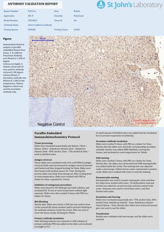
  • 1. Figure: Immunohistochemical analysis of paraffin embedded Human liver tissue. 1: E-cadherin Polyclonal Antibody was diluted at 1:200 (4 degree Celsius,overnight). 2: Sodium citrate pH 6.0 was used for antibody retrieval (>98 degree Celsius,20min). 3: Secondary antibody was diluted at 1:200 (room temperature, 30min). Negative control was used by secondary antibody only. Report Number 92819-a Host Rabbit Application IHC-P Clonality Polyclonal Model Number STJ92819 Clone ID NA Antibody Name Anti-E-cadherin antibody Testing Species HUMAN Testing Tissue LIVER ANTIBODY VALIDATION REPORT b. (A small amount of distilled water was added into the incubation box to prevent evaporation of antibody). 95. Secondary antibody incubation a. Slides were washed 3 times, with PBS on a shaker for 5min. Shortly after the slides were dried the corresponding secondary antibody solution was added (HRP labelled), covering the tissues, and incubated at room temperature for 30min. b. 96. DAB staining a. Slides were washed 3 times, with PBS on a shaker for 5min. b. Shortly after, the slides were dried and fresh DAB staining buffer was added inside the circles. The staining time was adjusted under a microscope. Yellow-brown colour represented a positive result. Slides were washed with water to stop the staining. c. 97. Haematoxylin staining a. Haematoxylin was used to counter-staining for 1min, and then the slides were washed with water. 1% Hydrochloric acid and alcohol was added for several seconds and then washed with water. Ammonia was used to reveal blue colour, and then flushed with water. b. 98. Desolation and Clearing i. Slides were incubated sequentially into: 75% alcohol 5min, 85% alcohol 5min, Anhydrous ethanol - 5min, Anhydrous ethanol - 5min & Xylene - 5min. Shortly after slides were dried and neutral gum was used to seal the slides. ii. 99. Visualization a. Results were validated with microscope, and the slides were scanned. Paraffin-Embedded Immunohistochemistry Protocol 89. 90. Tissue processing a. Slides were incubated sequentially into Xylene; 15min – Xylene, 15min - Anhydrous ethanol, 5min - Anhydrous ethanol, 5min - 85% alcohol, 5min - 75% alcohol & 5min – wash in distilled water. b. 91. Antigen retrieval a. Tissue slides were incubated with citric acid (PH6.0) antigen retrieval buffer and microwaved for antigen retrieval (heated until boiled and then stopped heating) for 8min. Slides were then heated with medium power for 7min. During this process slides were kept from drying out. After cooling down at room temperature, slides were washed with PBS on shaker for 5min, repeated for 3 times. b. 92. Inhibition of endogenous peroxidase a. Slides were placed in 3% Hydrogen peroxide solution, and incubated for 10 min at room temperature without light exposure. Slides were then washed 3 times with PBS on a shaker for 5mins. b. 93. BSA Blocking a. Shortly after slides were dried, a PAP pen was used to draw circles around the tissue sections (and to prevent draining of the antibody solution). Inside the circles, BSA was used to cover the tissue evenly, blocking for 30min. b. 94. Primary antibody incubation After blocking solution was removed a 1:200 solution of primary antibody/PBS was added on the slide, and incubated overnight at 4°C. St John's Laboratory Ltd. www.stjohnslabs.com
  • 2. Figure: Immunohistochemical analysis of paraffin embedded Human liver cancer tissue. 1: E- cadherin Polyclonal Antibody was diluted at 1:200 (4 degree Celsius,overnight). 2: Sodium citrate pH 6.0 was used for antibody retrieval (>98 degree Celsius,20min). 3: Secondary antibody was diluted at 1:200 (room temperature, 30min). Negative control was used by secondary antibody only. Report Number 92819-b Host Rabbit Application IHC-P Clonality Polyclonal Model Number STJ92819 Clone ID NA Antibody Name Anti-E-cadherin antibody Testing Species HUMAN Testing Tissue LIVER CANCER ANTIBODY VALIDATION REPORT b. (A small amount of distilled water was added into the incubation box to prevent evaporation of antibody). 84. Secondary antibody incubation a. Slides were washed 3 times, with PBS on a shaker for 5min. Shortly after the slides were dried the corresponding secondary antibody solution was added (HRP labelled), covering the tissues, and incubated at room temperature for 30min. b. 85. DAB staining a. Slides were washed 3 times, with PBS on a shaker for 5min. b. Shortly after, the slides were dried and fresh DAB staining buffer was added inside the circles. The staining time was adjusted under a microscope. Yellow-brown colour represented a positive result. Slides were washed with water to stop the staining. c. 86. Haematoxylin staining a. Haematoxylin was used to counter-staining for 1min, and then the slides were washed with water. 1% Hydrochloric acid and alcohol was added for several seconds and then washed with water. Ammonia was used to reveal blue colour, and then flushed with water. b. 87. Desolation and Clearing i. Slides were incubated sequentially into: 75% alcohol 5min, 85% alcohol 5min, Anhydrous ethanol - 5min, Anhydrous ethanol - 5min & Xylene - 5min. Shortly after slides were dried and neutral gum was used to seal the slides. ii. 88. Visualization a. Results were validated with microscope, and the slides were scanned. Paraffin-Embedded Immunohistochemistry Protocol 78. 79. Tissue processing a. Slides were incubated sequentially into Xylene; 15min – Xylene, 15min - Anhydrous ethanol, 5min - Anhydrous ethanol, 5min - 85% alcohol, 5min - 75% alcohol & 5min – wash in distilled water. b. 80. Antigen retrieval a. Tissue slides were incubated with citric acid (PH6.0) antigen retrieval buffer and microwaved for antigen retrieval (heated until boiled and then stopped heating) for 8min. Slides were then heated with medium power for 7min. During this process slides were kept from drying out. After cooling down at room temperature, slides were washed with PBS on shaker for 5min, repeated for 3 times. b. 81. Inhibition of endogenous peroxidase a. Slides were placed in 3% Hydrogen peroxide solution, and incubated for 10 min at room temperature without light exposure. Slides were then washed 3 times with PBS on a shaker for 5mins. b. 82. BSA Blocking a. Shortly after slides were dried, a PAP pen was used to draw circles around the tissue sections (and to prevent draining of the antibody solution). Inside the circles, BSA was used to cover the tissue evenly, blocking for 30min. b. 83. Primary antibody incubation After blocking solution was removed a 1:200 solution of primary antibody/PBS was added on the slide, and incubated overnight at 4°C. St John's Laboratory Ltd. www.stjohnslabs.com
  • 3. Figure: Immunohistochemical analysis of paraffin embedded Human stomach tissue. 1: E- cadherin Polyclonal Antibody was diluted at 1:200 (4 degree Celsius,overnight). 2: Sodium citrate pH 6.0 was used for antibody retrieval (>98 degree Celsius,20min). 3: Secondary antibody was diluted at 1:200 (room temperature, 30min). Negative control was used by secondary antibody only. Report Number 92819-c Host Rabbit Application IHC-P Clonality Polyclonal Model Number STJ92819 Clone ID NA Antibody Name Anti-E-cadherin antibody Testing Species HUMAN Testing Tissue STOMACH ANTIBODY VALIDATION REPORT b. (A small amount of distilled water was added into the incubation box to prevent evaporation of antibody). 73. Secondary antibody incubation a. Slides were washed 3 times, with PBS on a shaker for 5min. Shortly after the slides were dried the corresponding secondary antibody solution was added (HRP labelled), covering the tissues, and incubated at room temperature for 30min. b. 74. DAB staining a. Slides were washed 3 times, with PBS on a shaker for 5min. b. Shortly after, the slides were dried and fresh DAB staining buffer was added inside the circles. The staining time was adjusted under a microscope. Yellow-brown colour represented a positive result. Slides were washed with water to stop the staining. c. 75. Haematoxylin staining a. Haematoxylin was used to counter-staining for 1min, and then the slides were washed with water. 1% Hydrochloric acid and alcohol was added for several seconds and then washed with water. Ammonia was used to reveal blue colour, and then flushed with water. b. 76. Desolation and Clearing i. Slides were incubated sequentially into: 75% alcohol 5min, 85% alcohol 5min, Anhydrous ethanol - 5min, Anhydrous ethanol - 5min & Xylene - 5min. Shortly after slides were dried and neutral gum was used to seal the slides. ii. 77. Visualization a. Results were validated with microscope, and the slides were scanned. Paraffin-Embedded Immunohistochemistry Protocol 67. 68. Tissue processing a. Slides were incubated sequentially into Xylene; 15min – Xylene, 15min - Anhydrous ethanol, 5min - Anhydrous ethanol, 5min - 85% alcohol, 5min - 75% alcohol & 5min – wash in distilled water. b. 69. Antigen retrieval a. Tissue slides were incubated with citric acid (PH6.0) antigen retrieval buffer and microwaved for antigen retrieval (heated until boiled and then stopped heating) for 8min. Slides were then heated with medium power for 7min. During this process slides were kept from drying out. After cooling down at room temperature, slides were washed with PBS on shaker for 5min, repeated for 3 times. b. 70. Inhibition of endogenous peroxidase a. Slides were placed in 3% Hydrogen peroxide solution, and incubated for 10 min at room temperature without light exposure. Slides were then washed 3 times with PBS on a shaker for 5mins. b. 71. BSA Blocking a. Shortly after slides were dried, a PAP pen was used to draw circles around the tissue sections (and to prevent draining of the antibody solution). Inside the circles, BSA was used to cover the tissue evenly, blocking for 30min. b. 72. Primary antibody incubation After blocking solution was removed a 1:200 solution of primary antibody/PBS was added on the slide, and incubated overnight at 4°C. St John's Laboratory Ltd. www.stjohnslabs.com
  • 4. Figure: Immunohistochemical analysis of paraffin embedded Rat heart tissue. 1: E-cadherin Polyclonal Antibody was diluted at 1:200 (4 degree Celsius,overnight). 2: Sodium citrate pH 6.0 was used for antibody retrieval (>98 degree Celsius,20min). 3: Secondary antibody was diluted at 1:200 (room temperature, 30min). Negative control was used by secondary antibody only. Report Number 92819-d Host Rabbit Application IHC-P Clonality Polyclonal Model Number STJ92819 Clone ID NA Antibody Name Anti-E-cadherin antibody Testing Species RAT Testing Tissue HEART ANTIBODY VALIDATION REPORT b. (A small amount of distilled water was added into the incubation box to prevent evaporation of antibody). 62. Secondary antibody incubation a. Slides were washed 3 times, with PBS on a shaker for 5min. Shortly after the slides were dried the corresponding secondary antibody solution was added (HRP labelled), covering the tissues, and incubated at room temperature for 30min. b. 63. DAB staining a. Slides were washed 3 times, with PBS on a shaker for 5min. b. Shortly after, the slides were dried and fresh DAB staining buffer was added inside the circles. The staining time was adjusted under a microscope. Yellow-brown colour represented a positive result. Slides were washed with water to stop the staining. c. 64. Haematoxylin staining a. Haematoxylin was used to counter-staining for 1min, and then the slides were washed with water. 1% Hydrochloric acid and alcohol was added for several seconds and then washed with water. Ammonia was used to reveal blue colour, and then flushed with water. b. 65. Desolation and Clearing i. Slides were incubated sequentially into: 75% alcohol 5min, 85% alcohol 5min, Anhydrous ethanol - 5min, Anhydrous ethanol - 5min & Xylene - 5min. Shortly after slides were dried and neutral gum was used to seal the slides. ii. 66. Visualization a. Results were validated with microscope, and the slides were scanned. Paraffin-Embedded Immunohistochemistry Protocol 56. 57. Tissue processing a. Slides were incubated sequentially into Xylene; 15min – Xylene, 15min - Anhydrous ethanol, 5min - Anhydrous ethanol, 5min - 85% alcohol, 5min - 75% alcohol & 5min – wash in distilled water. b. 58. Antigen retrieval a. Tissue slides were incubated with citric acid (PH6.0) antigen retrieval buffer and microwaved for antigen retrieval (heated until boiled and then stopped heating) for 8min. Slides were then heated with medium power for 7min. During this process slides were kept from drying out. After cooling down at room temperature, slides were washed with PBS on shaker for 5min, repeated for 3 times. b. 59. Inhibition of endogenous peroxidase a. Slides were placed in 3% Hydrogen peroxide solution, and incubated for 10 min at room temperature without light exposure. Slides were then washed 3 times with PBS on a shaker for 5mins. b. 60. BSA Blocking a. Shortly after slides were dried, a PAP pen was used to draw circles around the tissue sections (and to prevent draining of the antibody solution). Inside the circles, BSA was used to cover the tissue evenly, blocking for 30min. b. 61. Primary antibody incubation After blocking solution was removed a 1:200 solution of primary antibody/PBS was added on the slide, and incubated overnight at 4°C. St John's Laboratory Ltd. www.stjohnslabs.com
  • 5. Figure: Immunohistochemical analysis of paraffin embedded Rat lung tissue. 1: E-cadherin Polyclonal Antibody was diluted at 1:200 (4 degree Celsius,overnight). 2: Sodium citrate pH 6.0 was used for antibody retrieval (>98 degree Celsius,20min). 3: Secondary antibody was diluted at 1:200 (room temperature, 30min). Negative control was used by secondary antibody only. Report Number 92819-e Host Rabbit Application IHC-P Clonality Polyclonal Model Number STJ92819 Clone ID NA Antibody Name Anti-E-cadherin antibody Testing Species RAT Testing Tissue LUNG ANTIBODY VALIDATION REPORT b. (A small amount of distilled water was added into the incubation box to prevent evaporation of antibody). 51. Secondary antibody incubation a. Slides were washed 3 times, with PBS on a shaker for 5min. Shortly after the slides were dried the corresponding secondary antibody solution was added (HRP labelled), covering the tissues, and incubated at room temperature for 30min. b. 52. DAB staining a. Slides were washed 3 times, with PBS on a shaker for 5min. b. Shortly after, the slides were dried and fresh DAB staining buffer was added inside the circles. The staining time was adjusted under a microscope. Yellow-brown colour represented a positive result. Slides were washed with water to stop the staining. c. 53. Haematoxylin staining a. Haematoxylin was used to counter-staining for 1min, and then the slides were washed with water. 1% Hydrochloric acid and alcohol was added for several seconds and then washed with water. Ammonia was used to reveal blue colour, and then flushed with water. b. 54. Desolation and Clearing i. Slides were incubated sequentially into: 75% alcohol 5min, 85% alcohol 5min, Anhydrous ethanol - 5min, Anhydrous ethanol - 5min & Xylene - 5min. Shortly after slides were dried and neutral gum was used to seal the slides. ii. 55. Visualization a. Results were validated with microscope, and the slides were scanned. Paraffin-Embedded Immunohistochemistry Protocol 45. 46. Tissue processing a. Slides were incubated sequentially into Xylene; 15min – Xylene, 15min - Anhydrous ethanol, 5min - Anhydrous ethanol, 5min - 85% alcohol, 5min - 75% alcohol & 5min – wash in distilled water. b. 47. Antigen retrieval a. Tissue slides were incubated with citric acid (PH6.0) antigen retrieval buffer and microwaved for antigen retrieval (heated until boiled and then stopped heating) for 8min. Slides were then heated with medium power for 7min. During this process slides were kept from drying out. After cooling down at room temperature, slides were washed with PBS on shaker for 5min, repeated for 3 times. b. 48. Inhibition of endogenous peroxidase a. Slides were placed in 3% Hydrogen peroxide solution, and incubated for 10 min at room temperature without light exposure. Slides were then washed 3 times with PBS on a shaker for 5mins. b. 49. BSA Blocking a. Shortly after slides were dried, a PAP pen was used to draw circles around the tissue sections (and to prevent draining of the antibody solution). Inside the circles, BSA was used to cover the tissue evenly, blocking for 30min. b. 50. Primary antibody incubation After blocking solution was removed a 1:200 solution of primary antibody/PBS was added on the slide, and incubated overnight at 4°C. St John's Laboratory Ltd. www.stjohnslabs.com
  • 6. Figure: Immunohistochemical analysis of paraffin embedded Rat kidney tissue. 1: E-cadherin Polyclonal Antibody was diluted at 1:200 (4 degree Celsius,overnight). 2: Sodium citrate pH 6.0 was used for antibody retrieval (>98 degree Celsius,20min). 3: Secondary antibody was diluted at 1:200 (room temperature, 30min). Negative control was used by secondary antibody only. Report Number 92819-f Host Rabbit Application IHC-P Clonality Polyclonal Model Number STJ92819 Clone ID NA Antibody Name Anti-E-cadherin antibody Testing Species RAT Testing Tissue KIDNEY ANTIBODY VALIDATION REPORT b. (A small amount of distilled water was added into the incubation box to prevent evaporation of antibody). 40. Secondary antibody incubation a. Slides were washed 3 times, with PBS on a shaker for 5min. Shortly after the slides were dried the corresponding secondary antibody solution was added (HRP labelled), covering the tissues, and incubated at room temperature for 30min. b. 41. DAB staining a. Slides were washed 3 times, with PBS on a shaker for 5min. b. Shortly after, the slides were dried and fresh DAB staining buffer was added inside the circles. The staining time was adjusted under a microscope. Yellow-brown colour represented a positive result. Slides were washed with water to stop the staining. c. 42. Haematoxylin staining a. Haematoxylin was used to counter-staining for 1min, and then the slides were washed with water. 1% Hydrochloric acid and alcohol was added for several seconds and then washed with water. Ammonia was used to reveal blue colour, and then flushed with water. b. 43. Desolation and Clearing i. Slides were incubated sequentially into: 75% alcohol 5min, 85% alcohol 5min, Anhydrous ethanol - 5min, Anhydrous ethanol - 5min & Xylene - 5min. Shortly after slides were dried and neutral gum was used to seal the slides. ii. 44. Visualization a. Results were validated with microscope, and the slides were scanned. Paraffin-Embedded Immunohistochemistry Protocol 34. 35. Tissue processing a. Slides were incubated sequentially into Xylene; 15min – Xylene, 15min - Anhydrous ethanol, 5min - Anhydrous ethanol, 5min - 85% alcohol, 5min - 75% alcohol & 5min – wash in distilled water. b. 36. Antigen retrieval a. Tissue slides were incubated with citric acid (PH6.0) antigen retrieval buffer and microwaved for antigen retrieval (heated until boiled and then stopped heating) for 8min. Slides were then heated with medium power for 7min. During this process slides were kept from drying out. After cooling down at room temperature, slides were washed with PBS on shaker for 5min, repeated for 3 times. b. 37. Inhibition of endogenous peroxidase a. Slides were placed in 3% Hydrogen peroxide solution, and incubated for 10 min at room temperature without light exposure. Slides were then washed 3 times with PBS on a shaker for 5mins. b. 38. BSA Blocking a. Shortly after slides were dried, a PAP pen was used to draw circles around the tissue sections (and to prevent draining of the antibody solution). Inside the circles, BSA was used to cover the tissue evenly, blocking for 30min. b. 39. Primary antibody incubation After blocking solution was removed a 1:200 solution of primary antibody/PBS was added on the slide, and incubated overnight at 4°C. St John's Laboratory Ltd. www.stjohnslabs.com
  • 7. Figure: Immunohistochemical analysis of paraffin embedded Rat spleen tissue. 1: E-cadherin Polyclonal Antibody was diluted at 1:200 (4 degree Celsius,overnight). 2: Sodium citrate pH 6.0 was used for antibody retrieval (>98 degree Celsius,20min). 3: Secondary antibody was diluted at 1:200 (room temperature, 30min). Negative control was used by secondary antibody only. Report Number 92819-g Host Rabbit Application IHC-P Clonality Polyclonal Model Number STJ92819 Clone ID NA Antibody Name Anti-E-cadherin antibody Testing Species RAT Testing Tissue SPLEEN ANTIBODY VALIDATION REPORT b. (A small amount of distilled water was added into the incubation box to prevent evaporation of antibody). 29. Secondary antibody incubation a. Slides were washed 3 times, with PBS on a shaker for 5min. Shortly after the slides were dried the corresponding secondary antibody solution was added (HRP labelled), covering the tissues, and incubated at room temperature for 30min. b. 30. DAB staining a. Slides were washed 3 times, with PBS on a shaker for 5min. b. Shortly after, the slides were dried and fresh DAB staining buffer was added inside the circles. The staining time was adjusted under a microscope. Yellow-brown colour represented a positive result. Slides were washed with water to stop the staining. c. 31. Haematoxylin staining a. Haematoxylin was used to counter-staining for 1min, and then the slides were washed with water. 1% Hydrochloric acid and alcohol was added for several seconds and then washed with water. Ammonia was used to reveal blue colour, and then flushed with water. b. 32. Desolation and Clearing i. Slides were incubated sequentially into: 75% alcohol 5min, 85% alcohol 5min, Anhydrous ethanol - 5min, Anhydrous ethanol - 5min & Xylene - 5min. Shortly after slides were dried and neutral gum was used to seal the slides. ii. 33. Visualization a. Results were validated with microscope, and the slides were scanned. Paraffin-Embedded Immunohistochemistry Protocol 23. 24. Tissue processing a. Slides were incubated sequentially into Xylene; 15min – Xylene, 15min - Anhydrous ethanol, 5min - Anhydrous ethanol, 5min - 85% alcohol, 5min - 75% alcohol & 5min – wash in distilled water. b. 25. Antigen retrieval a. Tissue slides were incubated with citric acid (PH6.0) antigen retrieval buffer and microwaved for antigen retrieval (heated until boiled and then stopped heating) for 8min. Slides were then heated with medium power for 7min. During this process slides were kept from drying out. After cooling down at room temperature, slides were washed with PBS on shaker for 5min, repeated for 3 times. b. 26. Inhibition of endogenous peroxidase a. Slides were placed in 3% Hydrogen peroxide solution, and incubated for 10 min at room temperature without light exposure. Slides were then washed 3 times with PBS on a shaker for 5mins. b. 27. BSA Blocking a. Shortly after slides were dried, a PAP pen was used to draw circles around the tissue sections (and to prevent draining of the antibody solution). Inside the circles, BSA was used to cover the tissue evenly, blocking for 30min. b. 28. Primary antibody incubation After blocking solution was removed a 1:200 solution of primary antibody/PBS was added on the slide, and incubated overnight at 4°C. St John's Laboratory Ltd. www.stjohnslabs.com
  • 8. Figure: Immunohistochemical analysis of paraffin embedded Mouse testis tissue. 1: E-cadherin Polyclonal Antibody was diluted at 1:200 (4 degree Celsius,overnight). 2: Sodium citrate pH 6.0 was used for antibody retrieval (>98 degree Celsius,20min). 3: Secondary antibody was diluted at 1:200 (room temperature, 30min). Negative control was used by secondary antibody only. Report Number 92819-h Host Rabbit Application IHC-P Clonality Polyclonal Model Number STJ92819 Clone ID NA Antibody Name Anti-E-cadherin antibody Testing Species MOUSE Testing Tissue TESTIS ANTIBODY VALIDATION REPORT b. (A small amount of distilled water was added into the incubation box to prevent evaporation of antibody). 18. Secondary antibody incubation a. Slides were washed 3 times, with PBS on a shaker for 5min. Shortly after the slides were dried the corresponding secondary antibody solution was added (HRP labelled), covering the tissues, and incubated at room temperature for 30min. b. 19. DAB staining a. Slides were washed 3 times, with PBS on a shaker for 5min. b. Shortly after, the slides were dried and fresh DAB staining buffer was added inside the circles. The staining time was adjusted under a microscope. Yellow-brown colour represented a positive result. Slides were washed with water to stop the staining. c. 20. Haematoxylin staining a. Haematoxylin was used to counter-staining for 1min, and then the slides were washed with water. 1% Hydrochloric acid and alcohol was added for several seconds and then washed with water. Ammonia was used to reveal blue colour, and then flushed with water. b. 21. Desolation and Clearing i. Slides were incubated sequentially into: 75% alcohol 5min, 85% alcohol 5min, Anhydrous ethanol - 5min, Anhydrous ethanol - 5min & Xylene - 5min. Shortly after slides were dried and neutral gum was used to seal the slides. ii. 22. Visualization a. Results were validated with microscope, and the slides were scanned. Paraffin-Embedded Immunohistochemistry Protocol 12. 13. Tissue processing a. Slides were incubated sequentially into Xylene; 15min – Xylene, 15min - Anhydrous ethanol, 5min - Anhydrous ethanol, 5min - 85% alcohol, 5min - 75% alcohol & 5min – wash in distilled water. b. 14. Antigen retrieval a. Tissue slides were incubated with citric acid (PH6.0) antigen retrieval buffer and microwaved for antigen retrieval (heated until boiled and then stopped heating) for 8min. Slides were then heated with medium power for 7min. During this process slides were kept from drying out. After cooling down at room temperature, slides were washed with PBS on shaker for 5min, repeated for 3 times. b. 15. Inhibition of endogenous peroxidase a. Slides were placed in 3% Hydrogen peroxide solution, and incubated for 10 min at room temperature without light exposure. Slides were then washed 3 times with PBS on a shaker for 5mins. b. 16. BSA Blocking a. Shortly after slides were dried, a PAP pen was used to draw circles around the tissue sections (and to prevent draining of the antibody solution). Inside the circles, BSA was used to cover the tissue evenly, blocking for 30min. b. 17. Primary antibody incubation After blocking solution was removed a 1:200 solution of primary antibody/PBS was added on the slide, and incubated overnight at 4°C. St John's Laboratory Ltd. www.stjohnslabs.com
  • 9. Figure: Immunohistochemical analysis of paraffin embedded Mouse lung tissue. 1: E-cadherin Polyclonal Antibody was diluted at 1:200 (4 degree Celsius,overnight). 2: Sodium citrate pH 6.0 was used for antibody retrieval (>98 degree Celsius,20min). 3: Secondary antibody was diluted at 1:200 (room temperature, 30min). Negative control was used by secondary antibody only. Report Number 92819-i Host Rabbit Application IHC-P Clonality Polyclonal Model Number STJ92819 Clone ID NA Antibody Name Anti-E-cadherin antibody Testing Species MOUSE Testing Tissue LUNG ANTIBODY VALIDATION REPORT b. (A small amount of distilled water was added into the incubation box to prevent evaporation of antibody). 7. Secondary antibody incubation a. Slides were washed 3 times, with PBS on a shaker for 5min. Shortly after the slides were dried the corresponding secondary antibody solution was added (HRP labelled), covering the tissues, and incubated at room temperature for 30min. b. 8. DAB staining a. Slides were washed 3 times, with PBS on a shaker for 5min. b. Shortly after, the slides were dried and fresh DAB staining buffer was added inside the circles. The staining time was adjusted under a microscope. Yellow-brown colour represented a positive result. Slides were washed with water to stop the staining. c. 9. Haematoxylin staining a. Haematoxylin was used to counter-staining for 1min, and then the slides were washed with water. 1% Hydrochloric acid and alcohol was added for several seconds and then washed with water. Ammonia was used to reveal blue colour, and then flushed with water. b. 10. Desolation and Clearing i. Slides were incubated sequentially into: 75% alcohol 5min, 85% alcohol 5min, Anhydrous ethanol - 5min, Anhydrous ethanol - 5min & Xylene - 5min. Shortly after slides were dried and neutral gum was used to seal the slides. ii. 11. Visualization a. Results were validated with microscope, and the slides were scanned. Paraffin-Embedded Immunohistochemistry Protocol 1. 2. Tissue processing a. Slides were incubated sequentially into Xylene; 15min – Xylene, 15min - Anhydrous ethanol, 5min - Anhydrous ethanol, 5min - 85% alcohol, 5min - 75% alcohol & 5min – wash in distilled water. b. 3. Antigen retrieval a. Tissue slides were incubated with citric acid (PH6.0) antigen retrieval buffer and microwaved for antigen retrieval (heated until boiled and then stopped heating) for 8min. Slides were then heated with medium power for 7min. During this process slides were kept from drying out. After cooling down at room temperature, slides were washed with PBS on shaker for 5min, repeated for 3 times. b. 4. Inhibition of endogenous peroxidase a. Slides were placed in 3% Hydrogen peroxide solution, and incubated for 10 min at room temperature without light exposure. Slides were then washed 3 times with PBS on a shaker for 5mins. b. 5. BSA Blocking a. Shortly after slides were dried, a PAP pen was used to draw circles around the tissue sections (and to prevent draining of the antibody solution). Inside the circles, BSA was used to cover the tissue evenly, blocking for 30min. b. 6. Primary antibody incubation After blocking solution was removed a 1:200 solution of primary antibody/PBS was added on the slide, and incubated overnight at 4°C. St John's Laboratory Ltd. www.stjohnslabs.com
  • 10. Figure: Immunohistochemical analysis of paraffin embedded Mouse kidney tissue. 1: E- cadherin Polyclonal Antibody was diluted at 1:200 (4 degree Celsius,overnight). 2: Sodium citrate pH 6.0 was used for antibody retrieval (>98 degree Celsius,20min). 3: Secondary antibody was diluted at 1:200 (room temperature, 30min). Negative control was used by secondary antibody only. Report Number 92819-j Host Rabbit Application IHC-P Clonality Polyclonal Model Number STJ92819 Clone ID NA Antibody Name Anti-E-cadherin antibody Testing Species MOUSE Testing Tissue KIDNEY ANTIBODY VALIDATION REPORT b. (A small amount of distilled water was added into the incubation box to prevent evaporation of antibody). 100. Secondary antibody incubation a. Slides were washed 3 times, with PBS on a shaker for 5min. Shortly after the slides were dried the corresponding secondary antibody solution was added (HRP labelled), covering the tissues, and incubated at room temperature for 30min. b. 101. DAB staining a. Slides were washed 3 times, with PBS on a shaker for 5min. b. Shortly after, the slides were dried and fresh DAB staining buffer was added inside the circles. The staining time was adjusted under a microscope. Yellow-brown colour represented a positive result. Slides were washed with water to stop the staining. c. 102. Haematoxylin staining a. Haematoxylin was used to counter-staining for 1min, and then the slides were washed with water. 1% Hydrochloric acid and alcohol was added for several seconds and then washed with water. Ammonia was used to reveal blue colour, and then flushed with water. b. 103. Desolation and Clearing i. Slides were incubated sequentially into: 75% alcohol 5min, 85% alcohol 5min, Anhydrous ethanol - 5min, Anhydrous ethanol - 5min & Xylene - 5min. Shortly after slides were dried and neutral gum was used to seal the slides. ii. 104. Visualization a. Results were validated with microscope, and the slides were scanned. Paraffin-Embedded Immunohistochemistry Protocol 105. 106. Tissue processing a. Slides were incubated sequentially into Xylene; 15min – Xylene, 15min - Anhydrous ethanol, 5min - Anhydrous ethanol, 5min - 85% alcohol, 5min - 75% alcohol & 5min – wash in distilled water. b. 107. Antigen retrieval a. Tissue slides were incubated with citric acid (PH6.0) antigen retrieval buffer and microwaved for antigen retrieval (heated until boiled and then stopped heating) for 8min. Slides were then heated with medium power for 7min. During this process slides were kept from drying out. After cooling down at room temperature, slides were washed with PBS on shaker for 5min, repeated for 3 times. b. 108. Inhibition of endogenous peroxidase a. Slides were placed in 3% Hydrogen peroxide solution, and incubated for 10 min at room temperature without light exposure. Slides were then washed 3 times with PBS on a shaker for 5mins. b. 109. BSA Blocking a. Shortly after slides were dried, a PAP pen was used to draw circles around the tissue sections (and to prevent draining of the antibody solution). Inside the circles, BSA was used to cover the tissue evenly, blocking for 30min. b. 110. Primary antibody incubation After blocking solution was removed a 1:200 solution of primary antibody/PBS was added on the slide, and incubated overnight at 4°C. St John's Laboratory Ltd. www.stjohnslabs.com
  • 11. Figure: Immunohistochemical analysis of paraffin embedded Mouse spleen tissue. 1: E- cadherin Polyclonal Antibody was diluted at 1:200 (4 degree Celsius,overnight). 2: Sodium citrate pH 6.0 was used for antibody retrieval (>98 degree Celsius,20min). 3: Secondary antibody was diluted at 1:200 (room temperature, 30min). Negative control was used by secondary antibody only. Report Number 92819-k Host Rabbit Application IHC-P Clonality Polyclonal Model Number STJ92819 Clone ID NA Antibody Name Anti-E-cadherin antibody Testing Species MOUSE Testing Tissue SPLEEN ANTIBODY VALIDATION REPORT b. (A small amount of distilled water was added into the incubation box to prevent evaporation of antibody). 111. Secondary antibody incubation a. Slides were washed 3 times, with PBS on a shaker for 5min. Shortly after the slides were dried the corresponding secondary antibody solution was added (HRP labelled), covering the tissues, and incubated at room temperature for 30min. b. 112. DAB staining a. Slides were washed 3 times, with PBS on a shaker for 5min. b. Shortly after, the slides were dried and fresh DAB staining buffer was added inside the circles. The staining time was adjusted under a microscope. Yellow-brown colour represented a positive result. Slides were washed with water to stop the staining. c. 113. Haematoxylin staining a. Haematoxylin was used to counter-staining for 1min, and then the slides were washed with water. 1% Hydrochloric acid and alcohol was added for several seconds and then washed with water. Ammonia was used to reveal blue colour, and then flushed with water. b. 114. Desolation and Clearing i. Slides were incubated sequentially into: 75% alcohol 5min, 85% alcohol 5min, Anhydrous ethanol - 5min, Anhydrous ethanol - 5min & Xylene - 5min. Shortly after slides were dried and neutral gum was used to seal the slides. ii. 115. Visualization a. Results were validated with microscope, and the slides were scanned. Paraffin-Embedded Immunohistochemistry Protocol 116. 117. Tissue processing a. Slides were incubated sequentially into Xylene; 15min – Xylene, 15min - Anhydrous ethanol, 5min - Anhydrous ethanol, 5min - 85% alcohol, 5min - 75% alcohol & 5min – wash in distilled water. b. 118. Antigen retrieval a. Tissue slides were incubated with citric acid (PH6.0) antigen retrieval buffer and microwaved for antigen retrieval (heated until boiled and then stopped heating) for 8min. Slides were then heated with medium power for 7min. During this process slides were kept from drying out. After cooling down at room temperature, slides were washed with PBS on shaker for 5min, repeated for 3 times. b. 119. Inhibition of endogenous peroxidase a. Slides were placed in 3% Hydrogen peroxide solution, and incubated for 10 min at room temperature without light exposure. Slides were then washed 3 times with PBS on a shaker for 5mins. b. 120. BSA Blocking a. Shortly after slides were dried, a PAP pen was used to draw circles around the tissue sections (and to prevent draining of the antibody solution). Inside the circles, BSA was used to cover the tissue evenly, blocking for 30min. b. 121. Primary antibody incubation After blocking solution was removed a 1:200 solution of primary antibody/PBS was added on the slide, and incubated overnight at 4°C. St John's Laboratory Ltd. www.stjohnslabs.com