Downloaded 16 times
![International Journal of Dental and Health Sciences
Volume 01, Issue 04
Case Report
EXTRACTION,IMMEDIATE IMPLANT-A
CASE REPORT
Nezar Watted1,Kareem Trabih2,Qawasmeh Nour3, Abdulgani Azzaldeen4,Abu-
Hussein Muhamad5
1-5.Center for Dentistry research and Aesthetics, Jatt/Israel
ABSTRACT:
Background: This case report describes extraction of a fractured right maxillary lateral
incisor tooth, followed by immediate placement of a dental implant in the prepared socket
and temporization by a bonded restoration.
Materials and Methods: The tooth was extracted with minimal hard and soft tissue trauma
and without flap reflection. The socket was prepared to the required depth and a
Biohorizons Implant was inserted. An impression was made 4 months after implant
insertion, and a definitive restoration was placed.
Results: The atraumatic operating technique and the immediate insertion of the Implant
resulted in the preservation of the hard and soft tissues at the extraction site. The patient
exhibited no clinical or radiologic complications through two years of clinical monitoring
after loading.
Conclusion: The dental implant and provisional restoration provided the patient with
immediate esthetics, function, comfort and most importantly preservation of tissues.
Key words: Implant, Immediate placement, Temporization,crown
INTRODUCTION:
Missing teeth and supporting oral
tissues have traditionally been replaced
with dentures or bridges permitting
restoration of chewing function, speech,
and aesthetics [1]. Dental implants offer an
alternative. These implants are inserted
into the jawbones to support a dental
prosthesis and are retained because of
the intimacy of bone growth on to their
surface [2]. This direct structural and
functional connection between living
bone and implant surface, termed
osseointegration, was first described by
Branemark 1977 [2,3]and has undoubtedly
been one of the most significant scientific
breakthroughs in dentistry over the past
30 years. Teeth may have been lost
through dental disease or trauma or they
may be congenitally absent. However in
many clinical situations compromised
teeth or roots may still be present in the
patient’s mouth. Traditionally, before
placing dental implants, compromised
teeth were removed and the extraction
sockets were left to heal for between
several months and 1 year. However, the
great majority of patients are interested
in shortening the treatment time between
*Corresponding Author Address:Dr.Abdulgani Azzaldeen,Nahef Village,Upper Galille,ISRAEL
Email:azzaldeenabdulgani@gmail.com](https://image.slidesharecdn.com/dm0326-140922032626-phpapp02/85/EXTRACTION-IMMEDIATE-IMPLANT-A-CASE-REPORT-1-320.jpg)
![International Journal of Dental and Health Sciences
Volume 01, Issue 04
Case Report
EXTRACTION,IMMEDIATE IMPLANT-A
CASE REPORT
Nezar Watted1,Kareem Trabih2,Qawasmeh Nour3, Abdulgani Azzaldeen4,Abu-
Hussein Muhamad5
1-5.Center for Dentistry research and Aesthetics, Jatt/Israel
ABSTRACT:
Background: This case report describes extraction of a fractured right maxillary lateral
incisor tooth, followed by immediate placement of a dental implant in the prepared socket
and temporization by a bonded restoration.
Materials and Methods: The tooth was extracted with minimal hard and soft tissue trauma
and without flap reflection. The socket was prepared to the required depth and a
Biohorizons Implant was inserted. An impression was made 4 months after implant
insertion, and a definitive restoration was placed.
Results: The atraumatic operating technique and the immediate insertion of the Implant
resulted in the preservation of the hard and soft tissues at the extraction site. The patient
exhibited no clinical or radiologic complications through two years of clinical monitoring
after loading.
Conclusion: The dental implant and provisional restoration provided the patient with
immediate esthetics, function, comfort and most importantly preservation of tissues.
Key words: Implant, Immediate placement, Temporization,crown
INTRODUCTION:
Missing teeth and supporting oral
tissues have traditionally been replaced
with dentures or bridges permitting
restoration of chewing function, speech,
and aesthetics [1]. Dental implants offer an
alternative. These implants are inserted
into the jawbones to support a dental
prosthesis and are retained because of
the intimacy of bone growth on to their
surface [2]. This direct structural and
functional connection between living
bone and implant surface, termed
osseointegration, was first described by
Branemark 1977 [2,3]and has undoubtedly
been one of the most significant scientific
breakthroughs in dentistry over the past
30 years. Teeth may have been lost
through dental disease or trauma or they
may be congenitally absent. However in
many clinical situations compromised
teeth or roots may still be present in the
patient’s mouth. Traditionally, before
placing dental implants, compromised
teeth were removed and the extraction
sockets were left to heal for between
several months and 1 year. However, the
great majority of patients are interested
in shortening the treatment time between
*Corresponding Author Address:Dr.Abdulgani Azzaldeen,Nahef Village,Upper Galille,ISRAEL
Email:azzaldeenabdulgani@gmail.com](https://image.slidesharecdn.com/dm0326-140922032626-phpapp02/75/EXTRACTION-IMMEDIATE-IMPLANT-A-CASE-REPORT-1-2048.jpg)
![Watted N. et al., Int J Dent Health Sci 2014; 1(4): 430-435
tooth extraction and implant placement
or even better in having the implants
inserted during the same session as the
teeth are extracted (immediate implants)
[4,5] . This would result Interventions for
replacing missing teeth: dental implants in
fresh extraction sockets. Implant
placement in fresh extraction sockets is
well documented [5] . Animal and human
studies have demonstrated attainment of
osseointegration following immediate
placement of implant in freshly extracted
tooth/teeth at a light microscopic level. In
addition, numerous human clinical studies
have documented high levels of success of
implant placed at the time of tooth
extraction and subsequently restored and
in function [2,5,6] .
CASE DETAIL:
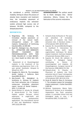
A 53 year old boy reported of root stump in
upper right front region and gave history of
trauma to maxillary Left Lateral incisor due to
accidental fall on floor during Cricket game
three years back in which he had fractured
crown and patient went for root canal
treatment but left it incomplete due to
negligence and slowly tooth crown was worn
out(Fig.1,2). He was referred to Implantology
for needful. Patient’s general and medical
history was taken and was non significant.
Patient was examined clinically and
Orthopantomograph was taken. After
thorough analysis clinically and
radiographically it was evaluated that there is
no underlying pathology and tooth root was
unrestorable but was surrounded by healthy
bone. It was there and then decided to do
extraction and place the implant immediately
and so was desired by the patient.(Fig.3)
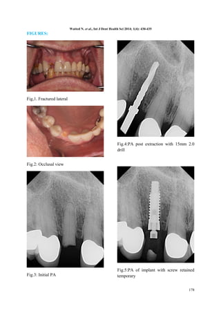
Tooth root was extracted atraumatically
under local anaesthesia after carefully raising
the flap The pilot drill (D-2.O mm) was put to
use for creating a osteotomy site of the
appropriate depth i.e. 15mm for implant
placement (Biohorizon-implant system) . It is
indexed with various markings corresponding
to the desired implant lengths.
When appropriate depth was reached with
the pilot drill, the implant depth probe was
used for tactile perception of intact bony
plates & or any perforations & the
confirmation of desired osteotomy depth.
Once desired depth was confirmed,
paralleling pins were placed to check the
proper alignment of the implant with adjacent
teeth & opposing occlusion. After
confirmation of depth & angulation, the
osteotomy site was prepared by a series of
gradually larger drills to the requisite width
with a speed of 1400-1600 r.p.m at 1: 16
reduction torque as per manufacturer
instructions. The Implant site was generously
irrigated with sterile saline to remove any
residual bone chips/other residue following
preparation. The depth of the osteotomy site
was ascertained with Implant depth probe.
The implant was removed from the sterile vial
using the insertion tool and delivered directly
into the osteotomy site. Contamination by
touching the implant with instruments made
of a dissimilar metal or by contact with soft
tissue, cloth or even surgical gloves may affect
the degree of osseointegration. The implant
was then pressed into the prepared site with
manual pressure aided by the insertion mount
& insertion tool attached to the implant head.
Following which, the insertion mount was
removed and hex driver was placed into the
implant internal hex & ratcheted with torque-controlled
implant ratchet. Implant was never
forced into the socket with excessive force as
this might lead to micro cracks in the surface
bone resulting in improper
osseointegration(Fig.,4,5). Implant that was
175](https://image.slidesharecdn.com/dm0326-140922032626-phpapp02/85/EXTRACTION-IMMEDIATE-IMPLANT-A-CASE-REPORT-2-320.jpg)
![Watted N. et al., Int J Dent Health Sci 2014; 1(4): 430-435
placed was checked for stability by applying
gentle pressure to determine if it could be
depressed or rotated. Also, primary implant
stability was assessed with the torque
controlled ratchet. The cover screw, provided
with the implant package was then placed
using the hex-driver using finger pressure
(Fig.6,7). At this point, implant was confirmed
to be immobile, which re-affirmed primary
implant stability.
The flap margins were then repositioned &
sutured tension free using 3-0 mersilk in
interrupted fashion (Fig.8). A radiograph was
taken post operatively to evaluate the implant
angulation & position (7).(Fig.9)
The patient was on regular recall and under
strict oral hygine measures.
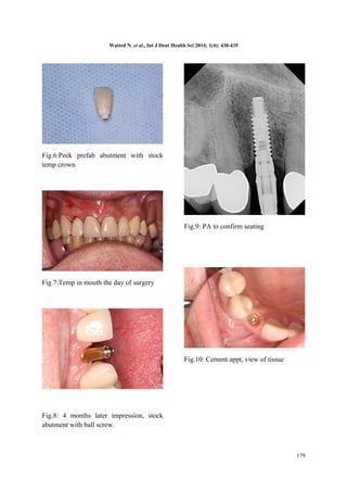
The patient was recalled after 12 weeks
radiograph was taken and prosthetic phase
was carried out under the opinion &
supervision of a prosthodontist. After soft
tissue healing (Fig.10,11),
Impressions were made with the impression
post attached to the implant using the closed
tray impression technique. Shade selection
was also done during this appointment. Casts
with impression post-implant analogue
complex, abutment, lab drivers and selected
shade were sent to laboratory for preparation
of cement retained porcelain fused to metal
crown. Healing abutment/ gingival former
was replaced till the time taken for laboratory
manufacture of prosthesis. After approx. 4-7
days, the healing abutments were removed
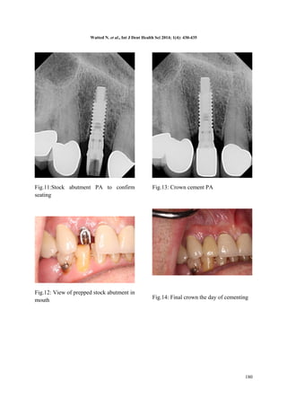
and replaced with final abutment. The PFM
crown was checked for its passive fitting to
abutment and non-interference with adjacent
teeth. Crown was then cemented with glass
ionomer cement (Fig12,13,14). The patient
was dispatched with a reminder of oral
hygiene instructions & the recall programme.
DISCUSSION:
Many clinical reports and experimental
studies in the animal model demonstrated
the favorable outcome of dental implants
immediately inserted in freshly extraction
socket, without the use of any
regenerative materials [2,3,5,8.9,10] . Our data
show a survival rate of immediately
implantation carried out on this patient at
one year after immediate implantation
and does not differ from the cases in
which implant was placed in healed sites.
These data agree with those from other
authors who evaluated the clinical success
rate of immediate implantation without
use of any membrane or graft material in
both humans and animals [11] . It must be
kept in mind that the present study is
related to immediate implant not
subjected to functional loading and
therefore not fully comparable with the
results from loaded implants. However, it
has been demonstrated that functional
loading does not impair, but rather
enhances, bone maturation. [5,12]
CONCLUSION:
This case report describes a technique to
preserve and augment anterior aesthetics
by combining atraumatic teeth extraction,
hard and soft tissue augmentation,
immediate provsionalization and using the
platform switching concept to preserve
the buccaI plate. The gingivaI tissue
surrounding the implants has remained
stable with no recession one year
following final crowns placement.
The implant therapy must fulfill both
functional and esthetic requirements to
176](https://image.slidesharecdn.com/dm0326-140922032626-phpapp02/85/EXTRACTION-IMMEDIATE-IMPLANT-A-CASE-REPORT-3-320.jpg)

This case report describes the immediate placement of a dental implant into a fresh extraction socket. A 53-year-old patient had a fractured maxillary lateral incisor extracted. The socket was prepared and a dental implant was immediately placed. Four months later, an impression was taken and a definitive crown placed. The patient exhibited no clinical or radiographic complications over two years of follow-up. Immediate implant placement and provisionalization preserved the hard and soft tissues and provided the patient with immediate aesthetics, function, and comfort.


