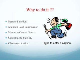
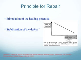
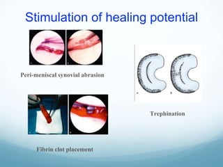
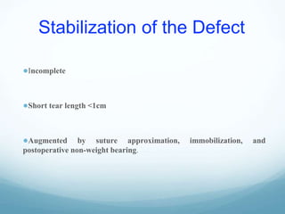
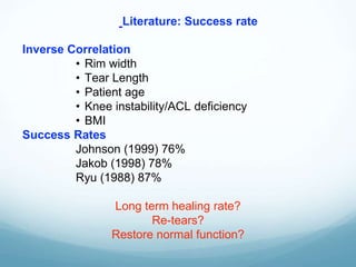
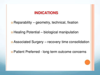
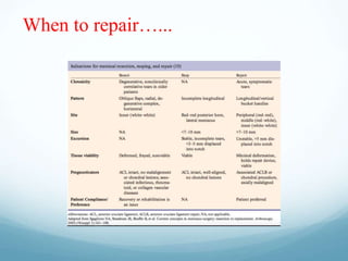
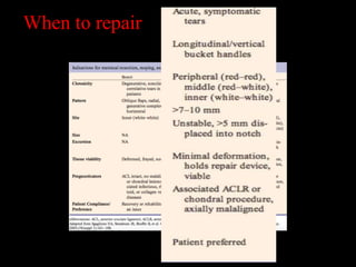
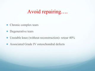
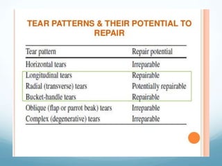
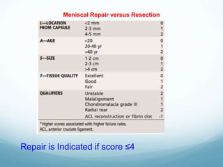
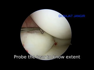
Ideal indications for
Meniscus Repair
Dr.Rajat Jangir
Consultant Arthroscopy Surgeon, Jaipur
Zones
Poulsen M, Johnson D. Meniscal injuries in the young, athletically active patient. The Physician and sportsmedicine1870.
Why to do it ??
● Restore Function
● Maintain Load transmission
● Minimize Contact Stress
● Contribute to Stability
● Chondroprotection Type to enter a caption.
Question ????
..….But then should every
TEAR be repaired ?
No
“Appropiate & informed decision required”
Only 20% repairable
*Fairbank et al. JBJS 1948;30B:664-70 *Chatain et al. Knee Surg 2001;9:15-18.
Principle for Repair
• Stimulation of the healing potential
• Stabilization of the defect *
*Newman A, Daniels A, Burks R. Principles and Decision Making in Meniscal Surgery. Arthroscopy: The Journal of
Arthroscopic and Related Surgery. 1993;9(1):33-51.
Stimulation of healing potential
Peri-meniscal synovial abrasion
Trephination
Fibrin clot placement
Stabilization of the Defect
●Incomplete
●Short tear length <1cm
●Augmented by suture approximation, immobilization, and
postoperative non-weight bearing.
Inverse Correlation
• Rim width
• Tear Length
• Patient age
• Knee instability/ACL deficiency
• BMI
Success Rates
Johnson (1999) 76%
Jakob (1998) 78%
Ryu (1988) 87%
Long term healing rate?
Re-tears?
Restore normal function?
Literature: Success rate
When to repair…...
When to repair
Avoid repairing….
● Chronic complex tears
● Degenerative tears
● Unstable knee (without reconstruction)- retear 40%
● Associated Grade IV osteochondral defects
Meniscal Repair versus Resection
Repair is Indicated if score ≤4
Dr.RAJAT JANGIR
Approach meniscal tears like fracture fixation !
● Perpendicular placement of implants every 5 mm
● Grab circumferential fiber bundle to ensure optimal purchase
strength
● Ensure that implants are not proud (intra-or extraarticular)
● Avoid stuffing the meniscus with stress riser inducing
implants
● Rehabilitation: Individualized
Ideal Indications Meniscus Repair  I Dr.RAJAT JANGIR JAIPUR

Ideal Indications Meniscus Repair I Dr.RAJAT JANGIR JAIPUR

  • 1.
    Ideal indications for MeniscusRepair Dr.Rajat Jangir Consultant Arthroscopy Surgeon, Jaipur
  • 3.
    Zones Poulsen M, JohnsonD. Meniscal injuries in the young, athletically active patient. The Physician and sportsmedicine1870.
  • 4.
    Why to doit ?? ● Restore Function ● Maintain Load transmission ● Minimize Contact Stress ● Contribute to Stability ● Chondroprotection Type to enter a caption.
  • 5.
    Question ???? ..….But thenshould every TEAR be repaired ? No “Appropiate & informed decision required” Only 20% repairable *Fairbank et al. JBJS 1948;30B:664-70 *Chatain et al. Knee Surg 2001;9:15-18.
  • 6.
    Principle for Repair •Stimulation of the healing potential • Stabilization of the defect * *Newman A, Daniels A, Burks R. Principles and Decision Making in Meniscal Surgery. Arthroscopy: The Journal of Arthroscopic and Related Surgery. 1993;9(1):33-51.
  • 7.
    Stimulation of healingpotential Peri-meniscal synovial abrasion Trephination Fibrin clot placement
  • 8.
    Stabilization of theDefect ●Incomplete ●Short tear length <1cm ●Augmented by suture approximation, immobilization, and postoperative non-weight bearing.
  • 9.
    Inverse Correlation • Rimwidth • Tear Length • Patient age • Knee instability/ACL deficiency • BMI Success Rates Johnson (1999) 76% Jakob (1998) 78% Ryu (1988) 87% Long term healing rate? Re-tears? Restore normal function? Literature: Success rate
  • 11.
  • 12.
  • 13.
    Avoid repairing…. ● Chroniccomplex tears ● Degenerative tears ● Unstable knee (without reconstruction)- retear 40% ● Associated Grade IV osteochondral defects
  • 15.
    Meniscal Repair versusResection Repair is Indicated if score ≤4
  • 16.
  • 17.
    Approach meniscal tearslike fracture fixation !
  • 18.
    ● Perpendicular placementof implants every 5 mm ● Grab circumferential fiber bundle to ensure optimal purchase strength ● Ensure that implants are not proud (intra-or extraarticular) ● Avoid stuffing the meniscus with stress riser inducing implants ● Rehabilitation: Individualized

Editor's Notes

  • #4 The “red-red zone” is located on the very outer edge of the meniscus and receives the most blood. The “red-white” zone is located in the middle 1/3 of the meniscus at the junction between the vascular and avascular portions of the meniscus. The “white-white” zone is the innermost aspect of the meniscus and comprises the avascular portion. The further out the lesion, the more likely the meniscus will heal
  • #6 Following removal - Cartilage overloading and aggravated degeneration THE BIG QUESTION
  • #7 Stimulation is accomplished by perimeniscal synovial abrasion and/or fibrin clot placement. Stability can be present inherently on the basis of incompleteness (the tear not extending the full thickness of the meniscus) or short tear length and can also be augmented by suture approximation, immobilization, and postoperative non-weight bearing.
  • #14 Flap tears, radial tears, or vertical tears with secondary lesions that extend into avascular inner 2/3 of meniscus, except in young teenagers Ligamentous ligamentous instability  (w/ ACL insufficiency, the rate or re-tearing approaches 40%, especially in younger active individuals, and therefore ACL reconstruction should be performed at the same surgery; ) Chronic complex tears. ¢ Degenerative tears. ¢ Unstable knee (without reconstruction). — Rule of thirds: ¢ ACL deficient knee – 30%. ¢ ACL stable knee – 60%. ¢ ACL reconstructed knee – 80-90%. ¢ Associated Gd. IV osteochondral defects. ¢ Old age.
  • #18 Nondisplaced meniscal tears in the peripheral vascular zone, especially when identified in a subacute set- ting, may already have undergone some early healing response. Often this includes fibrovascular scar formation lining the tear margins. Similar to debridement of bony edges in fractures and nonunions, the meniscal tear margins should be prepared by rasping these surfaces to improve apposition and optimize the healing surfaces. A variety of mechanical rasps are available, including forward-angle, back-angle, double-sided, and 90-degree double- sided instruments. An arthroscopic motorized shaver can also be used for gentle debridement of these edges. The goal is to establish a surface free of interposed material, ideally demonstrating punctate bleeding, which indicates vascular supply sufficient for reliable healing. This can usually be demonstrated by restricting the arthroscopic inflow prior to tourniquet inflation
  • #19 INDIVIDUALIZE – protection, WB, Motion, Return to Sport