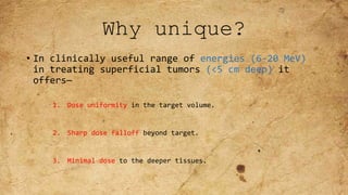
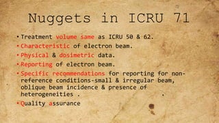
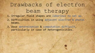
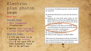
Radiosurgery combines stereotactic techniques and radiation beams to precisely deliver a high dose of radiation to tumors and lesions. The Exactrac system allows for frameless radiosurgery using infrared cameras and x-ray imaging to track internal anatomy and automatically position the patient for treatment without an invasive head frame. Treatment planning involves delineating the target and nearby organs at risk, specifying dose constraints, and optimizing the plan to conform the high dose region to the tumor while sparing surrounding healthy tissue.
This document discusses various methods used to evaluate radiotherapy treatment plans, including physical and biological parameters. Physically, plans are evaluated using isodose curves, dose distribution statistics, differential and cumulative dose-volume histograms (DVHs). Target coverage should be within 95-100% of the prescribed dose. Biologically, tumor control probability (TCP) and normal tissue complication probability (NTCP) models are used. The therapeutic ratio and index compare the dose required for tumor control versus normal tissue complications. NTCP models include Lyman-Kutcher-Burman and critical element/volume models. Plan evaluation ensures target doses are adequate while respecting organ tolerance doses.
This document provides information about total body irradiation (TBI). It discusses that TBI uses megavoltage photon beams to destroy the recipient's bone marrow and tumor cells prior to bone marrow transplantation. It is used to treat various diseases like leukemia, lymphoma, and multiple myeloma. TBI can be delivered at high or low doses, to half the body, or total nodes. Techniques include parallel opposed beams from linear accelerators or cobalt-60 machines. Dosimetry and in vivo dosimetry are important due to the large fields and difficulty achieving uniform dose. Complications can include sterility, secondary cancers, and growth issues.
TBI is the radiotherapy technique to irradiate whole body before doing stem cell transplant. The main purpose of doing TBIB is to condition the immune system of body so that there will be maximum chance of transplant acceptance.
This document summarizes key aspects of the International Commission on Radiation Units and Measurements (ICRU) Report 83 from 2010 on prescribing, recording, and reporting photon beam intensity-modulated radiation therapy (IMRT). The ICRU Report 50 from 1993 and Report 62 from 1999 established guidelines for defining target volumes like gross tumor volume, clinical target volume, and planning target volume. ICRU Report 83 aimed to update these guidelines for IMRT, which uses non-uniform fluence and dose distributions compared to earlier conformal radiation techniques. Key changes included separating the planning target volume into internal and setup margins, classifying organs at risk, and defining new metrics like the planning organ at risk volume and conformity index for evaluating IM
This document summarizes the process of simulation for radiation therapy treatment planning from CT imaging to treatment verification. It describes how patient positioning is done using lasers during CT scanning and how the CT images are imported into the treatment planning system. It also explains how the treatment planning system localizes CT markers and defines the isocenter in machine coordinates for treatment. Finally, it summarizes the verification process of aligning the patient using digital reconstructed radiographs and portal images to ensure accurate treatment delivery.
1. ICRU Report 83 provides guidelines for prescribing, recording, and reporting intensity-modulated radiation therapy (IMRT). It emphasizes using dose-volume histograms and statistics like median dose to describe dose distributions.
2. The report outlines three levels of prescribing and reporting with increasing complexity. Level 1 involves basic 2D dose distributions while Level 3 incorporates more advanced metrics like tumor control probability.
3. Key volumes discussed include gross tumor volume, clinical target volume, planning target volume, and organs at risk. The report standardized how to account for uncertainties and patient motion when defining these volumes.
This document discusses electron beam therapy and recommendations for prescribing, recording, and reporting it. Some key points:
- Electron beam therapy delivers a uniform dose to a specific depth below the skin, sparing deeper tissues. It is used to treat cancers within 6 cm of the surface.
- Factors like energy, field size, and tissue inhomogeneities affect dose distribution and must be considered in treatment planning.
- The ICRU reference point for dose reporting should be in the central part of the planning target volume at the peak dose level.
- Treatment planning involves choosing beam energy and field size based on target depth and calculating dose using factors, pencil beam, or Monte Carlo algorithms.
multiple filed arrangement in Radiotherapy, Medical College KolkataKazi Manir
The document discusses various radiation therapy techniques for dose distribution in matter using multiple fields and wedge fields. It covers:
1) Using multiple fields allows more uniform dose distribution in the tumor compared to a single field, while limiting dose to normal tissues.
2) Parallel opposed fields provide simplicity but can excessively dose normal tissues above and below the tumor. Larger field sizes are needed for adequate coverage.
3) Patient thickness, beam energy, and field size must be considered to minimize lateral tissue effects and ensure uniform dose distribution.
4) Multiple techniques like wedges, isocentric beams, and field matching seek to further optimize dose distribution while sparing critical structures. Proper planning and verification is important.
Permanent brachytherapy, also called seed implantation, involves placing tiny radioactive seeds directly into the prostate tumor. The seeds remain permanently and give a continuous low dose of radiation over months to treat early stage prostate cancer. Common seeds used are iodine-125 or palladium-103. Precise pre-planning and ultrasound guidance ensures correct seed placement to maximize dose to the tumor while minimizing dose to nearby organs. Potential side effects include urinary symptoms and erectile dysfunction, though risks are lower than other treatments. Seed implantation provides high radiation directly to the tumor with less impact on healthy tissue compared to external beam radiation.
The vmat vs other recent radiotherapy techniquesM'dee Phechudi
VMAT is a new type of intensity-modulated radiation therapy (IMRT) treatment technique that uses the same hardware (i.e. a digital linear accelerator) as used for IMRT or conformal treatment, but delivers the radiotherapy treatment using a rotational or arc geometry rather than several static beams.
This technique uses continuous modulation (i.e. moving the collimator leaves) of the multileaf collimator (MLC) fields, continuous change of the fluence rate (the intensity of the X rays) and gantry rotation speed across a single or multiple 360 degree rotations
This document discusses radiotherapy techniques for lymphoma, including:
1. It describes the radiotherapy fields used to treat different lymph node regions, such as cervical, supraclavicular, mediastinal, axillary, abdominal, and inguinal regions.
2. It provides details on the dose of radiotherapy used in combined modality treatment with chemotherapy, ranging from 20-36 Gy depending on disease stage and bulkiness.
3. It outlines both the acute and late side effects of radiotherapy, such as fatigue, dermatitis, hypothyroidism, infertility, and increased risk of secondary cancers. Reducing radiation volumes and doses over time has helped lower long-term risks.
This document discusses Image Guided Radiation Therapy (IGRT). It begins by explaining that radiotherapy has traditionally used imaging for treatment planning and execution when the target is not on the surface. It then describes various IGRT technologies, dividing them into non-radiation based systems like ultrasound, cameras, electromagnetic tracking and MRI; and radiation based systems like EPID, CBCT, fan beam KVCT and MVCT. These systems provide improved target localization and allow for corrections. IGRT aims to reduce errors and improve precision of radiotherapy.
This document discusses hemi-body and total body irradiation techniques. Total body irradiation (TBI) delivers a uniform whole body radiation dose and is used as a conditioning regimen before bone marrow transplantation. It was developed in the early 1900s and is now used to treat various cancers and blood disorders. TBI can be delivered using dedicated or modified conventional irradiators. Dosimetry and compensators are used to ensure uniform dose delivery. Adverse effects include nausea, vomiting, pneumonitis and cataracts. Hemi-body irradiation treats only the upper or lower half of the body and has fewer side effects than total body irradiation.
This document discusses Intra-cavitary Brachytherapy (ICBT) for treating cervical cancer. It describes different historical ICBT systems like Paris, Stockholm, and Manchester systems. It also discusses modern techniques like remote afterloaders and recommendations for reporting absorbed doses and volumes in ICBT. Key points include different dose rates (LDR, MDR, HDR), advantages of remote afterloaders in maintaining geometry and dose distribution, and recommending specifying absorbed dose to the target volume rather than at a single point for ICBT.
The document discusses the use of Tomotherapy for radiation treatment planning and delivery. It provides examples of how Tomotherapy allows for:
1) Highly conformal radiation plans that sculpt dose around complex tumor target shapes while minimizing dose to nearby organs.
2) Daily image guidance that enables adjustment of targets to account for changes in patient anatomy and tumor size during treatment.
3) Delivery of simultaneous integrated boosts to multiple tumor sites.
Isodose curves depict absorbed dose distributions and variations in volume and planes. They join points of equal dose. Isodose charts show the variation in dose as a function of depth and transverse distance from the central beam axis. Factors like beam energy, field size, and distance affect isodose curve shape through penumbra and dose deposition. Multiple beams are often needed to adequately treat tumors while sparing surrounding tissues. Beam arrangements, weights, and modifiers must be optimized for each plan.
Simultaneously integrated boost (SIB) allows different doses to be delivered simultaneously to the planning target volume (PTV) and gross tumor volume (GTV), reducing the number of fractions needed. SIB provides a greater biological effective dose while allowing individual dose optimization to both targets in a single plan, overcoming limitations of conventional fractionation. An institutional study compared SIB to conventional 3D conformal radiation therapy in 30 patients with brain, breast, or bladder cancer, finding SIB reduced maximum doses to targets and organs at risk while shortening treatment duration by about a week.
1.Aim of Radiotherapy
The goal of radiotherapy is to deliver a prescribed dose of radiation to the Target while sparing surrounding Healthy tissues to the largest extent possible
2.Organ Motion
Intra-fraction motion
during the fraction
Heartbeat
Swallowing
Coughing
Eye movement
Inter-fraction motion
- in between the fractions
Tumour change
Weight gain/loss
Positioning deviation
Breathing
Bowel and rectal filling
Bladder filling
Muscle relaxation/tension
3. Respiratory motion affects:
Respiratory motion affects all tumour sites in the thorax, abdomen and Pelvis. Tumours in the Lung, Liver, Pancreas, Oesophagus, Breast, Kidneys, prostate
Tumour displacement varies depending on the site and organ Location
Lung tumours can move several cm in any direction during irradiation
It is most prevalent and prominent in Lung cancers
4. Problems associated with respiratory motion during RT
Image acquisition limitations
Treatment planning limitations
Radiation delivery limitations
5. Methods to Account for Respiratory Motion
1. Motion encompassing methods
2. Respiratory gating methods
3. Breath hold methods
4. Forced shallow breathing with abdominal compression
5. Real-time tumor tracking methods
Summary:
The management of respiratory motion in radiation oncology is an evolving field
IGRT provides a solution for combating organ motion in radiotherapy
Delivering higher dose to tumor and less dose to normal tissue.
Limited clinical studies, needs to be studied further
IGRT – the future of radiotherapy
Stereotactic radiosurgery (SRS) is a non-invasive technique that uses precisely targeted radiation to treat tumors and abnormalities in the brain. It can deliver high doses of radiation to tumors while minimizing damage to surrounding healthy tissue. SRS uses stereotactic guidance systems and imaging to pinpoint tumors for radiation targeting in fewer treatments than traditional therapy. Common applications of SRS include treatment of brain metastases, acoustic neuromas, meningiomas, arteriovenous malformations, and pituitary tumors. Control rates for these indications are high, often over 90% depending on factors like tumor size and location.
This document discusses lung stereotactic body radiotherapy (SBRT) for the treatment of early stage non-small cell lung cancer (NSCLC). It covers treatment indications for SBRT, methods used to account for tumor motion including 4DCT planning and respiratory gating, treatment planning guidelines, evidence from studies showing high rates of local control and survival, and results from RTOG trials of SBRT for lung cancer. In particular, it highlights that SBRT achieves local control rates of 85-95% and overall survival rates of 50-95% at 3-5 years for early stage NSCLC.
Total skin electron therapy (TSET) is used to treat cutaneous T-cell lymphoma by delivering a uniform dose of radiation to the entire skin surface while sparing underlying organs. It requires large electron fields over the entire body and precise dosimetry. The most common technique uses six electron beams arranged at 60 degree intervals to provide circumferential coverage. Proper field design and calibration are needed to achieve uniform dose across irregular body surfaces and minimize dose from bremsstrahlung x-rays.
CyberKnife: Radiosurgery System Introductionduttaradio
Radiation source is mounted on a precisely controlled industrial robot.
- Image guidance system(continuous tracking system)
- Eliminates the need of gating techniques and restrictive head frames
Several institutions have studied stereotactic body radiation therapy (SBRT) for primary lung cancer. Indiana University studies showed a maximum tolerated dose of 66 Gy for T2 lesions delivered over 3 fractions, with 1-year local control rates of 98%. Other studies from Aarhus University, Kyoto University, Air Force General Hospital in Beijing, and University of Marburg demonstrated 1-2 year local control rates ranging from 85-95% using SBRT dose fractions between 30-60 Gy delivered over 1 to 10 fractions.
This document discusses various particle beams used in radiation therapy, including their properties and effectiveness. It states that proton beams have superior dose distribution compared to photon beams but lower LET. Neutron beams have high LET properties but poor dose distribution. Heavy charged particle beams like carbon ions have both superior distribution and high LET. BNCT uses boron compounds and neutrons to specifically target tumor cells but is limited by availability and cost. Overall, the document provides an overview of different particle therapies and their advantages over conventional photon radiation.
Electron beam therapy uses electrons to deliver radiation to treat cancers close to the surface of the body. Electrons deposit most of their dose in the first few centimeters, sparing deeper tissues. Key factors in electron beam planning include selecting an appropriate electron energy to cover the tumor volume while minimizing dose beyond it, shaping the electron field using collimators or bolus material, and techniques for field junctions and irregular surfaces. Examples of clinical applications include treatment of skin cancers or chest wall irradiation after mastectomy.
This document discusses the importance of treatment verification in radiotherapy and outlines the process. It notes that even small errors can have negative consequences so treatment verification is essential to ensure the right dose is delivered to the right area. The key aspects of treatment verification are machine setup, monitor units, patient positioning and imaging by comparing images to references. Errors can be systematic from planning or random from daily variations; various methods are described to reduce errors and ensure treatments are accurately delivered.
Electron beam therapy uses megavoltage electron beams to treat superficial tumors within 6 cm of the skin surface, sparing deeper tissues. The dose distribution of electron beams provides a uniform dose in the target region followed by a rapid dose fall-off. Treatment planning for electron beams requires consideration of electron energy, air gaps, tissue inhomogeneities, and adjacent fields to determine the optimal dose distribution. Electron beams can effectively treat many superficial cancers of the skin, limbs, and surgical beds.
CONTENTS
Electron arc therapy.
Introduction to electron arc therapy
Calibration of electron arc therapy
field shaping
beam energy
Treatment planning
location of the isocentre
scanning field width
collimation used in electron arc therapy.
summary
1) Megavoltage electron beams represent an important treatment modality for superficial tumors up to 5 cm deep, sparing deeper tissues. They are commonly used to treat skin lesions and boost areas previously treated with photon irradiation.
2) Electron beams have high surface dose that decreases with depth, reaching half the maximum dose around the practical range. Bremsstrahlung production increases with energy. Beam energy is selected based on the required depth of treatment.
3) Tissue heterogeneities like air cavities or bone can shift dose distributions. Bolus and beam obliquity are used to compensate. Treatment planning considers target coverage and critical structure sparing.
This document discusses electron beam therapy. It begins by explaining that electron beams allow for uniform dose delivery to within approximately 6 cm of the surface, sparing deeper tissues. Electrons are useful for treating various cancers of the skin, head and neck, breast, and other sites. It then covers topics like electron beam production, interactions in tissue, depth dose curves, effects of field size and obliquity, tissue heterogeneities, treatment planning, and future directions like intensity-modulated electron therapy.
1) Electron beam therapy uses high-energy electrons between 6-20MeV to treat superficial tumors less than 5cm deep. It is useful for cancers of the skin, eye, breast, head and neck, and gastrointestinal tract.
2) Electron beams have distinct advantages over x-rays and brachytherapy in minimizing dose to deeper tissues and providing dose uniformity.
3) The depth that receives 90% of the maximum dose, called R90, is typically one-third to one-fourth of the electron energy in MeV. This determines the maximum treatment depth.
1. Isodose curves represent the dose distribution from radiation beams and are lines connecting points of equal percentage depth dose. They are used to depict the volumetric and planar variations in absorbed dose.
2. The parameters that affect the shape of isodose curves include beam quality, source size, SSD, SDD, field size, and beam modifiers like wedges and flattening filters. Lower beam energy results in greater lateral scatter and more bulging curves.
3. Multiple radiation fields can be combined using appropriate beam weights, sizes, angles and modifiers to deliver a more uniform dose to the tumor while sparing surrounding tissues. Parameters like setup accuracy and plan practicality are also considered.
Electron beam radiotherapy uses megavoltage electron beams ranging from 6-20 MeV to treat superficial tumors within 6 cm of the skin surface. It provides a uniform dose at a specified depth with rapid dose fall-off, sparing deeper tissues. Common tumors treated include skin, lymphomas, and breast cancer. Electrons deposit dose via interactions like collision and scattering. Dose distribution is characterized by a rapid buildup to a maximum within 1 cm followed by a rapid falloff beyond the treatment depth.
Electron beam radiotherapy uses megavoltage electron beams ranging from 6-20 MeV to treat superficial tumors within 6 cm of the skin surface. It provides a uniform dose at a specified depth with rapid dose fall-off, sparing deeper tissues. Common tumors treated include skin, lymphomas, and breast tissue. Electrons deposit dose via interactions with atomic electrons and nuclei, with dose decreasing rapidly beyond the 90% depth. Beam characteristics like central axis depth dose, isodose curves, and field size must be carefully considered for treatment planning.
1) Megavoltage photon therapy uses high energy photon beams, typically above 1 MV, generated via linear accelerators or cobalt-60 sources.
2) The three main interactions between photons and tissue are the photoelectric effect, Compton scattering, and pair production. Compton scattering is the most common interaction for megavoltage beams between 6-20 MV.
3) Key characteristics of megavoltage photon beams include percentage depth dose curves, beam profiles, isodose charts, and considerations for tissue heterogeneities. Wedge filters can also be used to modify beam profiles for uniform target coverage.
Electron beam therapy uses electrically charged particles called electrons that are generated by a linear accelerator to treat superficial cancers. It deposits dose uniformly from the surface to a specific depth before rapidly falling off, sparing deeper tissues. Electron energies up to 20 MeV can treat disease within 6 cm of the surface. Accessories like applicators, cutouts, bolus and internal shields are used to shape the beam for treatment fields and protect healthy tissues. Precise dose specification and reporting is important for electron therapy due to the rapid dose fall-off and higher skin doses compared to prescription depth.
Electron beam therapy uses electrically charged particles called electrons to treat superficial cancers. It delivers a uniform dose to a specified depth before rapidly falling off, sparing deeper tissues. Common treatment sites include skin, head and neck, breast, and other areas within 6 cm of the surface. Electron beams are shaped using applicators and cutouts. Dose is often prescribed at the depth of maximum dose to provide homogeneous coverage while sparing healthy tissue beyond the tumor. Bolus material can be used to modify the electron range and shape dose distributions for irregular surfaces.
This document provides an overview of interstitial brachytherapy principles and concepts. It discusses the history and evolution of brachytherapy sources from radium to modern radioactive sources like iridium-192. Key concepts covered include dose rate calculations, implant systems like the Paris system, and factors that influence dose distribution from a radioactive source like distance, absorption and scattering. The document also describes temporary and permanent brachytherapy sources and different methods of source application including preloading, afterloading and remote afterloading.
1. The Manchester system was one of the first standardized methods for interstitial brachytherapy implantation using radium sources. It provided rules for determining the amount of radium needed, its distribution, and dose specification based on the treatment area, distance, and desired total dose.
2. The amount of radium was calculated using exposure rates and tables that specified milligram hours of radium per 1000 rads of exposure for different areas and distances. The distribution of radium sources followed the inverse square law to achieve uniform dosing within 10% across the target area.
3. The system defined geometric terms and provided specific rules for distributing radium sources in planar moulds that were circular, square,
The document discusses techniques for measuring resistivity and mapping resistivity variations across semiconductor wafers. It begins by defining resistivity and listing typical resistivity values for different materials. It then describes two common measurement techniques: two-point probe and four-point probe. Four-point probe is more accurate as it eliminates lead and contact resistance. Factors that affect measurement accuracy like sample size, carrier injection, and probe spacing are also covered. The document concludes by explaining techniques for wafer mapping like double implant, modulated photoreflectance, and optical densitometry.
4. Isodose Distribution In Radiation Oncology .pptxAbhishekMewara2
The document provides an overview of isodose distribution and related concepts. It discusses isodose charts which characterize radiation dose distribution in 3D volumes. Measurements are done using ion chambers in water phantoms. Parameters like beam quality and field size influence isodose curve shape. Wedge filters tilt isodose curves to modify dose distribution. Combining parallel opposed or multiple fields optimizes dose to the tumor. The isocenter is the point of intersection for machine axes. Different target volumes like GTV, CTV and PTV are defined to account for tumor extent and uncertainties.
Total body irradiation (TBI) is a form of radiotherapy used prior to bone marrow transplants to reduce the risk of transplant rejection and destroy any remaining cancer cells. TBI techniques use large photon fields, usually from cobalt-60 machines or LINACs, to irradiate the entire body. Common techniques include opposing anterior-posterior beams or lateral beams. Precise dosimetry is required due to the large fields and total body exposure, with dose uniformity targets of within ±10% across the body. In vivo dosimetry using TLD or diodes is also employed to verify accurate dose delivery. Early side effects from TBI include fatigue, nausea, hair loss and skin irritation due to the whole body irradiation
This document discusses various concepts related to the measurement of absorbed radiation dose. It defines key terms like fluence, kerma, exposure, absorbed dose and stopping power. It describes different methods of absorbed dose measurement including calorimetry, chemical dosimetry and solid state methods. The Bragg-Gray cavity theory relating dose in a dosimeter to dose in surrounding medium is also explained.
Medical uses of ionizing radiation include radiotherapy, medical imaging like CT scans and X-rays, and nuclear medicine. Radiotherapy uses radiation to treat cancer and can involve external beam techniques like 3D conformal radiation therapy (3D CRT), intensity modulated radiation therapy (IMRT), volumetric modulated arc therapy (VMAT), stereotactic radiosurgery, and brachytherapy. Emerging techniques like proton beam therapy further improve radiation targeting and dose distribution. Precise imaging guidance and computer planning help deliver high radiation doses safely and effectively to tumors while avoiding nearby healthy tissues.
- Proton therapy uses protons rather than photons to treat cancer as protons deposit most of their energy at the end of their range, known as the Bragg peak, allowing for higher doses to the tumor and reduced doses to surrounding healthy tissues.
- Protons are produced from hydrogen gas and accelerated using either a cyclotron or synchrotron before being delivered to the patient in either a scattering or scanning beam technique.
- Treatment planning involves CT imaging of the patient to determine proton stopping powers, delineating the tumor, selecting beam directions, and optimizing the plan using pencil beam algorithms to calculate dose.
- Potential applications of proton therapy include central nervous system cancers, head and neck cancers, lung cancers, and some
Updates on Electron Beam Therapy
I) Introduction
II) Central Axis Depth dose distribution
III) Dosimetric parametrics of electron beam
IV) Clinical Considerations of Electron beam therapy
Promoting Wellbeing - Applied Social Psychology - Psychology SuperNotesPsychoTech Services
A proprietary approach developed by bringing together the best of learning theories from Psychology, design principles from the world of visualization, and pedagogical methods from over a decade of training experience, that enables you to: Learn better, faster!
8 Surprising Reasons To Meditate 40 Minutes A Day That Can Change Your Life.pptxHolistified Wellness
We’re talking about Vedic Meditation, a form of meditation that has been around for at least 5,000 years. Back then, the people who lived in the Indus Valley, now known as India and Pakistan, practised meditation as a fundamental part of daily life. This knowledge that has given us yoga and Ayurveda, was known as Veda, hence the name Vedic. And though there are some written records, the practice has been passed down verbally from generation to generation.
Cell Therapy Expansion and Challenges in Autoimmune DiseaseHealth Advances
There is increasing confidence that cell therapies will soon play a role in the treatment of autoimmune disorders, but the extent of this impact remains to be seen. Early readouts on autologous CAR-Ts in lupus are encouraging, but manufacturing and cost limitations are likely to restrict access to highly refractory patients. Allogeneic CAR-Ts have the potential to broaden access to earlier lines of treatment due to their inherent cost benefits, however they will need to demonstrate comparable or improved efficacy to established modalities.
In addition to infrastructure and capacity constraints, CAR-Ts face a very different risk-benefit dynamic in autoimmune compared to oncology, highlighting the need for tolerable therapies with low adverse event risk. CAR-NK and Treg-based therapies are also being developed in certain autoimmune disorders and may demonstrate favorable safety profiles. Several novel non-cell therapies such as bispecific antibodies, nanobodies, and RNAi drugs, may also offer future alternative competitive solutions with variable value propositions.
Widespread adoption of cell therapies will not only require strong efficacy and safety data, but also adapted pricing and access strategies. At oncology-based price points, CAR-Ts are unlikely to achieve broad market access in autoimmune disorders, with eligible patient populations that are potentially orders of magnitude greater than the number of currently addressable cancer patients. Developers have made strides towards reducing cell therapy COGS while improving manufacturing efficiency, but payors will inevitably restrict access until more sustainable pricing is achieved.
Despite these headwinds, industry leaders and investors remain confident that cell therapies are poised to address significant unmet need in patients suffering from autoimmune disorders. However, the extent of this impact on the treatment landscape remains to be seen, as the industry rapidly approaches an inflection point.
Hiranandani Hospital in Powai, Mumbai, is a premier healthcare institution that has been serving the community with exceptional medical care since its establishment. As a part of the renowned Hiranandani Group, the hospital is committed to delivering world-class healthcare services across a wide range of specialties, including kidney transplantation. With its state-of-the-art facilities, advanced medical technology, and a team of highly skilled healthcare professionals, Hiranandani Hospital has earned a reputation as a trusted name in the healthcare industry. The hospital's patient-centric approach, coupled with its focus on innovation and excellence, ensures that patients receive the highest standard of care in a compassionate and supportive environment.
- Video recording of this lecture in English language: https://youtu.be/kqbnxVAZs-0
- Video recording of this lecture in Arabic language: https://youtu.be/SINlygW1Mpc
- Link to download the book free: https://nephrotube.blogspot.com/p/nephrotube-nephrology-books.html
- Link to NephroTube website: www.NephroTube.com
- Link to NephroTube social media accounts: https://nephrotube.blogspot.com/p/join-nephrotube-on-social-media.html
One health condition that is becoming more common day by day is diabetes.
According to research conducted by the National Family Health Survey of India, diabetic cases show a projection which might increase to 10.4% by 2030.
Rasamanikya is a excellent preparation in the field of Rasashastra, it is used in various Kushtha Roga, Shwasa, Vicharchika, Bhagandara, Vatarakta, and Phiranga Roga. In this article Preparation& Comparative analytical profile for both Formulationon i.e Rasamanikya prepared by Kushmanda swarasa & Churnodhaka Shodita Haratala. The study aims to provide insights into the comparative efficacy and analytical aspects of these formulations for enhanced therapeutic outcomes.
3. Why unique?
• In clinically useful range of energies (6-20 MeV)
in treating superficial tumors (<5 cm deep) it
offers—
1. Dose uniformity in the target volume.
2. Sharp dose falloff beyond target.
3. Minimal dose to the deeper tissues.
4. MAIN USES
a) Treatment of skin & lip cancers.
b) Chest wall irradiation for breast cancer.
c) Boost dose to nodes.
d) Head & Neck irradiation.
5. Nuggets in ICRU 71
• Treatment volume same as ICRU 50 & 62.
• Characteristic of electron beam.
• Physical & dosimetric data.
• Reporting of electron beam.
• Specific recommendations for reporting for non-
reference conditions-small & irregular beam,
oblique beam incidence & presence of
heterogeneities .
• Quality assurance
6. Physical
characteristi
cs of
electron beam
• R p-The point at which the
tangent at the steepest
point(the inflection point)on
the almost straight descending
portion of the depth-dose curve
meets the extrapolated
bremsterlung background(Dₓ)
• E p,o=C1+C₂Rp+C₃R²p
• Rt=depth on the beam axis, of a
given isodose relevant for the
treatment.
• Broad Beam-central axis depth
dose distribution can be considered
independent of the field size, when
further increased.
Lax & Brahme-Field size
diameter>Rp represent broad
beam situation.
7. Effect of field size on central-
axis depth-dose curves
7MeV 13MeV 20MeV
8. DOSE
GRADIENT
• G- A measure of dose fall-off.
• G=
𝑅𝑝
𝑅𝑟−𝑅𝑞
• Lower value of G(2.5)is observed
in most accelerators.
• Higher value of G (3.3) –an
accelerator with optimized design
for the treatment head.
• G<2.3 for broad beams (5-30
MeV) indicates the flattening &
the collimating system is of poor
design & that unnecessary large
volume of normal tissues are
irradiated in a Single beam
technique.
• G decreases for small fields.
9. Oblique
beam
incidence
Changes-
1. ↑Surface dose.
2. ↑dose at the
maximum along the
beam axis.
3. ↓penetration of the
therapeutic depth
dose.
4. ↑range of penetration
of a low dose
component.
Isodose distribution has a
“wedge” form & the
10. collimation
Electron beam is
generally
collimated with
external
applicators, placed
directly on the
skin or close to
skin.
Without using
external
applicators large
penumbra results.
11. Collimation
contd..
a) OLD TYPE-
1 scattering foil.
Large energy & angular spread.
b) CONVENTIONAL TYPE-
2 scattering foils.
Much less energy & angular
spread.
c) NEW DEVELOPMENT-
A scanning beam.
Leaf collimator is used(for both
photon & electron).
Air is replaced by He.
It is seen (karlsson et al 1992)
using He in place of air reduces
penumbra by about 40%.
a. b. c.
12. Drawbacks of electron
beam therapy
1. Irregular field shapes are laborious to set up.
2. Difficulties in using adjacent electron & photon
beams.
3. Dose determination & computation difficult,
particularly in case of heterogenicities.
13. REPORTING e-BEAM
• In LEVEL 1- 3 values constitute the minimum
information to be reported.
1) Dose @ ICRU Reference Point.
2) Maximum & 3) minimum dose to PTV.
• In LEVEL 2&3-
1) More accurate information regarding 2) & 3).
2) DVH for PTV, Some cases CTV,GTV, PRV to be
reported.
14. REPORTING e-BEAM
• Reporting is very similar
to ICRU 50 & 62.
• ICRU Reference Point
(general criteria)-
1. Dose @this point should be
clinically relevant.
2. Point should be easy to
define in a clear &
unambiguous way.
3. Where the dose can be
accurately determined.
4. In a region where there is
no steep dose gradient.
• To fulfill these criteria…
a) Always @the center (or in
the central part) of the
PTV.
b) If 1 beam is used in e
beam therapy, whenever
possible,the point should
be @ the beam axis,@ the
level of the peak dose.
• Dose @ ICRU Reference
point is ICRU Reference
dose.
15. ICRU Reference point @max depth
dose
ICRU Reference point & max depth
dose are @ different area.
16. contd…
• Ideally the PTV should be covered by the TV.
• As it is not be the case, proportion PTV enclosed
by the TV should be recorded…like
Portion of the PTV receiving 95,90 & 85% of the ICRU
Reference dose is referred as PTV95, PTV90, PTV85.
17. Dose at Reference
Points
Issues to be considered:-
• Geometric factors.
• Dose computation
techniques.
ICRU Reference Point is at the
center/at the central part of PTV-
• No steep dose gradient.
• Relatively homogenous
dose around Reference
Point.
• Dose @ the point is
equal /close to the dose
delivered to large
portion of PTV.
18. • Depending on the algorithm used & voxel size there
may be statistically significant fluctuation in
dose calculation.
• In such cases average dose value within a sphere
(usually 1cm in diameter) centered around the ICRU
Reference point is calculated,
19. Reporting dose
distribution to OAR
• Probability of late effects depends on-
a.Dose level.
b.Fractionation.
c.Absolute volume &/or fration of the OAR
irradiated.
• For each OAR the Maximum dose should be
reported.
21. Small & irregularly
shaped beams
Specialities
• Depth of max dose moves
towards surface.
• ↓Depth of therapeutic range.
• ↑Relative surface dose.
• Dose fall-off shallower.
• ↓Dose rate.
Changes ↑with ↓energy
Level 1,2 & 3
• Same as single e beam
22. Extended SSD
Specialities
• ↑SSD leads to-
o↑Surface dose.
oModerate change in
depth-dose region,
except build up
region.
oLoss in beam
flatness.
oWider penumbra.
Level 1-3
• same
23. Heterogeneities
Effect of heterogeneities Level 1
• If only level 1 is available,
use of e beam to be
reconsidered.
LEVEL 2
• Peak absorbed dose in water to
be determined for an incident
beam in a homogeneous phantom.
• An accurate dose distribution
(corrected for
heterogeneities) should be
obtained, so that radiation
oncologist can decide the ICRU
Reference Point.
24. Bolus
Used to..
• ↑Skin dose.
• Compensate for surface
irregularities/oblique
beam incidence.
• Match the beam
penetration with the
shape of the PTV.
LEVEL 1-3
• SSD should be clearly
stated.
• ICRU Reference Point
should be in the tissue,
not in the bolus.
• Others being same.
25. Electron
plus photon
beam
Used for-
Coaxial beams
1.More homogenous
irradiation.
2.Better skin sparing &
normal tissue sparing.
Adjacent beams-
• A CTV may have to be
covered by several
PTVs & for each PTV an
ICRU Reference Point
has to be defined.
26. Combination of e beams
Adjacent parallel
• Used when PTV is too large.
• Special care to be taken at the
field junctions.
Parallel opposed
• Less frequently used.
• Usualy beams of high energy is
(about 40 MeV) is used.
• Significant over/under dosing
can result from beam energy
<30 MeV.