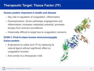
November 12, 2015
Breakthrough Opportunities
in Retinal Disease and Cancer
William L. Greene, MD
Chief Executive Officer
Ophthalmology Innovation Summit
Snapshot
Tissue Factor: central to pathophysiology of retinal disease and cancer
– Targeting an “undrugged” pathway
– Leveraging advances in Tissue Factor biology, new scientific understandings
Addressing significant therapeutic needs in large markets
– Wet AMD: critical need for disease modifying agents
– Cancer: opportunity to treat tumor, vasculature, and microenvironment
with a single targeted agent
Lead clinical candidate in Phase 2a for wet AMD
Expanding portfolio of development programs
– Ocular Melanoma, IND 1H 2016
Positioned for success
– World-class management team, advisors, Board
– Strong financial position and IP
2
Product Pipeline
Indication Research Preclinical Phase 1 Phase 2 Phase 3
Near Term
Milestones
Wet AMD
Top line data
2H 2016
Additional
Retinal
Indications
Ocular
Melanoma
IND
1H 2016
Solid
Tumors
3
Therapeutic Target: Tissue Factor (TF)
Human protein important in health and disease
– Key role in regulation of coagulation, inflammation
– Overexpression: drives pathologic angiogenesis and
inflammation; increases metastatic potential, promotes
escape from immune surveillance
– Historically difficult to target due to coagulation concerns
ICON-1: First-in-class human immunoconjugate
fusion protein
– Engineered to safely bind TF by replacing its
natural ligand without significant effect on
coagulation function
– Acts similar to a therapeutic mAb
4
Tissue Factor: Central Role in Wet AMD
5
Macula with Wet AMD
CNV:
TF overexpressed
on vessels and in vitreous
Accessible to bind to anti-TF agent
Angiogenesis
Wet AMD
Hypoxia Inflammatory
Cytokines
Increased
Levels of TF
Macrophages
(Inflammatory)
Inflammation
Increased
VEGF
Release
Amplification
of Angiogenic
Cytokines
Release of
Proinflammatory
Cytokines
(e.g., IL-6, IL-8)
ICON-1 for Wet AMD: Preclinical Data
A. Micrograph of control lesion: CNV
growth prominent
6
B. Micrograph of a lesion treated with
mI-con1 (mouse version of ICON-
1): little evidence of CNV
Bora et al. Proc Natl Acad Sci 2003.
• Murine models: prevention and regression of CNV after single dose
• Porcine model: dose response observed
• Excellent safety profile
Wet AMD Mouse Model
ICON-1 for Wet AMD: Phase 1 Study
First in human single dose trial: focused on safety
– Broad inclusion criteria; 9/18 subjects newly diagnosed
– 3 dose groups: 0.06 mg, 0.15 mg or 0.30 mg
Safety: no ocular or systemic safety issues; no immunogenicity seen
Signals of potential dose-related biologic activity
– Visual acuity increases, reduced retinal fluid, reduced leakage on angiograms
7
58
+19
+4
+16
+11
+16 +21
467
-236
-57
-148
-175
-215
-238
Week 5 Week 8 Week 12 Week 16
-300
-250
-200
-150
-100
-50
0
50
100
150
200
250
300
30
35
40
45
50
55
60
65
70
75
80
Screen Wk 1 Wk 2 Wk 5 Wk 8 Wk 12 Wk 24
Microns(μm)
Letters(ETDRS)
BCVA
OCT: Center
Subfield Change
Anti-VEGF
Treatment
Treatment-naïve patient, 0.3mg dose group
ICON-1 Phase 1: Subject Case Study
8
Treatment Naïve
Patient Screening
Follow Up
Week 24
Single Dose ICON-1
Week 5
Central Retinal Thickness (CRT) on Optical Coherence Tomography (OCT)
and CNV leakage on Fundus Fluorescein Angiography (FFA)
VA (Letters)
58 (20/63)
VA (Letters)
69 (20/40)
VA (Letters)
79 (20/25)
EMERGE Study: Data Expected 2H 2016
Phase 2a, Double-Masked, Active-Controlled, Multicenter (U.S.), Initiated 2015
9
“Rescue treatment” with Lucentis is available at any time during the study based on
protocol-defined criteria.
ICON-1 300μg
1:1:1 Randomization
Lucentis 0.5mg
ICON-1 300μg
Lucentis 0.5mg
End of Study: Month 6
Primary Endpoint: Month 3
Treatment-Naïve
Primary, Active CNV
N=90
ClinicalTrials.gov Identifier: NCT02358889
ICON-1 for Ocular Melanoma
Broadly applicable cancer mechanism
– TF widely expressed on tumor cells, tumor vasculature, and in microenvironment
– Marker for aggressive tumor growth, poor prognosis; central to process of
metastasis
TF overexpressed in cutaneous and ocular melanoma
Positive ICON-1 data in xenograft (cutaneous) melanoma model
Plan: initiate IND in OM 1H 2016
Ongoing research in other solid tumors
10
Melanoma mouse xenograft treated with
“murine version” of ICON-1
Hu Z, Sun Y & Garen A, Proc Natl Acad Sci (USA), 1999.
Leadership Team
11
William Greene, MD
Chief Executive Officer
Gabriela Burian, MD MPH
Chief Medical Officer
Kirk Dornbush
Co-Founder and President
K. Peter Hirth, PhD
Senior Scientific Advisor
Thi-Sau Migone, PhD
Head of Research
George Montgomery
SVP Financial Strategy
Board of Directors and Advisors
Board of Directors Scientific and Clinical Advisory Board
Kirk Dornbush, Iconic Founder
Todd Foley, MPM Capital
William Greene, MD, Iconic
K. Peter Hirth, PhD (Independent)
Johan Kördel, PhD, Lundbeckfond Ventures
Bruce Robertson, PhD, H.I.G. BioVentures
E. Jonathan Soderstrom, PhD, Yale
Evangelos S. Gragoudas, MD, Massachusetts
Eye & Ear Infirmary
Christine R. Gonzales, MD, Retina and
Vitreous Center
Darius M. Moshfeghi, MD, Stanford University
Elias Reichel, MD, Tufts University
Carl D. Regillo, MD, Wills Eye Hospital
Wolfram Ruf, MD, Scripps Research Institute
Steven D. Schwartz, MD, Jules Stein Eye
Institute, UCLA
12
Investors
Summary
Breakthrough opportunities in retinal diseases and cancer
– Deep knowledge of TF biology
– Unique therapeutic applications based on known and emerging TF science
– Addressing critical therapeutic needs in large markets
Multiple product platform in development
– ICON-1: IND OM 1H 2016; Phase 2a Wet AMD data 2H 2016
– Additional targeted oncology molecules in development
Poised for execution, strong financial position
– World-class advisors and team members with proven track record
– $40 million raised to date; Series C financing in 2015
– Substantial near-term milestones
13
THANK YOU
www.IconicTherapeutics.com
14

Iconic Therapeutics

  • 1.
    November 12, 2015 BreakthroughOpportunities in Retinal Disease and Cancer William L. Greene, MD Chief Executive Officer Ophthalmology Innovation Summit
  • 2.
    Snapshot Tissue Factor: centralto pathophysiology of retinal disease and cancer – Targeting an “undrugged” pathway – Leveraging advances in Tissue Factor biology, new scientific understandings Addressing significant therapeutic needs in large markets – Wet AMD: critical need for disease modifying agents – Cancer: opportunity to treat tumor, vasculature, and microenvironment with a single targeted agent Lead clinical candidate in Phase 2a for wet AMD Expanding portfolio of development programs – Ocular Melanoma, IND 1H 2016 Positioned for success – World-class management team, advisors, Board – Strong financial position and IP 2
  • 3.
    Product Pipeline Indication ResearchPreclinical Phase 1 Phase 2 Phase 3 Near Term Milestones Wet AMD Top line data 2H 2016 Additional Retinal Indications Ocular Melanoma IND 1H 2016 Solid Tumors 3
  • 4.
    Therapeutic Target: TissueFactor (TF) Human protein important in health and disease – Key role in regulation of coagulation, inflammation – Overexpression: drives pathologic angiogenesis and inflammation; increases metastatic potential, promotes escape from immune surveillance – Historically difficult to target due to coagulation concerns ICON-1: First-in-class human immunoconjugate fusion protein – Engineered to safely bind TF by replacing its natural ligand without significant effect on coagulation function – Acts similar to a therapeutic mAb 4
  • 5.
    Tissue Factor: CentralRole in Wet AMD 5 Macula with Wet AMD CNV: TF overexpressed on vessels and in vitreous Accessible to bind to anti-TF agent Angiogenesis Wet AMD Hypoxia Inflammatory Cytokines Increased Levels of TF Macrophages (Inflammatory) Inflammation Increased VEGF Release Amplification of Angiogenic Cytokines Release of Proinflammatory Cytokines (e.g., IL-6, IL-8)
  • 6.
    ICON-1 for WetAMD: Preclinical Data A. Micrograph of control lesion: CNV growth prominent 6 B. Micrograph of a lesion treated with mI-con1 (mouse version of ICON- 1): little evidence of CNV Bora et al. Proc Natl Acad Sci 2003. • Murine models: prevention and regression of CNV after single dose • Porcine model: dose response observed • Excellent safety profile Wet AMD Mouse Model
  • 7.
    ICON-1 for WetAMD: Phase 1 Study First in human single dose trial: focused on safety – Broad inclusion criteria; 9/18 subjects newly diagnosed – 3 dose groups: 0.06 mg, 0.15 mg or 0.30 mg Safety: no ocular or systemic safety issues; no immunogenicity seen Signals of potential dose-related biologic activity – Visual acuity increases, reduced retinal fluid, reduced leakage on angiograms 7 58 +19 +4 +16 +11 +16 +21 467 -236 -57 -148 -175 -215 -238 Week 5 Week 8 Week 12 Week 16 -300 -250 -200 -150 -100 -50 0 50 100 150 200 250 300 30 35 40 45 50 55 60 65 70 75 80 Screen Wk 1 Wk 2 Wk 5 Wk 8 Wk 12 Wk 24 Microns(μm) Letters(ETDRS) BCVA OCT: Center Subfield Change Anti-VEGF Treatment Treatment-naïve patient, 0.3mg dose group
  • 8.
    ICON-1 Phase 1:Subject Case Study 8 Treatment Naïve Patient Screening Follow Up Week 24 Single Dose ICON-1 Week 5 Central Retinal Thickness (CRT) on Optical Coherence Tomography (OCT) and CNV leakage on Fundus Fluorescein Angiography (FFA) VA (Letters) 58 (20/63) VA (Letters) 69 (20/40) VA (Letters) 79 (20/25)
  • 9.
    EMERGE Study: DataExpected 2H 2016 Phase 2a, Double-Masked, Active-Controlled, Multicenter (U.S.), Initiated 2015 9 “Rescue treatment” with Lucentis is available at any time during the study based on protocol-defined criteria. ICON-1 300μg 1:1:1 Randomization Lucentis 0.5mg ICON-1 300μg Lucentis 0.5mg End of Study: Month 6 Primary Endpoint: Month 3 Treatment-Naïve Primary, Active CNV N=90 ClinicalTrials.gov Identifier: NCT02358889
  • 10.
    ICON-1 for OcularMelanoma Broadly applicable cancer mechanism – TF widely expressed on tumor cells, tumor vasculature, and in microenvironment – Marker for aggressive tumor growth, poor prognosis; central to process of metastasis TF overexpressed in cutaneous and ocular melanoma Positive ICON-1 data in xenograft (cutaneous) melanoma model Plan: initiate IND in OM 1H 2016 Ongoing research in other solid tumors 10 Melanoma mouse xenograft treated with “murine version” of ICON-1 Hu Z, Sun Y & Garen A, Proc Natl Acad Sci (USA), 1999.
  • 11.
    Leadership Team 11 William Greene,MD Chief Executive Officer Gabriela Burian, MD MPH Chief Medical Officer Kirk Dornbush Co-Founder and President K. Peter Hirth, PhD Senior Scientific Advisor Thi-Sau Migone, PhD Head of Research George Montgomery SVP Financial Strategy
  • 12.
    Board of Directorsand Advisors Board of Directors Scientific and Clinical Advisory Board Kirk Dornbush, Iconic Founder Todd Foley, MPM Capital William Greene, MD, Iconic K. Peter Hirth, PhD (Independent) Johan Kördel, PhD, Lundbeckfond Ventures Bruce Robertson, PhD, H.I.G. BioVentures E. Jonathan Soderstrom, PhD, Yale Evangelos S. Gragoudas, MD, Massachusetts Eye & Ear Infirmary Christine R. Gonzales, MD, Retina and Vitreous Center Darius M. Moshfeghi, MD, Stanford University Elias Reichel, MD, Tufts University Carl D. Regillo, MD, Wills Eye Hospital Wolfram Ruf, MD, Scripps Research Institute Steven D. Schwartz, MD, Jules Stein Eye Institute, UCLA 12 Investors
  • 13.
    Summary Breakthrough opportunities inretinal diseases and cancer – Deep knowledge of TF biology – Unique therapeutic applications based on known and emerging TF science – Addressing critical therapeutic needs in large markets Multiple product platform in development – ICON-1: IND OM 1H 2016; Phase 2a Wet AMD data 2H 2016 – Additional targeted oncology molecules in development Poised for execution, strong financial position – World-class advisors and team members with proven track record – $40 million raised to date; Series C financing in 2015 – Substantial near-term milestones 13
  • 14.