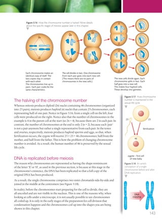
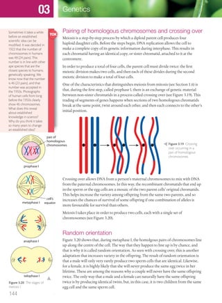
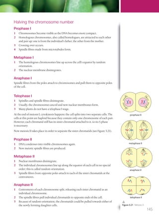
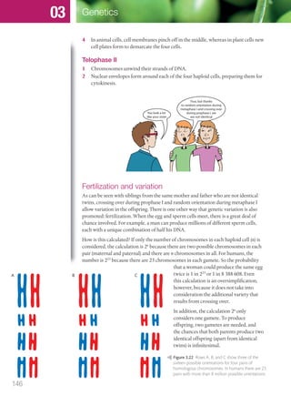
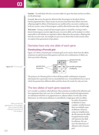
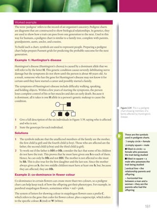
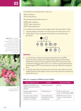
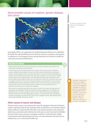
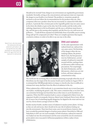
Genetics
03
M03_BIO_SB_IBDIP_9045_U03.indd 118 26/09/2014 12:09
Essential ideas
3.1 Every living organism inherits a blueprint for life from its parents.
3.2 Chromosomes carry genes in a linear sequence that is shared by
members of a species.
3.3 Alleles segregate during meiosis allowing new combinations to be
formed by the fusion of gametes.
3.4 The inheritance of genes follows patterns.
3.5 Biologists have developed techniques for artificial manipulation of
DNA, cells, and organisms.
Chromosomes carry genes in a linear sequence that is shared by
Alleles segregate during meiosis allowing new combinations to be
Biologists have developed techniques for artificial manipulation of
•What will my first baby look like?
•Will my children be able to see the difference between red and green, even though I
cannot?
•How can we find out who was at a crime scene by analysing their DNA?
•How can crops be genetically changed to improve their quality and quantity?
•Is it possible to clone humans?
•How many genes do I have?
•If I find a gene that has medical value, can I patent it and make money from my
discovery?
In order to answer these questions, the mechanisms of genetics must be understood.
Genetics is the science of how inherited information is passed on from one generation
to the next using the genetic material of genes and deoxyribonucleic acid (DNA).
One of the most famous
experiments in biology was
Gregor Mendel’s pea-breeding
investigations, which revealed
important insights into the
secrets of genetics.
NATURE OF SCIENCE
Developments in
scientific research
follow improvements
in technology: gene
sequencers are used for
the sequencing of genes.
3.1 Genes
Understandings:
● A gene is a heritable factor that consists of a length of DNA and influences a specific characteristic.
● A gene occupies a specific position on a chromosome.
● The various specific forms of a gene are alleles.
● Alleles differ from each other by one or only a few bases.
● New alleles are formed by mutation.
● The genome is the whole of the genetic information of an organism.
● The entire base sequence of human genes was sequenced in the Human Genome Project.
Applications and skills:
● Application: The causes of sickle cell anaemia, including a base substitution mutation, a change to
the base sequence of mRNA transcribed from it, and a change to the sequence of a polypeptide in
haemoglobin.
● Application: Comparison of the number of genes in humans with other species.
● Skill: Use of a database to determine differences in the base sequence of a gene in two species.
119
M03_BIO_SB_IBDIP_9045_U03.indd 119 26/09/2014 12:09
Guidance
● Students should be able to recall one specific base substitution that causes glutamic acid to be
substituted by valine as the sixth amino acid in the haemoglobin polypeptide.
● The number of genes in a species should not be referred to as genome size as this term is used for the
total amount of DNA. At least one plant and one bacterium should be included in the comparison,
and at least one species with more genes and one with fewer genes than a human.
● The GenBank® database can be used to search for DNA base sequences. The cytochrome c gene
sequence is available for many different organisms and is of particular interest because of its use in
reclassifying organisms into three domains.
● Deletions, insertions, and frame shift mutations do not need to be included.
What is a gene?
Have you ever heard people say ‘she looks just like her mum’ or ‘that kind of thing
skips a generation’? Although those people might not have known it, they were talking
about genetics.
CHALLENGE YOURSELF
1 Look at the list of characteristics below and think about which ones are determined by DNA
and which are not. Are there some that can be influenced by both DNA and a person’s
environment? For example, most people who have inherited a light skin colour can darken
their skin by tanning in the sun.
• Skin colour
• Freckles
• Number of fingers on
each hand
• Blood type
• Colour blindness
• Sex (male/female)
• Ability to digest lactose
• Reflexes
• Type of ear wax (wet
or dry)
• A scar from an accident
• Ability to speak
• Ability to speak Spanish
• Height
• Personality
• Intelligence
What about this: if a man had to have his left foot removed because of a war injury, would his
future children be born with only one foot? Before scientists understood the mechanisms of
genetics, it was believed that acquired characteristics could be passed on from one generation
to the next. This idea has been refuted. The classic debate of nature versus nurture is a good
topic for a Theory of knowledge discussion.
nucleus
Chromosomes are found in
the nuclei of cells. Humans
have 46 chromosomes, in
23 pairs. For simplicity, the
diagram above only shows
12 pairs.
Chromosomes are made of
very long DNA molecules.
Each chromosome contains
many smaller sections
called genes. Genes are
made of DNA.
Genes
A gene is a heritable factor that consists of a length of DNA and influences a
specific characteristic. ‘Heritable’ means passed on from parent to offspring, and
‘characteristic’ refers to genetic traits such as your hair colour or your blood type. The
estimated 21 000 genes that you possess are organized into chromosomes.
Whenever a definition is
given for a major concept
in biology, in this instance
the term ‘gene’, be sure to
memorize its definition
word for word. Such
definitions have been
phrased carefully so that
all the important details
are included.
Figure 3.1 Zooming into a cell
reveals where DNA is found.
120
Genetics03
M03_BIO_SB_IBDIP_9045_U03.indd 120 26/09/2014 12:09
A gene is found at a particular locus on a chromosome
A gene for a specific trait occupies a corresponding place, called a locus (plural loci), on
a chromosome (see Figure 3.2; there will be more about chromosomes in Section 3.2).
When geneticists map out the sequences of DNA, they carefully map the locus of each
sequence. When further research reveals that a particular sequence controls a certain
heritable factor, the locus of the gene is noted for further reference. For example,
scientists now know that the locus of the gene controlling a protein called transducin
that enables colour vision is found on chromosome 1. A mutation of this gene stops
a person from being able to make the protein transducin properly, which is necessary
to transmit information about colour from the eye to the brain; as a result, the person
will not see in colour. This is an extremely rare genetic condition called complete
achromatopsia. When we say ‘the ability to see in colour is a genetic trait’ we mean one
of two things is happening with someone’s DNA: either that person has the DNA code
for making colour vision possible or that person does not have it. This is illustrated in
Figure 3.3.
Where the gene is
found:
Sequence of
bases (starting at
position 229):
Protein and trait:
. . . CACCAGCAACCC . . . Transducin protein
synthesized, colour vision
possible
Locus for
transducin gene on
chromosome 1
. . . CACCAGTAACCC . . . Protein not formed
correctly, colour vision
not possible
You will recall that you possess two copies of each gene in your body: one copy from
your mother and one from your father. As a result, if you could look at the locus of the
transducin gene on one of the two copies of your first chromosome, for example, you
would find the same gene at the same locus on the other copy of chromosome 1. One
copy would be the one your mother gave you and the other would be the copy your
father gave you. Would those genes be identical? Not necessarily, because genes can
come in different forms.
Alleles: versions of genes
Variations or versions of a gene are called alleles. An allele is one specific form of a
gene, differing from other alleles by one or a few bases. In the example of transducin
and colour vision above, a single base pair difference between the most common allele
(with a C at position 235) and the rare mutated allele (with a T at position 235) is all
that is takes to determine whether you can distinguish colours or not. These different
forms allow for a single trait, such as the trait for the ability to see in colour, to have
variants, in this example either colour or grey-scale vision.
Figure 3.2 The locus is the
specific position of a gene on
a chromosome.
locus
Figure 3.3 The presence of a
C or a T makes a big difference
in colour vision.
121
M03_BIO_SB_IBDIP_9045_U03.indd 121 26/09/2014 12:09
Table 3.2 Examples of possible alleles
Gene or trait Examples of possible alleles
Colour vision Allele to produce transducin (the protein necessary for colour
vision); allele that does not code correctly for transducin
Skin pigmentation Allele to make pigments; allele that cannot make pigments
(in other words an allele for albinism, see Section 3.4)
Blood type Allele for type A; allele for type B; allele for type O
Another example of the difference between two alleles of the same trait is the
difference that causes the genetic condition cystic fibrosis.
Cystic fibrosis
Maintaining a proper balance of fluids in the body is essential for good health. One
such fluid is mucus, a thick, slippery, substance used in many parts of the body,
including the lungs and intestines. A gene called CFTR, found on chromosome 7,
plays a key role in the production of mucus. The standard version of this gene (the
CHALLENGE YOURSELF
Table 3.1 Comparison of the number of genes in humans and other species
Organism Scientific name Number of bases Number of genes
Virus
(bacteriophage)
phiX174 * 5400 11
Bacterium Escherichia coli (type
K-12)
4639000 4377
Nematode
(roundworm)
Caenorhabditis
elegans
100292000 20000
Human Homo sapiens 3000000000 21000
Asian rice Oryza sativa 430000000 up to 56000
Baker’s yeast Saccharomyces
cerevisiae
12495000 5770
Mouse-ear cress Arabidopsis thaliana 135000000 25000
Fruit fly Drosophila
melanogaster
122654000 27407
Japanese canopy
plant
Paris japonica** 150000000000 Unknown
*First genome ever sequenced (in 1977).
**Largest plant genome sequenced so far.
2
(a) Which species has the largest number of genes?
(b) Which species has the smallest number of genes?
(c) Which species has the most similar number of genes to humans?
(d) Some people are tempted to say that the more genes an organism has, the more advanced it
is. Discuss this idea: what kinds of arguments support it and what arguments refute it?
122
Genetics03
M03_BIO_SB_IBDIP_9045_U03.indd 122 26/09/2014 12:09
standard allele) allows a person’s mucus-producing cells to function properly, whereas
an allele generated by a mutation of the CFTR gene causes cystic fibrosis. People with
this genetic condition produce abnormally excessive quantities of mucus in various
organs and have difficulties with their respiratory and digestive systems, among other
complications. In this example, the trait is for mucus production; one allele is for a
balanced mucus production, the other for excessive mucus production that leads to
cystic fibrosis. We will see later how to calculate the chances of a child inheriting this
condition from his or her parents.
One base can make a big difference
From the sections on transcription and translation of DNA, you will remember
how important it is for each letter in the genetic code to be in a specific place. If, for
whatever reason, one or more of the bases (A, C, G, or T) is misplaced or substituted for
a different base, the results can be dramatic. As we have seen with cystic fibrosis, the
difference between one version of a gene and another (the mutated and non-mutated
alleles of the CFTR gene) can mean the difference between healthy organs and organs
hampered by an overproduction of mucus.
Another example of a change of bases can be seen in the gene ABCC11, which
determines several things, one of them being whether or not the cerumen (ear wax)
that you produce is wet or dry. Some people produce dry cerumen, which is flaky and
crumbly with a grey colour, while others produce earwax that is more fluid and has
an amber colour. The gene that determines this is on chromosome 16 and has two
alleles: the G variant codes for dry cerumen, the A variant codes for wet cerumen. The
allele containing G for wet earwax is much more common in European and African
populations, while the allele containing A is much more common among Asians. Why
is this of interest to geneticists? For one thing, it can reveal a lot about how populations
have migrated and interbred in the past, but it can also reveal other things about
our health. As curious as it may seem, the ABCC11 gene is also partly responsible
for the smell of underarm sweat, as well as the production of breast milk, and could
potentially have a link to breast cancer. Most women probably would not care whether
or not they have the gene for dry or fluid earwax, but if they could find out whether
they had an allele that could reduce their chances of having breast cancer, they might
be much more interested.
How are such differences in genes generated in populations? We will now look at how
mutations work.
How new alleles are produced
Look at the two sequences of DNA below, which are from the coding strand of a
section of genetic information that helps in the formation of haemoglobin, found in
red blood cells. Look carefully at the two sequences. Identify the difference between
the two and complete the following phrase.
DNA sequence 1: GTG CAC CTG ACT CCT GAG GAG
DNA sequence 2: GTG CAC CTG ACT CCT GTG GAG
‘Codon number __ along the first sequence has the letter __ in position number __,
whereas the codon in the same position in sequence 2 has the letter __ instead.’
Worked example
123
M03_BIO_SB_IBDIP_9045_U03.indd 123 26/09/2014 12:09
Solution
Codon number 6 along the first sequence has the letter A in position number 2,
whereas the codon in the same position in sequence 2 has the letter T instead.
Now look at the effect this has on the mRNA sequences produced from the template
strand that is found opposite the coding strand when the DNA is unzipped for
transcription:
mRNA sequence 1: GUG CAC CUG ACU CCU GAG GAG
mRNA sequence 2: GUG CAC CUG ACU CCU GUG GAG
Using Figure 3.4 and the mRNA sequences above, showing which codons are
associated with which amino acids, fill in the names of the missing amino acids (a) to
(h) in Figure 3.5.
UUU
UUC
Phenyl-
alanine
UUA
UUG
Leucine
UAU
UAC
Tyrosine
Second base
Firstbase
Thirdbase
UAA
UAG
Stop codon
Stop codon
UGU
UGC
Cysteine
UGA
UGG
Stop codon
Tryptophan
AAU
AAC
Asparagine
AAA
AAG
Lysine
AGU
AGC
Serine
AGA
AGG
Arginine
CAU
CAC
Histidine
CAA
CAG
Glutamine
GAU
GAC
Aspartic
acid
GAA
GAG
Glutamic
acid
CUU
CUC
CUA
CUG
Leucine
UCU
UCC
UCA
UCG
Serine
CCU
CCC
CCA
CCG
Proline
CGU
CGC
CGA
CGG
Arginine
GGU
GGC
GGA
GGG
Glycine
ACU
ACC
ACA
ACG
Threonine
GCU
GCC
GCA
GCG
Alanine
GUU
GUC
GUA
GUG
Valine
AUU
AUC
AUA
AUG
Isoleucine
Methionine
start codon
U
U C A G
C
A
G
U
C
A
G
U
C
A
G
U
C
A
G
U
C
A
G
Sequence 1: valine – histidine – (a)______ – (b)______ – (c)______ – (d)______ –
glutamic
acid
Sequence 2: valine – histidine – (e)______ – (f)______ – (g)______ – (h)______ –
glutamic
acid
Solution
Sequence 1: valine – histidine – leucine – threonine – proline –
glutamic
acid
–
glutamic
acid
Sequence 2: valine – histidine – leucine – threonine – proline – valine –
glutamic
acid
Notice how the error of only one letter in the original DNA code changed the
composition of amino acids in sequence 2. This would change the composition and
the structure of the resulting protein, in the same way that changing the shapes and
compositions of some of the bricks used to build a house would change the shape
(and therefore the structural integrity) of the house. This kind of change in the DNA
code is produced by a mutation.
Figure 3.5 Using Figure 3.4
and the mRNA sequences
above, can you find the
missing amino acids for boxes
(a) to (h)?
Figure 3.6 Is this what you
found? We will need these
sequences later when we
explore sickle cell disease.
Figure 3.4 Codons and their
associated amino acids.
For example, RNA coding for
lysine is AAA.
124
Genetics03
M03_BIO_SB_IBDIP_9045_U03.indd 124 26/09/2014 12:09
Mutations
A mutation is a random, rare change in genetic material. One type involves a change
of the sequence of bases in DNA. If DNA replication works correctly, this should not
happen (see Section 2.7). But nature sometimes makes mistakes. For example, the base
thymine (T) might be put in the place of adenine (A) along the DNA sequence. When
this happens, the corresponding bases along the messenger RNA (mRNA) are altered
during transcription.
As we have seen with the example of cystic fibrosis, mutated genes can have a negative
effect on a person’s health. Sometimes, however, mutations can have a positive effect
that is beneficial to an organism’s survival.
Are mutations good or bad for us?
LRP5 is a gene that helps immune system cells make a certain type of protein that
acts as a receptor on their surfaces. Research indicates that this receptor is used by
the human immunodeficiency virus (HIV) to infect the cells (see Section 6.3 for
a description of HIV). People with a mutation of LRP5 cannot make this receptor
protein on their immune system’s cells and, as a result, HIV cannot infect them. This
means that people with a mutated allele of LRP5 are naturally immune to HIV. Such a
mutation is very rare in the human population.
A mutation that provides an individual or a species with a better chance for survival
is considered to be a beneficial mutation, and there is a good chance that it will be
passed on to the next generation. In contrast, mutations that cause disease or death are
detrimental mutations, and they are less likely to be passed on to future generations,
because they decrease the chances of an individual’s survival. In addition to beneficial
and harmful mutations, there are neutral mutations that do not have an effect on a
species’ survival.
When a mutation is successfully passed on from one generation to the next, it
becomes a new allele: it is a new version of the original gene. This is how new alleles
are produced. You and everyone you know possess many mutations. Whether they are
harmful, beneficial or neutral depends on what they are and what kind of environment
you need to survive in.
On the left, a white-eyed
mutant fruit fly, and on the
right the kind of fruit fly
typically found in nature,
called the wild-type.
125
M03_BIO_SB_IBDIP_9045_U03.indd 125 26/09/2014 12:10
Table 3.3 A summary of different types of mutations: not all mutations are negative
Type of mutation Example in plants Example in animals
Detrimental A mutation that does not allow a plant to
produce a chemical that it secretes against
insect attacks, thereby making it vulnerable
A mutation that makes it impossible to
produce a certain digestive enzyme, thereby
making digestion difficult and therefore
compromising the ability of the organism to
survive
Neutral A single base substitution in a gene for
chlorophyll that does not change the amino
acid coded for, and therefore does not modify
the chlorophyll (e.g. GUA and GUG both code
for valine)
A single base substitution in a gene for
haemoglobin that does not change the amino
acid coded for, and therefore does not modify
the haemoglobin (e.g. GAG and GAA both
code for glutamic acid)
Beneficial A mutation giving a plant the ability to make
a protein that increases its frost resistance,
which would be beneficial in colder climates
A mutation changing the shape or size of the
lens of the eye, giving the animal more acute
vision for finding prey
Gene therapy is the process of taking a beneficial gene from a person who possesses it
and putting it into a person who does not have it, but who needs it to stay healthy. The
challenge is that it is very difficult to get the DNA into the sick person’s cells. One way
is to force the gene into the patient’s cells using a virus to deliver it. Partly because of
a lack of understanding of how to use viruses safely to deliver genes, the decision was
made to stop all testing of gene therapy on human patients in the USA in 1999, when
an 18-year-old patient died after a virus had been injected into his body. However,
gene therapy trials are coming back, little by little, notably in helping blind children to
regain their eyesight.
Who decides whether an experiment is safe? Is the loss of life for some patients
participating in trials necessary in order to find a cure? If years of research had not been
delayed because human trials had been stopped, wouldn’t we have made much more
progress by now in curing genetic diseases?
Base substitution mutation
The type of mutation that results in a single letter being changed is called a base
substitution mutation. The consequence of changing one base could mean that a
A gene to help digestion
For most of our existence, humans have been hunter-gatherers and our genes are
generally well adapted for this lifestyle. Originally, as for all mammals, the only age at
which we drank milk was when we were infants. By the time our ancestors reached
adulthood, their bodies had stopped being able to digest milk; more precisely, humans
could not break down the disaccharide in milk called lactose. This continues to be the
case for most of the human population today: more than half of the human population
has lactose intolerance and those people can only digest lactose in their infancy. In the
past 10 000 years, however, many human populations have adopted an agricultural-
based lifestyle, raising animals for milk and consuming dairy products on a daily
basis. In their genetic makeup, many agricultural societies show a higher frequency
of the genetic code that allows humans to digest lactose throughout adulthood. From
an evolutionary point of view, this advantage has increased humans’ ability to survive
harsh climatic conditions. As European human populations spread out and established
populations outside Europe, notably in North America, they brought their lactose
tolerance (and their livestock) with them.
126
Genetics03
M03_BIO_SB_IBDIP_9045_U03.indd 126 26/09/2014 12:10
different amino acid is placed in the growing polypeptide chain. This may
have little or no effect on the organism, or it may have a major influence on
the organism’s physical characteristics.
Sickle cell disease
In humans, a mutation is sometimes found in the gene that codes for
haemoglobin in red blood cells. This mutation gives a different shape to the
haemoglobin molecule. The difference leads to red blood cells that look very
different from the usual flattened disc with a hollow in the middle.
The mutated red blood cell, with a characteristic curved shape, made its
discoverers think of a sickle (a curved knife used to cut tall plants). The
condition that results from this mutation is therefore called sickle cell
disease, also known as sickle cell anaemia.
The kind of mutation that causes sickle cells is a base substitution mutation. If you
look back at the two sequences given previously in the worked example on page 123,
the first is for the section of the haemoglobin gene’s DNA that codes for standard-
shaped red blood cells, whereas the second sequence shows the mutation that leads to
the sickle shape. In this case, one base is substituted for another so that the sixth codon
in this sequence of haemoglobin, GAG, becomes GTG. As a result, during translation,
instead of adding glutamic acid, which is the intended amino acid in the sixth position
of the sequence, valine is added there instead. Again, refer back to the worked example
to see this mutation.
Because valine has a different shape and different properties compared with glutamic
acid, the shape of the resulting polypeptide chain is modified. As a result of this, the
haemoglobin molecule has different properties that cause the complications associated
with sickle cell disease.
The symptoms of sickle cell disease are weakness, fatigue, and shortness of breath.
Oxygen cannot be carried as efficiently by the irregularly shaped red blood cells. In
addition, the haemoglobin tends to crystallize within the red blood cells, causing them
to be less flexible. The affected red blood cells can get stuck in capillaries, so blood flow
can be slowed or blocked, a condition that is painful for the sufferer.
People affected by sickle cell anaemia are at risk of passing the mutated gene on to their
offspring. From a demographic point of view, the mutated gene is mostly found in
populations originating from West Africa or from the Mediterranean.
The advantages of sickle cell disease
Although sickle cell disease is a debilitating condition, those who have it are very
resistant to malaria infection. Malaria is an infectious disease that occurs in tropical
regions. A parasite called Plasmodium is transmitted to human blood by an infected
female Anopheles mosquito feeding on the blood. The parasite attacks the person’s red
blood cells and produces symptoms of high fever and chills, and can result in death.
In terms of the shapes of human red blood cells, we all carry two copies of the gene for
the shape of our red blood cells, one copy that we inherited from our mother and the
other that we inherited from our father. People born with two copies for standard disc-
shaped cell have only disc-shaped cells and are highly susceptible to malaria infection.
People who have one gene that is for disc-shaped cells and one for sickle-shaped cells
G
A
G
G
T
G
Three standard, disc-shaped
red blood cells, and one
sickle-shaped cell.
Figure 3.7 A mutation where
base A is replicated by base T;
GAG codes for glutamic acid
and GTG codes for valine.
127
M03_BIO_SB_IBDIP_9045_U03.indd 127 26/09/2014 12:10
have what is called sickle cell trait. They have some sickle-shaped cells
and some disc-shaped cells in their bloodstream but in most cases they
do not suffer from anaemia. Anaemia is the result of low red blood cell
levels and is characterized by a paleness of skin and low energy levels.
People with sickle cell trait have a better resistance to malaria because
of chemical imbalances that make the survival of Plasmodium in their
blood more difficult. The insufficient quantities of potassium in sickle-
shaped cells cause Plasmodium to die. Lastly, people who inherit a sickle
cell gene from both their mother and their father can produce only
sickle-shaped cells and suffer from severe anaemia that can sometimes
be fatal. On the other hand, they have the highest resistance to malaria.
Malaria can be transmitted
by the female Anopheles
mosquito, which is therefore
one of the deadliest animals
on Earth.
Table 3.4 Different types of genetic conditions
Condition Example
1 Chromosomal
anomaly
Caused by an extra chromosome, such as Down syndrome
(chromosome 21) (see page 147)
2 Autosomal
genetic disease
Caused by a gene found on one of the 22 non-sex chromosomes,
such as cystic fibrosis (chromosome 1) (see page 140)
3 Sex-linked
genetic disease
Caused by a gene found on the X or Y chromosome, such as
colour blindness found on the X chromosome (see page 159)
The first type is generally considered to be a one-time error in the distribution of
chromosomes, whereas the second and third types are passed on generation after
generation.
A genome
How do we know all that we do about genes? How do we know where they are and
what they do? Before answering these questions, it is important to appreciate the
point that, although we have made considerable progress in the past few decades,
our maps of human chromosomes are still far from complete, and there are many
DNA sequences for which we do not know the function. As an analogy, think of the
maps produced by cartographers and explorers in the Middle Ages; many parts of the
globe remained uncharted and had the words terra incognita (Latin for ‘unknown land’)
inscribed on them.
Sequencing DNA
In order to find out which gene does what, a list must be made showing the order of
all the nucleotides in the DNA code. Researchers use highly specialized laboratory
When we look at where sickle cell disease is most common in the world,
there appears to be a significant overlap with the places where malaria
occurs. Is this just a coincidence? Or is there a reason for this? Scientists
and statistics experts often say that ‘correlation does not mean causality’,
meaning that just because two things occur in the same place at the same
time does not necessarily mean that one causes the other. How can we tell
the difference between causality and correlation? The answer is that there
must be some kind of mechanism that could explain how one could cause
the other. From what you have read about sickle cell disease and malaria in
this chapter, what do you think? Are they merely correlated or is there also
causality?
128
Genetics03
M03_BIO_SB_IBDIP_9045_U03.indd 128 26/09/2014 12:10
equipment including sequencers to locate and identify
sequences of bases. The complete set of an organism’s
base sequences is called its genome.
A short fragment of a sequence looks like this:
GTGGACCTGACTCCTGAGGAG. Each letter
represents one of the four bases in the DNA code.
This short fragment contains seven codons with a
total of 21 bases represented by letters. Now imagine
3 billion of those letters: what would that look like? If
you printed out 3000 base letters per page, it would
need 1 million pages, which would stack about 100
m high. That’s an impressive quantity of information,
especially considering that you can keep it all in the
nucleus of a typical cell in your body.
The complete genomes of some organisms have been
worked out. Among those organisms are the fruit fly,
Drosophila melanogaster, and the bacterium, Escherichia
coli, because these two organisms have been used
extensively in genetics experiments for decades.
How do geneticists work out the complete genome?
Many steps are necessary. Here is a summary of one way of doing it: the Sanger
technique (see Figure 3.8).
•Once a DNA sample has been taken, it is chopped up into fragments and copies are
made of the fragments. A primer sequence is added to help start the process.
•To determine the sequence, a DNA polymerase enzyme attaches to one copy of
the first fragment (let’s call it fragment 1). Then it will start to add free nucleotides
following the principle of complementary base pairing. Two kinds of nucleotides will
be added.
•Some free nucleotides are standard ones and others are special dideoxynucleotide
triphosphates (ddNTPs; labelled ddA, ddT, ddC, and ddG in Figure 3.8) added as DNA
chain terminators, meaning that when one is reached, the elongation of the strand
is stopped. These have been previously marked with fluorescent markers to identify
them. Sometimes the chain termination happens all the way at the end of fragment 1,
but most of the time the process stops before it reaches the end. This process happens
on each of the many copies of fragment 1.
•The result is a series of new strands, some dozens of bases long, others only a handful
of bases long, and some that have all the bases of fragment 1.
•Now everything is ready for the sequencing: the multiple chains of varying lengths
(each with a fluorescently marked end) are placed in order from longest to shortest.
This is done using a technique called gel electrophoresis, which will be explained in
Section 3.5.
•To recognize each letter, a laser activates the fluorescent markers on the nucleotides
as they go through the process. A sensor hooked up to a computer analyses the
wavelength of the light and determines whether it represents an A, T, C, or G.
•The process must be repeated many times: for A, T, C, and G. Repetitions make sure
there are no errors. Fortunately, many copies of fragment 1 were made, so this is easy
to check.
Computers are used to speed
up the sequencing process.
Thanks to modern
communication
technologies, it is possible
for scientists working
all over the world to
collaborate and contribute
to a scientific endeavour
such as sequencing the
genome of plants that help
feed the world. Rice is one
example: biologists from
10 countries contributed
to sequencing the first rice
genome.
129
M03_BIO_SB_IBDIP_9045_U03.indd 129 26/09/2014 12:10
•Once fragment 1 is done, the lab technicians must process fragment 2, fragment 3,
and so on, until all the fragments of the original sequence have been processed.
•At this stage, the challenge is to put all the sequenced fragments of code together.
When the original sequence was chopped up to make all these fragments, they
became mixed up and out of order. Now that we know what their sequences are,
we need to know the order in which to put them. This daunting task has been made
easier by computers, but it consists of lining up any overlapping segments until they
all match.
Since the Sanger technique was invented, many techniques have been developed to
analyse each fragment only once, making it unnecessary to make multiple copies
of each. This reduces the time and the cost of sequencing a genome. The objective
of developing new sequencing techniques is to have a fast, inexpensive way to map
anyone’s genome.
The Human Genome Project
In 1990, an international cooperative venture called the Human Genome Project set
out to sequence the complete human genome. Because the genome of an organism is a
catalogue of all the bases it possesses, the Human Genome Project hoped to determine
the order of all the bases A, T, C, and G in human DNA. In 2003, the Project announced
that it had succeeded in achieving its goal. Now, scientists are working on deciphering
which sequences represent genes and which genes do what. The human genome can
be thought of as a map that can be used to show the locus of any gene on any one of
the 23 pairs of chromosomes.
In the 1997 science
fiction film GATTACA, one
of the main characters
brings a sample of cells
to a walk-up window at
an establishment that
provides anonymous
genome services. Within
seconds, she gets a full
printout and analysis of
the genome of the man
she wants to know about.
He is not aware that she is
doing this. One objective
of science fiction as an art
form is to warn society
of what might happen in
the future if we are not
careful. This film raises
serious questions about
how far technology will
lead us and whether or
not we want to go in that
direction. Our society
will need to make some
difficult decisions in the
coming years concerning
our genomes and who has
access to the information
contained within them.
? ? ? ? ? ? ? ? ? ? ? ?Primer
A T C G
Standard free nucleotides
ddA ddT ddGddC
Fluorescently marked chain terminator nucleotides
Fragment 1 with a primer and 12 unknown bases
1. DNA polymerase adds free nucleotides and
one chain terminator nucleotide at random to each copy
of fragment 1. Example: ddG attached to 4th position.
? ? ?Primer ddG
? ?Primer
2. Several copies made of
different lengths ending
in ddG.ddG
? ? ? ? ? ? ? ?Primer ddG
?Primer ddG?
? ? ? ? ? ? ? ?Primer ddG? ? ?
? ? ?Primer ddG
? ?Primer ddG
? ? ? ? ? ? ? ?Primer ddG
?Primer ddG?
? ? ? ? ? ? ? ?Primer ddG? ? ?
? ?
? ?
3. Gel electrophoresis puts the
fragments in order from longest to
shortest. Now we know that there
are Gs in positions 2, 3, 6, 9 and 12.
Primer
G G G G G
Primer
G G G G GC C
Primer
G G G G GC CT T
Primer
G G G G GC CT T A A A
4. The same process is repeated for ddC, ddT and ddA.
5. It is now possible to determine fragment 1’s sequence:
G CG CC C CTTTAA
Figure 3.8 One method of
DNA sequencing is called the
Sanger technique.
130
Genetics03
M03_BIO_SB_IBDIP_9045_U03.indd 130 26/09/2014 12:10
Before the Human Genome was mapped, fewer than 100 loci were known for genetic
diseases. After the mapping was completed, more than 1400 were known, and today
the number is in the thousands and increasing.
In addition, by comparing the genetic makeup of populations around the world,
countless details can be revealed about our ancestries and how human populations
have migrated and mixed their genes with other populations over time. Without
knowing it, you are carrying around in each one of your cells a library of information
about your past.
There is another international connection with the Human Genome Project in the sense that
this project is a good example of scientists from all over the world working together. Dozens of
nationalities participated in the project, and the results are available for free access worldwide
thanks to online databases open to the public.
Delving into human genetics confirms two major themes:
• we are all the same
• we are all different.
On the one hand, the Human Genome Project has shown that there are only a very small
number of DNA bases that make one person different from any other person in the world. This
creates a feeling of unity, of oneness with all people. From peanut farmers in West Africa, to
computer technicians in California, to fishermen in Norway, to businesswomen in Hong Kong,
all humans carry inside them a common genetic heritage.
On the other hand, the Human Genome Project has shown that the small differences that do
exist are important ones that give each person his or her uniqueness in terms of skin colour,
facial features and resistance to disease, for example. These differences should be appreciated
and celebrated as strengths. Unfortunately, they are often the basis of discrimination and
misunderstanding.
Can one genetic group be considered genetically superior to another? History has shown that
many people think so, yet genetics shows that this is not the case. All human populations,
whatever slight differences their genomes may have, deserve equal esteem as human beings.
Dr Francis Collins, one of the
leaders of the Human Genome
Project team.
Dr Craig Venter, one of the
leaders of the Human Genome
Project team.
Using DNA to make medicines
Another advantageous use of the human genome is the production of new
medications. This process involves several steps:
•find beneficial molecules that are produced naturally in healthy people
•find out which gene controls the synthesis of a desirable molecule
•copy that gene and use it as instructions to synthesize the molecule in a laboratory
•distribute the beneficial therapeutic protein as a new medical treatment.
This is not science fiction: genetic engineering firms are finding such genes regularly.
One current line of research is dealing with genes that control ageing. How much
money do you think people would be willing to pay for a molecule that could reverse
the effects of ageing and prolong life by several decades?
Many companies offer genome sequencing for private citizens willing to pay the price. Some
of the products offered are revelations about ancient family origins and risk factors for some
health problems, such as the chances of developing certain types of cancer or heart disease.
Would you want to know if there was a chance that your life could be suddenly shortened by
the presence or absence of a certain gene? Would you tell your family and friends? Would you
want your parents to do such a test? Should people tell their employer or each other about
any health-related issues revealed by a genomic analysis? Or, on the contrary, is this a private,
personal thing that no one else needs to know about? How accurate and reliable are these
analyses? Should we believe everything they say?
131
M03_BIO_SB_IBDIP_9045_U03.indd 131 26/09/2014 12:10
CHALLENGE YOURSELF
3 Cytochrome c is a protein found in mitochondria and it plays a key role in cell respiration
by shuttling electrons from one place to another. If an organism did not have the genetic
code to make cytochrome c, it could not survive. By comparing the genetic sequence
used to produce this protein in various species, scientists were able to see how mutations
accumulate over time. The differences between a horse’s base sequence for this protein and
a zebra’s is much smaller than the differences between a horse’s and a lizard’s. Humans and
chimpanzees have identical cytochrome c amino acid sequences, whereas the yeast Candida
krusei has 51 amino acid differences compared with humans.
For this exercise, find the relevant PDF file in the hotlinks at the end of this section. Follow
the instructions to compare the genetic sequences for various organisms for the gene that
makes cytochrome c.
Use the hotlinks to find ancient sequences of base pairs to compare between species
of prokaryotes. Can you find a correlation between the numbers of mutations and the
evolutionary distance between species?
What if a biotech company finds a useful human gene in your body? For example, a
gene that produces a protein to help balance cholesterol levels in the body and prevent
heart problems. Can the company patent that gene in order to protect its discovery
and in order to earn money from it? With a patent, the company could charge
pharmaceutical manufacturers that wanted to use the gene to make new medicines. In
many countries there are few if any laws about such things because the techniques are
so new.
Isolate a
non-mutated
gene in a
healthy
person
Get an
organism to
produce the
healthy protein
using the
isolated gene
Find unhealthy
patients who
possess the
mutated gene
and cannot
make the
protein
Inject the
healthy protein
into the
patients to treat
them
Can human genes be patented?
In the spring of 2013, the United States Supreme Court heard a landmark case
between a biotech company, Myriad Genetics (the defendant), and the Association for
Molecular Pathology, AMP (the plaintiff), a group of genetics experts who specialize
in many things, including the diagnosis of genetic diseases and disorders. Myriad
Figure 3.9 The process of
protein therapy, a promising
use of genetically modified
organisms.
NATURE OF SCIENCE
Why are scientists interested in comparing the genetic codes of various species? For one thing,
when looking at a gene that every living thing should have, such as a gene for how to make
ribosomes, the number of mutations a species has in that gene compared with another species
gives insight into how closely they are related to each other.
Because of a certain number of differences in metabolism and genetic makeup in types of
single-celled organisms that looked similar to bacteria, the biologist Carl Woese proposed the
domain Archaea to distinguish them from bacteria (prokaryotes) and eukaryotes. Although
Archaea do not have a nucleus, they have enough differences compared with prokaryotes
to set them apart from other bacteria. Among the species in this group are single-celled
organisms that thrive in very salty conditions, some that live in hot springs at extreme
temperatures, and many others that live in the soil or in the ocean: some might be living in you
or on you right now.
It took decades for Woese’s proposal to be accepted, but the overwhelming evidence in
Archaea’s favour made it very difficult for opponents of the idea to argue against it.
132
Genetics03
M03_BIO_SB_IBDIP_9045_U03.indd 132 26/09/2014 12:10
had a patent on naturally occurring human genes called BRCA, which can be used to
tell whether a woman has a genetically increased chance of breast cancer or ovarian
cancer, two of the most common and deadly cancers in Western society today. AMP
was taking Myriad to court because they thought the BRCA gene sequences should
be available freely for diagnosing cancer. AMP thought that it was unfair that clinical
teams could not access the BRCA genes to do their own testing and diagnosis. They
argued that a company such as Myriad should not be able to put an industrial patent
on genes, because DNA sequences occur naturally and are not invented by a company:
therefore, they are not patentable objects.
Myriad’s argument was that, although DNA is found in nature, genes are all connected
to each other, whereas the isolated sequences for which they had patents could only
be the product of a biotech laboratory using sophisticated equipment to do the
separation and identification: therefore, the DNA sequences in question were not in
their natural form. The researchers at Myriad were the first to patent these fragments
of DNA and recognize their usefulness. They patented their BRCA genes just as any
pharmaceutical company would patent a new molecule that they thought would make
a useful medicine. These patents, and the diagnostic tests associated with them, have
made Myriad a very successful and profitable company. Because it is their intellectual
property, anyone who wants a genetic test for breast cancer or ovarian cancer must go
to them. Myriad argued that taking away their patents would take away their livelihood
because it would allow any company to develop and perform their own diagnostic
tests.
Another argument from the plaintiff was that, because Myriad was the sole company
to administer BRCA diagnostics, it was impossible for a patient to get a second opinion,
a key step in the diagnosis and treatment of a medical condition as serious as cancer.
Also, Myriad could charge high fees because they had no competition in the market.
Myriad’s justification for the cost of the tests was that biotech research requires very
expensive laboratory equipment and highly trained professionals, so the money
earned from the diagnostic tests helps the company invest in new developments to
advance scientific knowledge and continue putting new diagnostic tools in place.
In the end, the US Supreme Court found it unconstitutional to patent a DNA sequence
found in nature. Justice Clarence Thomas wrote ‘A naturally occurring DNA segment
is a product of nature and not patent eligible merely because it has been isolated.’
A patent is an
authorization for a person
or a company to make,
use, or sell an invention,
and it makes it illegal for
anyone else to make, use,
or sell it. For example,
Thomas Edison had
more than 1000 patents
for the many things he
invented, such as his
improved electric light
bulb and his phonograph.
In the medical field, it is
common to patent new
pharmaceutical molecules
developed in laboratories
so that only the initial
company that invented the
drug can manufacture and
sell it. Typically, a patent
filed today is limited to 20
years.
Biotechnology is posing a
rising number of challenges
to the legal institutions of the
world.
133
M03_BIO_SB_IBDIP_9045_U03.indd 133 26/09/2014 12:10
Section summary
•Living organisms get their DNA from their parents.
•The locus of a chromosome is a specific position where a gene (a heritable unit of
DNA that codes for a specific trait) is found.
•One allele (a version of a gene) can differ from another allele by one or more DNA
bases.
•The Human Genome Project has allowed scientists to map the positions of genes and
their mutations.
Exercises
1 What is the difference between an allele and a gene?
2 Give an example of a mutation in an eagle’s offspring that could be considered a beneficial mutation.
3 Explain why eukaryotic chromosomes always come in pairs.
To learn more about
genes, go to the hotlinks
site, search for the title
or ISBN, and click on
Chapter 3: Section 3.1.
3.2 Chromosomes
Understandings:
● Prokaryotes have one chromosome consisting of a circular DNA molecule.
● Some prokaryotes also have plasmids but eukaryotes do not.
● Eukaryote chromosomes are linear DNA molecules associated with histone proteins.
● In a eukaryote species there are different chromosomes that carry different genes.
● Homologous chromosomes carry the same sequence of genes but not necessarily the same alleles
of those genes.
● Diploid nuclei have pairs of homologous chromosomes.
● Haploid nuclei have one chromosome of each pair.
● The number of chromosomes is a characteristic feature of members of a species.
● A karyogram shows the chromosomes of an organism in homologous pairs of decreasing length.
● Sex is determined by sex chromosomes and autosomes are chromosomes that do not determine
sex.
Applications and skills:
● Application: Cairns’ technique for measuring the length of DNA molecules by autoradiography.
● Application: Comparison of genome size in T2 phage, Escherichia coli, Drosophila melanogaster,
Homo sapiens, and Paris japonica.
● Application: Comparison of diploid chromosome numbers of Homo sapiens, Pan troglodytes, Canis
familiaris, Oryza sativa, and Parascaris equorum.
● Application: Use of karyograms to deduce sex and diagnose Down syndrome in humans.
● Skill: Use of databases to identify the locus of a human gene and its polypeptide product.
Guidance
● The terms karyotype and karyogram have different meanings. Karyotype is a property of a cell: the
number and type of chromosomes present in the nucleus, not a photograph or diagram of them.
● Genome size is the total length of DNA in an organism. The examples of genome and chromosome
number have been selected to allow points of interest to be raised.
● The two DNA molecules formed by DNA replication prior to cell division are considered to be sister
chromatids until the splitting of the centromere at the start of anaphase. After this, they are individual
chromosomes.
NATURE OF SCIENCE
Developments in research
follow improvements
in techniques:
autoradiography was used
to establish the length
of DNA molecules in
chromosomes.
134
Genetics03
M03_BIO_SB_IBDIP_9045_U03.indd 134 26/09/2014 12:10
The chromosome in prokaryotes
You will recall from Chapter 1 that the nucleoid region of a bacterial cell contains a
single, long, continuous, circular thread of DNA. Therefore, this region is involved
with cell control and reproduction.
Notice how the presence of a single circular chromosome is a very different situation
from all the cells we looked at in Section 3.1, which always had chromosomes in pairs.
Why is this? Prokaryotes can reproduce using binary fission (dividing), whereas
organisms such as plants and animals more frequently use sexual reproduction
(involving a male and a female). Any time two parents are involved, the offspring will
have pairs of chromosomes rather than single chromosomes. Because prokaryotes
have only one parent, they have only one chromosome.
Plasmids
Escherichia coli, like many prokaryotes
(bacteria), have small loops of DNA that are
extra copies of some of the genetic material of
the organism. These loops are called plasmids.
These small, circular, DNA molecules are not
connected to the main bacterial chromosome.
The plasmids replicate independently of the
chromosomal DNA. Plasmid DNA is not
required by the cell under normal conditions,
but it may help the cell adapt to unusual
circumstances. Plasmids can be found in
Archaea as well as in bacteria.
As we will see later in Section 3.5, these loops can be used in genetic engineering.
Genetic manipulation using plasmids is not possible in eukaryotes such as plants
and animals, because they do not have plasmids. Other techniques must be used for
genetically modified (GM) crops and animals, which we will discuss later (also in
Section 3.5).
Eukaryote chromosomes
The DNA of eukaryotic cells most often occurs in the form of chromosomes.
Chromosomes carry information necessary for the cell to exist. This allows the
organism, whether unicellular or multicellular, to survive. DNA is the genetic material
of the cell. It enables certain traits to be passed on to the next generation. When the cell
is not dividing, the chromosomes are not visible structures. During this phase, the
cell’s DNA is in the form of chromatin. Chromatin is formed of strands of DNA and
proteins called histones.
soft cell wall holds
the cell together
and protects it
cytoplasm – where the cell's
reactions occur
plasmids – small circles
of DNA that also contain
genes to make proteins
cell membrane controls
what goes into and out
of the cell
circular chromosome
made of DNA
Figure 3.10 The DNA in
prokaryotes can be found in
the circular chromosome or in
plasmids.
Figure 3.11 This drawing
shows how DNA is packaged
into chromosomes.
135
M03_BIO_SB_IBDIP_9045_U03.indd 135 26/09/2014 12:10
When looking at unfolded DNA with an electron microscope, you can see what looks
like beads on a string. Each of the beads is a nucleosome. A nucleosome consists of two
molecules of each of four different histones. The DNA wraps twice around these eight
protein molecules. The DNA is attracted to the histones because DNA is negatively
charged and the histones are positively charged. Between the nucleosomes is a single
string of DNA. There is often a fifth type of histone attached to the linking string of
DNA near each nucleosome. This fifth histone leads to further wrapping (packaging)
of the DNA molecule and eventually to the highly condensed or supercoiled
chromosomes.
When DNA is wrapped around the histones and then further wrapped in even
more elaborate structures, it is inaccessible to transcription enzymes. Therefore, the
wrapping or packaging of DNA regulates the transcription process. This allows only
certain areas of the DNA molecule to be involved in protein synthesis.
Multiple chromosomes
As shown in Table 3.5, eukaryotes have more than one chromosome. Most eukaryotes
have multiple pairs of chromosomes, and each chromosome will carry a different set
of instructions for the cell.
Table 3.5 A comparison of eukaryote chromosomes and prokaryote
chromosomes
Prokaryote Eukaryote
Number of chromosomes 1 2 or more*
Shape Circular Linear
Histones Not present** Present
Presence of plasmids Sometimes Never
Organized into pairs No Yes
* It is rare for eukaryotes to have one chromosome, but some can, such as male bees, wasps, and
ants.
** Among prokaryotes, archaeans have the same properties as bacteria (prokaryotes) in this table, with
the exception that histones are present in archaean DNA but not in bacterial DNA.
Homologous chromosomes: same genes but not
always the same alleles
In a typical human cell, the 46 chromosomes can
be grouped into 23 pairs of chromosomes called
homologous chromosomes. Homologous means
similar in shape and size, and it means that the two
chromosomes carry the same genes. The example in
Figure 3.12 shows one of the 23 pairs of homologous
chromosomes found in humans.
Remember that the reason there are two of each
chromosome is that one came from the father and the
other from the mother. Although a pair of homologous
Figure 3.12 Homologous
chromosomes. Although these
are the same size and shape,
and carry the same genes,
the different coloured bands
on the short arms of each
chromosome reveal that they
do not carry the same allele of
the gene at the locus shown.
136
Genetics03
M03_BIO_SB_IBDIP_9045_U03.indd 136 26/09/2014 12:10
chromosomes carries the same genes, they are not identical because the alleles for the
genes from each parent could be different. In Figure 3.12, we can see that the locus
shown contains different coloured bands, revealing that this individual got a different
allele from his or her mother than from his or her father for this particular gene.
It is important to note that the shapes you see in Figure 3.12 represent two
chromosomes together as a single pair, but that each chromosome has been doubled
as a result of DNA replication. Chromosomes only look like this when the cell they
are in is getting ready to divide. At this stage, the two blue-banded zones are part
of two connected sister chromatids forming a single chromosome attached at the
centromere. Likewise, the two red-banded zones belong to two sister chromatids. Each
chromatid includes the long arm as well as the short arm (the one that contains the
coloured bands in this example). This will be important to remember later, when we
watch the sister chromatids split during cell division. When the chromatids separate,
they become two identical chromosomes. But as long as they are attached at the
centromere, they are considered to be part of a single chromosome.
Examining chromosomes in root tips
*Safety alerts: The chemicals in this lab, as well as the risk of breaking glass during the
squashing process, require vigilance and caution. Ask your teacher what precautions to
consider.*
There are two options for doing this lab, depending on time and materials available. You can
either prepare your own root tip squashes from plant material grown in the laboratory, or you
can examine pre-made root tip preparations from a laboratory supply company.
For the first option, carry out the following.
• Over a beaker full of water, suspend a plant that will produce roots in the water, for example
garlic, onion, or potato. Use toothpicks to support it.
• Leave it for 2–5 days until little white roots have pushed their way down into the water. Top
up the water periodically if it gets low.
• Cut off the roots and place them first into ethanoic acid for 10 min, then into 1 M HCl for 10
min, then rinse them with water.
• Cut off 2mm of the tips, and place these segments on a microscope slide.
• Stain them with orcein, allowing it to soak in for a few minutes.
• To spread the cells out on the slide, use a mounted needle.
• Place a cover slip over the root tips, and place several layers of paper towel over the slide
and cover slip. Push down firmly to squash the tissue.
• If you have the time and materials, you can compare the chromosomes in your root tips with
professionally prepared slides.
Diploid and haploid cells
The term diploid is used to describe a nucleus that has chromosomes organized into
pairs of homologous chromosomes. Most cells in the human body are diploid cells,
and in such cells the nucleus contains a set of 23 chromosomes from the mother and
23 from the father. There is a category of cells that only contain 23 chromosomes in
total: the sex cells, also called gametes. Because the chromosomes in sperm and egg
cells do not come in pairs, but rather only have a single chromosome from each pair,
they are said to be haploid.
The adult form of animal cells is rarely haploid, but there are exceptions, for example
male bee, wasp, and ant cells are haploid. Generally speaking, the vast majority of cells
in sexually reproducing organisms are diploid, and only the gametes are haploid.
137
M03_BIO_SB_IBDIP_9045_U03.indd 137 26/09/2014 12:10
The variable n represents the haploid number, and it refers to the number of sets of
chromosomes that a nucleus can have. For a human egg cell, n = 23. When an egg
cell is fertilized by a sperm cell (a sperm is also haploid and therefore contains 23
chromosomes), a zygote is formed and the two haploid nuclei fuse together, matching
up their chromosomes into pairs. Hence humans generally have a total of 23 + 23 = 46
chromosomes. This means that in humans, 2n = 46, so diploid cells in humans have 23
pairs of chromosomes making a total of 46 chromosomes. Compare this number with
some of the other species in Table 3.6.
Table 3.6 A comparison of types of cells and chromosome numbers
Types of cells and chromosome numbers
Species Haploid = n Diploid = 2n
Human, Homo sapiens 23 46
Chimpanzee, Pan troglodytes 24 48
Domestic dog, Canis familiaris 39 78
Rice, Oryza sativa 12 24
Roundworm, Parascaris equorum 1 2
Chromosome number: a defining feature
As you can see, the number 46 for humans is very different compared with the number
for a worm. One of the best-studied worms in genetics laboratories is Caenorhabditis
elegans, whose genome was first sequenced in 1998. It has six chromosomes, meaning
its diploid number, 2n, is 6, and therefore its haploid number, n, is 3. It would be
expected that all the cells in C. elegans would have six chromosomes, and, likewise,
that all cells in humans would have 46. Although this is
true for most cells, we have already seen the exception of
haploid cells (n), and we will see later that some people
can be born with chromosomes missing (45 or fewer)
or with extra chromosomes (47 or greater), but these
remain exceptions. In addition, some cells do not contain
a nucleus and have no chromosomes to show, such as red
blood cells. Generally speaking, however, the number of
chromosomes is a characteristic feature of the cells of a
species.
Karyograms and karyotypes
A karyogram is a representation of the chromosomes
found in a cell arranged according to a standard format, as
in the example in the photo to the left. The chromosomes
are placed in order according to their size and shape. The
shape depends mainly on the position of the centromere.
A karyogram is used to show a person’s karyotype,
which is the specific number and appearance of the
chromosomes in his or her cells.
This is a karyogram showing
all 23 pairs of chromosomes.
What can we learn about the
individual’s karyotype from
this figure? This karyogram
was prepared using false
colour imagery.
CHALLENGE
YOURSELF
4 Use the karyogram in the
photo below to determine
whether the child is a boy
or a girl. How do you know?
Does the child’s karyotype
include any anomalies? If
so, describe what you see.
138
Genetics03
M03_BIO_SB_IBDIP_9045_U03.indd 138 26/09/2014 12:11
How is such an image obtained? Once the cells
of an organism have been collected and grown in
culture, a karyogram is made following the steps
below. For an explanation of how the cells are
collected, see Section 3.3.
1 The cells are stained and prepared on a glass
slide, to see their chromosomes under a light
microscope.
2 Photomicrograph images are obtained of the
chromosomes during a specific phase of cell
division called the mitotic metaphase (see
Section 1.6).
3 The images are cut out and separated, a process
that can be done using scissors or using a
computer.
4 The images of each pair of chromosomes
are placed in order by size and the position
of their centromeres. Generally speaking,
the chromosomes are arranged in order
by decreasing length. The exception is in
the 23rd pair of chromosomes, which can
contain one or two X chromosomes, which
are considerably larger than the chromosomes
in the 22nd pair (see the chromosome pair
marked X in the photo on page 138).
Sex determination
The 23rd pair of chromosomes are called the sex chromosomes because they
determine whether a person is a male or a female. The X chromosome is longer than
the Y chromosome, and contains many more genes. Unlike the other 22 pairs of
chromosomes, this is the only pair in which it is possible to find two chromosomes
that are very different in size and shape.
In human females there are two X chromosomes. When women produce
gametes, each egg will contain one X chromosome. Human males have
one X chromosome and one Y chromosome. When males produce sperm
cells, half of them contain one X chromosome and half contain one Y
chromosome. As a result, when an egg cell meets a sperm cell during
fertilization, there is always a 50% chance that the child will be a boy and a
50% chance that the child will be a girl (see Figure 3.13):
• XX = female
• XY = male.
The chances remain the same no matter how many boys or girls the family
already has.
Any chromosome that is not a sex chromosome is called an autosome, or autosomal
chromosome. Humans have 22 pairs of autosomes and one pair of sex chromosomes
(see Figure 3.14).
XX
XX XY
X X
XY
X Y
Parents:
Gametes:
Offspring:
Images of chromosomes are
cut out and pasted to make a
karyogram.
Figure 3.13 How sex is
determined: will the baby be a
boy or a girl?
139
M03_BIO_SB_IBDIP_9045_U03.indd 139 26/09/2014 12:11
If a trait or gene is described as autosomal, its locus is on one of the 22 pairs of
autosomes, not on the sex chromosomes. Where a gene is located determines whether
or not the trait it controls is more common in males or females. When a trait is more
common in one sex than the other, there is a good chance that the trait is sex-linked,
and that the locus of the gene is on either the X chromosome or the Y chromosome
(see sex linkage in Section 3.4). If there is no pattern to the frequency of a trait between
females and males, it is most likely to be an autosomal trait.
Autoradiography
Autoradiography is a technique in which radiation from a substance is captured on
photographic film or by a camera sensor. Unlike an X-ray, during which the film or
sensor is exposed to an external source of radioactivity, autoradiograms (the images
formed by autoradiography) are exposed to radioactive particles being given off by the
substance itself. This technique has been described as structures such as DNA being
able to ‘take their own pictures’. It is used in genetics work to obtain images of DNA
strands so that their lengths can be measured.
Cairns’ technique involves injecting radioactive materials into the DNA samples that
will expose the film faster. Such materials are called radio markers. In the case of
measuring the lengths of DNA strands, the DNA forming during replication is given
a radioactive form of a molecule called thymidine. Thymidine is a component of a
DNA nucleotide made up of a pentose sugar bonded to thymine; it is represented by
the letter T in the genetic code. The radioactive form added in the experiment is called
3
H-thymidine, in which the 3
H is the radioactive isotope of hydrogen. An isotope is a
version of an atom with a different atomic mass compared with other versions of the
same atom, usually because it has more neutrons. The radioactive 3
H molecule is used
as a radio marker to keep track of where those thymidine molecules are, because it
leaves traces of its presence on photographic film.
This technique was used by John Cairns in 1962 to demonstrate that a bacterium’s
chromosome is made up of a single circle of DNA and that it is replicated by being
Figure 3.14 Human
chromosomes: grey =
autosomes, purple = sex
chromosomes.
140
Genetics03
M03_BIO_SB_IBDIP_9045_U03.indd 140 26/09/2014 12:11
unzipped. The photos he took using autoradiography looked like the image below, and
Cairns called them theta structures because they were reminiscent of the Greek letter
theta (θ).
DNA being
unzipped
one strand of
DNA being
pulled away
from the other
two strands of
the DNA that
have not been
unzipped yet
CHALLENGE YOURSELF
5 Scientists have long dreamed of the moment when they can see the mysterious secret code of life.
For example, discovering the code behind something as wonderfully useful as insulin, the protein in
your blood that helps regulate blood sugar, was the dream for many decades. Wouldn’t it be nice to
be able to read the code, copy it, and use the copy to make insulin in a laboratory? In the early 1980s,
a small company called Genentech was the first to make laboratory-synthesized insulin available to
patients who needed it to treat their diabetes. The company went on to many other projects in the
field of biotechnology and, three decades later, the company was worth tens of billions of dollars.
Today, a lot of the discoveries that took months or years to make are just a few clicks away, because
they are available for everyone to consult. The online genetic database at the National Center for
Biotechnology Information (NCBI), for example, has many genes that you yourself can look up.
Interested in insulin or haemoglobin? Search for those words or their genes (INS for insulin or HBB
for one of the subunits of haemoglobin). At the end of this section, use the hotlinks to see if you
can find out at what position and on what chromosome you can find the secret code for these
valuable molecules of life. If you ask for the FASTA data (pronounced ‘Fast A’), you can see every
A, T, C, and G that makes up a gene coding for a protein. Also, check out the NCBI 1000 Genome
Browser, an online map of human genes chromosome by chromosome. If you get lost, they have
video tutorials to help.
Section summary
•Chromosomes are made of DNA, which contains a linear sequence of bases that code
for each organism’s traits.
•The chromosome number is characteristic of a certain species.
•Thanks to binary fission, a prokaryote has one parent cell and possesses a single
circular DNA molecule as its chromosome.
•Organisms that reproduce sexually have two parents and as a result tend to have
pairs of chromosomes in the nuclei of their cells. The pairs consist of homologous
chromosomes that carry the same genes but not necessarily the same versions of
those genes (alleles).
•Plasmids are circles of extra copies of DNA and can be found in some prokaryotes
but not in eukaryotes.
•Eukaryotes and archaeans have histones associated with their DNA, whereas bacteria
do not. Haploid = n, diploid = 2n.
Figure 3.15 A diagram of
what Cairns saw when the
circular DNA chromosome of
E. coli was being unzipped for
replication.
141
M03_BIO_SB_IBDIP_9045_U03.indd 141 26/09/2014 12:11
•Photos of homologous chromosomes can be lined up into pairs by size and shape
to construct a karyogram. One use of a karyogram is to see if a future baby will
be a girl (XX) or a boy (XY). Chromosomes that are not sex chromosomes are
called autosomes.
Exercises
4 Draw and label a chromosome. Include the following labels: chromatid, centromere. Indicate an
example of a locus.
5 Explain why prokaryotes are never diploid.
To learn more about
chromosomes, go to the
hotlinks site, search for the
title or ISBN, and click on
Chapter 3: Section 3.2.
3.3 Meiosis
Understandings:
● One diploid nucleus divides by meiosis to produce four haploid nuclei.
● The halving of the chromosome number allows a sexual life cycle with fusion of gametes.
● DNA is replicated before meiosis so that all chromosomes consist of two sister chromatids.
● The early stages of meiosis involve pairing of homologous chromosomes and crossing over
followed by condensation.
● Orientation of pairs of homologous chromosomes prior to separation is random.
● Separation of pairs of homologous chromosomes in the first division of meiosis halves the
chromosome number.
● Crossing over and random orientation promotes genetic variation.
● Fusion of gametes from different parents promotes genetic variation.
Applications and skills:
● Application: Non-disjunction can cause Down syndrome and other chromosome abnormalities.
● Application: Studies showing age of parents influences chances of non-disjunction.
● Application: Description of methods used to obtain cells for karyotype analysis, e.g. chorionic villus
sampling and amniocentesis, and the associated risks.
● Skill: Drawing diagrams to show the stages of meiosis resulting in the formation of four haploid
cells.
Guidance
● Preparation of microscope slides showing meiosis is challenging and permanent slides should be
available in case no cells in meiosis are visible in temporary mounts.
● Drawings of the stages of meiosis do not need to include chiasmata.
● The process of chiasmata formation need not be explained.
Producing four haploid nuclei
The vast majority of cells in a person’s body each contains 46 chromosomes. Gametes
(sperm cells and egg cells) cannot contain 46 chromosomes for the simple reason
that, if they did, when they fused together during fertilization, the baby that would
be formed would have a total of 92 chromosomes, and each new generation would
double its chromosome number, making an impossibly large amount of DNA to
deal with. To avoid this problem of accumulating too many chromosomes, humans
and other animals produce egg cells and sperm cells in such a way that the number
of chromosomes in their nuclei is halved. Hence, sperms and eggs only contain
23 chromosomes, one from each pair, rather than complete pairs. In order to make
such special cells with half the chromosomes, a special type of cell division is needed:
meiosis. Such a splitting is called a reduction division.
NATURE OF SCIENCE
Making careful
observations: meiosis was
discovered by microscope
examination of dividing
germ-line cells.
142
Genetics03
M03_BIO_SB_IBDIP_9045_U03.indd 142 26/09/2014 12:11
The halving of the chromosome number
Whereas mitosis produces diploid (2n) nuclei containing 46 chromosomes (organized
into 23 pairs), meiosis produces haploid (n) nuclei that contain 23 chromosomes, each
representing half of one pair. Notice in Figure 3.16, from a single cell on the left, four
cells were produced on the right. Notice also that the number of chromosomes in the
example is 4 in the parent cell at the start (so 2n = 4), because there are 2 in each pair. In
contrast, the number of chromosomes at the end is only 2 (n = 2), because each ‘pair’
is not a pair anymore but rather a single representative from each pair. In the testes
and ovaries, respectively, meiosis produces haploid sperms and eggs, so that, when
fertilization occurs, the zygote will receive 23 + 23 = 46 chromosomes; half from the
mother, and half from the father. This is how the problem of changing chromosome
number is avoided. As a result, the human number of 46 is preserved by the sexual
life cycle.
DNA is replicated before meiosis
The reason why chromosomes are represented as having the shape reminiscent
of the letter ‘X’ or ‘H’, as used in the previous section, is because at this stage in the
chromosome’s existence, the DNA has been replicated so that a full copy of the
original DNA has been produced.
As a result, the single chromosome comprises two sister chromatids side-by-side and
joined in the middle at the centromere (see Figure 3.18).
In reality, before the chromosomes start preparing for the cell to divide, they are
all uncoiled and are not visible in the nucleus. This is one of the reasons why, when
looking at cells under a microscope, it is not usually possible to see chromosomes
all coiled up. It is only in the early stages of the preparation for cell division that
condensation happens and the chromosomes coil up into the shapes you are being
shown in this chapter.
F
c
M
st
th
46
23 23
fertilization
zygote – first cell
of new baby
DNA
copying
Figure 3.18 An artist’s
conception of a single
chromosome before and after
DNA replication.
Each chromosome makes an
identical copy of itself. The
two copies stay in contact
with each other.
The chromosomes line up in
pairs. Each pair codes for the
same characteristics.
The new cells divide again. Each
chromosome splits in two. Each
half goes into a new cell.
This makes four haploid cells.
These develop into gametes.
The cell divides in two. One chromosome
from each pair goes into each new cell.
(This means there are no pairs of
chromosomes in the new cells.)
Figure 3.17 How chromosome
number is maintained in the
sexual life cycle.
Figure 3.16 How the chromosome number is halved. More details
about the specific stages of meiosis appear later in this chapter.
143
M03_BIO_SB_IBDIP_9045_U03.indd 143 26/09/2014 12:11
Pairing of homologous chromosomes and crossing over
Meiosis is a step-by-step process by which a diploid parent cell produces four
haploid daughter cells. Before the steps begin, DNA replication allows the cell to
make a complete copy of its genetic information during interphase. This results in
each chromatid having an identical copy, or sister chromatid, attached to it at the
centromere.
In order to produce a total of four cells, the parent cell must divide twice: the first
meiotic division makes two cells, and then each of these divides during the second
meiotic division to make a total of four cells.
One of the characteristics that distinguishes meiosis from mitosis (see Section 1.6) is
that, during the first step, called prophase I, there is an exchange of genetic material
between non-sister chromatids in a process called crossing over (see Figure 3.19). This
trading of segments of genes happens when sections of two homologous chromatids
break at the same point, twist around each other, and then each connects to the other’s
initial position.
Crossing over allows DNA from a person’s maternal chromosomes to mix with DNA
from the paternal chromosomes. In this way, the recombinant chromatids that end up
in the sperm or the egg cells are a mosaic of the two parent cells’ original chromatids.
This helps increase the variety among offspring from the same two parents, and so
increases the chances of survival of some offspring if one combination of alleles is
more favourable for survival than others.
Meiosis I takes place in order to produce two cells, each with a single set of
chromosomes (see Figure 3.20).
Random orientation
Figure 3.20 shows that, during metaphase I, the homolgous pairs of chromosomes line
up along the centre of the cell. The way that they happen to line up is by chance, and
that is why it is called random orientation. As seen with crossing over, this is another
adaptation that increases variety in the offspring. The result of random orientation is
that a male will only very rarely produce two sperm cells that are identical. Likewise,
for a female, it is highly likely that she will never produce the same egg twice in her
lifetime. These are among the reasons why a couple will never have the same offspring
twice. The only way that a male and a female can naturally have the same offspring
twice is by producing identical twins, but, in this case, it is two children from the same
egg cell and the same sperm cell.
Figure 3.19 Crossing
over occurring in a
pair of homologous
chromosomes.
Sometimes it takes a while
before an established
scientific idea can be
modified. It was decided in
1922 that the number of
chromosomes in humans
was 48 (24 pairs). This
number is in line with other
ape species that are the
closest species to humans,
genetically speaking. We
know now that the number
is 46 (23 pairs), and that
number was accepted in
the 1950s. Photographs
of human cells from long
before the 1950s clearly
show 46 chromosomes.
What does this reveal
about established
knowledge in science?
Why do you think it takes
so many years to change
an established idea?
Figure 3.20 The stages of
meiosis I.
pair of
homologous
chromosomes
prophase I
metaphase I
cell’s
equator
anaphase I
telophase I
144
Genetics03
M03_BIO_SB_IBDIP_9045_U03.indd 144 26/09/2014 12:11
Halving the chromosome number
Prophase I
1 Chromosomes become visible as the DNA becomes more compact.
2 Homologous chromosomes, also called homologues, are attracted to each other
and pair up: one is from the individual’s father, the other from the mother.
3 Crossing over occurs.
4 Spindle fibres made from microtubules form.
Metaphase I
1 The homologous chromosomes line up across the cell’s equator by random
orientation.
2 The nuclear membrane disintegrates.
Anaphase I
Spindle fibres from the poles attach to chromosomes and pull them to opposite poles
of the cell.
Telophase I
1 Spindles and spindle fibres disintegrate.
2 Usually, the chromosomes uncoil and new nuclear membranes form.
3 Many plants do not have a telophase I stage.
At the end of meiosis I, cytokinesis happens: the cell splits into two separate cells. The
cells at this point are haploid because they contain only one chromosome of each pair.
However, each chromatid still has its sister chromatid attached to it, so no S phase
is necessary.
Now meiosis II takes place in order to separate the sister chromatids (see Figure 3.21).
Prophase II
1 DNA condenses into visible chromosomes again.
2 New meiotic spindle fibres are produced.
Metaphase II
1 Nuclear membranes disintegrate.
2 The individual chromosomes line up along the equator of each cell in no special
order; this is called random orientation.
3 Spindle fibres from opposite poles attach to each of the sister chromatids at the
centromeres.
Anaphase II
1 Centromeres of each chromosome split, releasing each sister chromatid as an
individual chromosome.
2 The spindle fibres pull individual chromatids to opposite ends of the cell.
3 Because of random orientation, the chromatids could be pulled towards either of
the newly forming daughter cells. Figure 3.21 Meiosis II.
prophase II
metaphase II
anaphase II
telophase II
145
M03_BIO_SB_IBDIP_9045_U03.indd 145 26/09/2014 12:11
4 In animal cells, cell membranes pinch off in the middle, whereas in plant cells new
cell plates form to demarcate the four cells.
Telophase II
1 Chromosomes unwind their strands of DNA.
2 Nuclear envelopes form around each of the four haploid cells, preparing them for
cytokinesis.
You look a lot
like your sister
True, but thanks
to random orientation during
metaphase I and crossing over
during prophase I, we
are not identical
Fertilization and variation
As can be seen with siblings from the same mother and father who are not identical
twins, crossing over during prophase I and random orientation during metaphase I
allow variation in the offspring. There is one other way that genetic variation is also
promoted: fertilization. When the egg and sperm cells meet, there is a great deal of
chance involved. For example, a man can produce millions of different sperm cells,
each with a unique combination of half his DNA.
How is this calculated? If only the number of chromosomes in each haploid cell (n) is
considered, the calculation is 2n
because there are two possible chromosomes in each
pair (maternal and paternal) and there are n chromosomes in all. For humans, the
number is 223
because there are 23 chromosomes in each gamete. So the probability
that a woman could produce the same egg
twice is 1 in 223
or 1 in 8 388 608. Even
this calculation is an oversimplification,
however, because it does not take into
consideration the additional variety that
results from crossing over.
In addition, the calculation 2n
only
considers one gamete. To produce
offspring, two gametes are needed, and
the chances that both parents produce two
identical offspring (apart from identical
twins) is infinitesimal.
A B C
Figure 3.22 Rows A, B, and C show three of the
sixteen possible orientations for four pairs of
homologous chromosomes. In humans there are 23
pairs with more than 8 million possible orientations.
146
Genetics03
M03_BIO_SB_IBDIP_9045_U03.indd 146 26/09/2014 12:11
Extra or missing chromosomes
Sometimes errors occur during meiosis and a child can receive an atypical number
of chromosomes, such as 47 instead of 46. One such anomaly is called Down
syndrome, and it happens when there is an extra chromosome in the 21st pair. The
extra chromosome results from a phenomenon called non-disjunction, which can
happen at different times but most often occurs when the 21st pair of homologous
chromosomes fails to separate during anaphase I. Hence, the egg the woman produces
has two 21st chromosomes instead of one. And when a sperm cell fertilizes the egg,
the total number of 21st chromosomes is three.
NATURE OF SCIENCE
Researchers wanted to find out what influences affected the frequency of Down syndrome.
Studies were done by collecting statistics on the many different characteristics of the parents
and families of children born with Down syndrome. Such studies are called epidemiological
studies, and they look at trends in populations, often examining thousands of cases. Many
graphs were made to see if there was a correlation between various factors. The factor that
gave the most conclusive results was the age of the mother, as can be seen in the results of one
such study shown in Figure
3.23.
The error giving an extra
chromosome to the 21st
pair can happen during
meiosis I or meiosis II,
which is why the graph
shows both, but the
majority of cases are
meiosis I. Thanks to such
a graph, what advice can
doctors give women who
wish to avoid this syndrome
in their children?
7
6
5
4
3
2
1
0
numberofcasesper1000births
15 20 25 30
age
meiosis I cases
meiosis II cases
35 40 45
http://biomed, emory.edu/reproduced with permission
Figure 3.23 Correlation of age
of mother and occurrence of
Down syndrome in children.
The two boys in this photo are
fraternal twins. The one on the
right received an extra 21st
chromosome and has Down
syndrome.
147
M03_BIO_SB_IBDIP_9045_U03.indd 147 26/09/2014 12:11
Obtaining cells for
karyotyping
An unborn baby’s cells can be
extracted in one of two ways: either
by a process called amniocentesis or
by removing cells from the chorionic
villus. Amniocentesis involves using
a hypodermic needle to extract some
of the amniotic fluid around the
developing baby. Inside the liquid,
some of the baby’s cells can be found
and used for the preparation of a
karyotype. For the second method,
cells are obtained by chorionic villus
sampling, which involves obtaining
a tissue sample from the placenta’s
finger-like projections into the uterus
wall.
In either case, among the cells collected are foetal cells that are then grown in the
laboratory. The preparation of a karyotype is an expensive and invasive procedure. It is
usually used for seeing whether an unborn baby has any chromosomal anomalies, e.g.
45 or 47 chromosomes instead of 46. If the parents or doctors are concerned about the
chromosomal integrity of an unborn child (for example, if an expectant mother is over
the age of 35), a karyotype is recommended.
CHALLENGE
YOURSELF
10 Without looking back at the
drawings showing them,
can you draw the stages of
meiosis? Start with a single
cell that has two pairs of
chromosomes, each having
two sister chromatids. In
the end, you should have
four cells with two single
chromosomes in each.
Figure 3.24 Finding an
unborn baby’s chromosomes
by collecting cells from
the chorionic villus with a
hypodermic needle. The
probe on the lower part of
the future mother’s abdomen
is the sonogram probe. An
amniocentesis is similar
except that the needle would
extract cells from the liquid
surrounding the embryo.
CHALLENGE YOURSELF
Parents who are concerned that they might have a high risk of producing a baby with a chromosomal
anomaly (for example a karyotype with 45 or 47 chromosomes instead of 46) may be interested in
having a karyogram prepared of the unborn baby’s genetic material. This tool has allowed specialists
called genetic counsellors to advise parents about their future baby. Genetic counsellors can analyse
the karyogram and tell the parents about any chromosomal anomalies such as Down syndrome.
6 What would a genetic counsellor look for in a karyogram to find out if an unborn child has Down
syndrome?
Parents who find out that their future child will have a chromosomal disorder that would lead to
learning disabilities have a choice to make: some may choose to terminate the pregnancy and try for
another child without any anomalies, whereas other parents may decide that they will keep the child
no matter what.
7 What factors do parents use to make such a difficult decision?
Here is another difficult issue that raises ethical concerns: because a karyogram can be used to
determine whether the future baby is a boy or a girl, some parents use this to choose whether they will
have the baby. For example, in cultures where having a boy is considered to be more valuable than
having a girl (notably in countries where the law prohibits couples from having more than one child),
parents might be tempted to terminate pregnancies when the baby is not the sex they want.
8 What would a genetic counsellor look for to determine the gender of the unborn child?
9 In your country, is this an acceptable use of technology?
148
Genetics03
M03_BIO_SB_IBDIP_9045_U03.indd 148 26/09/2014 12:11
Section summary
•Meiosis allows for the shuffling of alleles, greatly increasing variety in the offspring.
Meiosis is a reduction division, producing four haploid sex cells (gametes) from
one diploid parent cell. During meiosis, the two sister chromatids that were formed
during DNA replication are pulled apart.
•Genetic variety can be maximized in three ways: (1) random orientation of
homologous pairs during metaphase I; (2) the process of crossing over during
prophase I; and (3) sexual reproduction, where there is a certain amount of chance in
which sperm cell encounters which egg.
•In the first division of meiosis, the chromosome number is halved (from 2n to n).
•Later, when the gametes fuse during fertilization, the typical chromosome number
will be re-established: n + n = 2n.
Exercises
6 Look at Figure 3.23. Although the graph clearly shows an increase in the risk of non-disjunction as the
mother’s age increases, many babies with Down syndrome are born to mothers under the age of 35.
Think about it. Can you explain why?
7 Why is meiosis referred to as a reduction division?
8 Explain why meiosis rather than mitosis is necessary for gamete production.
9 State the name of a type of cell in your body that is haploid.
10 Draw and label the stages of meiosis II.
3.4 Inheritance
Understandings:
● Mendel discovered the principles of inheritance with experiments in which large numbers of pea
plants were crossed.
● Gametes are haploid so contain only one allele of each gene.
● The two alleles of each gene separate into different haploid daughter nuclei during meiosis.
● Fusion of gametes results in diploid zygotes with two alleles of each gene that may be the same
allele or different alleles.
● Dominant alleles mask the effects of recessive alleles but co-dominant alleles have joint effects.
● Many genetic diseases in humans are due to recessive alleles of autosomal genes, although some
genetic diseases are due to dominant or co-dominant alleles.
● Some genetic diseases are sex linked. The pattern of inheritance is different with sex-linked genes
due to their location on sex chromosomes
● Many genetic diseases have been identified in humans but most are very rare.
● Radiation and mutagenic chemicals increase the mutation rate and can cause genetic diseases and
cancer.
Applications and skills:
● Application: Inheritance of ABO blood groups.
● Application: Red–green colour blindness and haemophilia as examples of sex-linked inheritance.
● Application: Inheritance of cystic fibrosis and Huntington’s disease.
● Application: Consequences of radiation after nuclear bombing of Hiroshima and accident at
Chernobyl.
● Skill: Construction of Punnett grids for predicting the outcomes of monohybrid genetic crosses.
● Skill: Comparison of predicted and actual outcomes of genetic crosses using real data.
● Skill: Analysis of pedigree charts to deduce the pattern of inheritance of genetic diseases.
NATURE OF SCIENCE
Making quantitative
measurements with
replicates to ensure
reliability: Mendel’s genetic
crosses with pea plants
generated numerical data.
149
M03_BIO_SB_IBDIP_9045_U03.indd 149 26/09/2014 12:11
Guidance
● Alleles carried on X chromosomes should be shown as superscript letters on an upper case X, such
as Xh
.
● The expected notation for ABO blood group alleles is:
Phenotypes
O
A
B
AB
Genotypes
ii
IA
IA
or IA
i
IB
IB
or IB
i
IA
IB
Mendel’s experiments with pea plants
Who was Gregor Mendel?
In 1865, an Austrian monk named Gregor Mendel published the results of his
experiments on how garden pea plants passed on their characteristics. At the time,
the term ‘gene’ did not exist (he used the term ‘factors’ instead) and the role that DNA
played would not be discovered for nearly another century. Some of the questions
Mendel asked were:
•How can I be sure that I will get only smooth peas and no wrinkled ones?
•How can I be sure that the resulting plants will be short or tall?
•How can I be sure to obtain only flowers of a certain colour?
NATURE OF SCIENCE
Gregor Mendel used artificial pollination in a series of experiments in which he carefully chose the pollen of various plants
to fertilize other individuals of the same species. He used a small brush to place the pollen on the reproductive parts of the
flowers, thus replacing the insects that do it naturally. This technique takes away the role of chance because the experimenter
knows exactly which plants are fertilized by which pollen.
In one cross, he wanted to see what would happen if he bred tall plants with short plants. The result was that he got all tall
plants (see the last row of Table 3.7). But then when he crossed the resulting tall plants with each other, some of the offspring
in the new generation were short.
Table 3.7 also shows some of the other characteristics he tried to cross. The × in the first column shows a cross between one
variety of pea plant and another. The expected ratio after two generations of crosses is 3:1 (for every 3 of the first type of
plant, we would expect 1 of the other type): look how close Mendel got.
Table 3.7 Mendel’s results
Characteristics in
parents
First generation
produced
Second generation
produced
Ratio of results seen
in second generation
Round × wrinkled seeds 100% round 5474 round
1850 wrinkled
2.96:1
Yellow × green seeds 100% yellow 6022 yellow
2001 green
3.01:1
Green × yellow pods 100% green 428 green
152 yellow
2.82:1
Tall × short plants 100% long 1787 long
277 short
2.84:1
Can you identify the independent variable and dependent variable in each experiment? What about the controlled variables:
which things did Mendel make sure were the same from one experiment to the other so that the investigation was a fair
test? Does this experiment have the expected characteristics of repeatability and verifiability? Could you do the exact same
experiments today, over a century and a half later, and get similar results?
Gregor Mendel (1822–1884)
studied the genetics of garden
pea plants.
150
Genetics03
M03_BIO_SB_IBDIP_9045_U03.indd 150 26/09/2014 12:11
Figure 3.25 This drawing
shows you a pair of
chromosomes showing a
homozygous state, AA.
Figure 3.26 This drawing
shows you a pair of
chromosomes showing a
heterozygous state, Aa.
Key terminology
In order to understand the science of genetics, you first need to know the following
terminology.
Genotype – The symbolic representation of the pair of alleles possessed by an
organism, typically represented by two letters.
Examples: Bb, GG, tt.
Phenotype – The characteristics or traits of an organism.
Examples: five fingers on each hand, colour blindness, type O blood.
Dominant allele – An allele that has the same effect on the phenotype whether it is
paired with the same allele or a different one. Dominant alleles are always expressed in
the phenotype.
Example: the genotype Aa gives the dominant A trait because the a allele is masked;
the a allele is not transcribed or translated during protein synthesis.
Recessive allele – An allele that has an effect on the phenotype only when present in
the homozygous state.
Example: aa gives rise to the recessive trait because no dominant allele is there to
mask it.
Co-dominant alleles – Pairs of alleles that both affect the phenotype when present in
a heterozygote.
Example: a parent with curly hair and a parent with straight hair can have children
with different degrees of hair curliness, because both alleles influence hair condition
when both are present in the genotype.
Locus – The particular position on homologous chromosomes of a gene (as seen
in Figure 3.2 and labelled in Figure 3.25). Each gene is found at a specific place on a
specific pair of chromosomes.
Homozygous – Having two identical alleles of a gene (see Figure 3.25).
Example: AA is a genotype that is homozygous dominant, whereas aa is the genotype
of someone who is homozygous recessive for that trait.
Heterozygous – Having two different alleles of a gene (see Figure 3.26). This results
from the fact that the paternal allele is different from the maternal one.
Example: Aa is a heterozygous genotype.
When Gregor Mendel proposed his ideas about ‘factors’ (genes) controlling inherited traits,
scientists were not eager to adopt his theories. It was not until many decades later, when a new
generation of scientists repeated his experiments, that the scientific community started to get
excited about genetics. What factors influence scientists in their decision to accept or reject
new theories?
Also, when examined closely by experts in statistics, some of Mendel’s results seem too good to
be true. His numbers do not show the expected variations that are typically found by farmers
and researchers when breeding plants. What happened? Did he think that the unexpected
results were mistakes and so omitted them from his findings? Or did he purposefully change
the numbers so they would fit with what he wanted to show? Such a practice is called fudging
the data, and it is considered to be unethical. No one knows why Mendel’s numbers are so
close to perfection, and the mystery may never be solved. How can we be sure that modern
scientific studies are free from fudged data?
A A
A a
151
M03_BIO_SB_IBDIP_9045_U03.indd 151 26/09/2014 12:11
Carrier – An individual who has a recessive allele of a gene that does not have an effect
on the phenotype.
Example: Aa carries the gene for albinism (like the penguin in the photo below)
but has pigmented skin, which means an ancestor must have been albino and some
offspring might be albino; if both parents are unaffected by a recessive condition yet
both are carriers, some of their progeny could be affected (because they would be aa).
Test cross – Testing a suspected heterozygote plant or animal by crossing it with a
known homozygous recessive (aa). Because a recessive allele can be masked, it is often
impossible to tell whether an organism is AA or Aa unless they produce offspring that
have the recessive trait. An example of a test cross is shown later in this section when
we explore three generations of pea plants.
Gametes have only one allele of each gene
Constructing a Punnett grid
Figure 3.27 shows a Punnett grid. A Punnett grid can be used to show how the alleles
of parents are split between their gametes and how new combinations of alleles can
show up in their offspring.
A a
A AA Aa
a Aa aa
gametes from
one parent
gametes from the
other parent
possible genotypes
of offspring
The purpose of a Punnett grid is to show all the possible combinations of genetic
information for a particular trait in a monohybrid cross. A monohybrid cross is one in
which the parents have different alleles and that shows the results for only one trait.
The two alleles of each gene separate
Let’s consider a condition called albinism. Most animals are unaffected by albinism and
have pigmented skin, hair, eyes, fur, or feathers. But some animals lack pigmentation.
An individual with little or no pigmentation is called an albino. For the sake of this
illustration, we will assume albinism is controlled by a single gene with two alleles.
In reality, the genetics
of albinism is more
complex, notably because
there are multiple types
of albinism. However,
using our simplification,
A will represent the allele
for pigmentation and a
will represent the allele
for albinism. We can
trace the inheritance of
albinism with a Punnett
grid.
Figure 3.27 A Punnett grid.
Albino animals lack
pigmentation, so this penguin
does not have the black
markings characteristic of
most penguins.
152
Genetics03
M03_BIO_SB_IBDIP_9045_U03.indd 152 26/09/2014 12:11
In order to set up a Punnett grid, the following steps must be followed.
1 Choose a letter to show the alleles.
Use the capital and lower case versions of the letter to represent the different alleles.
Usually, a capital letter represents the dominant allele and the lower case letter
represents the recessive allele. For example:
•A = dominant allele, allows pigments to form
•a = recessive allele, albinism, allows few or no pigments to form.
Get used to saying ‘big A’ and ‘little a’ when reading alleles and genotypes. Also, do not
mix letters: for example, you cannot use P for pigmented and a for albino. Once you
have chosen a letter, write down what it means so that it is clear which allele is which.
2 Determine the parents’ genotypes.
To be sure that no possibilities are forgotten, write out all three possibilities and decide
by a process of elimination which genotype or genotypes fit each parent.
The three possibilities here are:
•homozygous dominant (AA) – in this case, the phenotype shows pigmentation
•heterozygous (Aa) – in this case, the phenotype shows pigmentation but the
heterozygote is a carrier of the albino allele
•homozygous recessive (aa) – in this case, the phenotype shows albinism.
The easiest genotype to determine by simply looking at a person or animal is aa. The
other two are more of a challenge. To determine whether an individual is AA or Aa,
we have to look for evidence that the recessive gene was received from an albino parent
or was passed on to the individual’s offspring. In effect, the only way to produce an
albino is for each parent to donate one a.
3 Determine the gametes that the parents could produce.
An individual with a genotype AA can only make gametes with the allele A in them.
Heterozygous carriers can make A-containing gametes or a-containing gametes.
Obviously, individuals whose genotype is aa can only make gametes that contain the a
allele. So you can record and label with A or a all the possible gametes.
4 Draw a Punnett grid.
Once all the previous steps have been completed, drawing the actual grid is simple. The
parents’ gametes are placed on the top and side of the grid. As an example, consider a
cross involving a female carrier Aa crossed with a male albino aa.
You might guess that, because there are three a alleles and only one A, there should be
a three out of four chance of seeing offspring with the recessive trait. But this is not the
case. Figure 3.28 is a grid with the parents’ gametes.
a a
A
a
Now you can fill in the empty squares with each parent’s possible alleles by copying
the letters from the top down and from left to right. When letters of different sizes end
up in the same box, the big one goes first.
Be careful when choosing
letters. Nearly half the
letters of the alphabet
should in fact be avoided
because they are too
similar in their capital and
lower case forms. Don’t
use Cc, Ff, Kk, Oo, Pp, Ss,
Uu, Vv, Ww, Xx, Yy, Zz.
Figure 3.28 Punnett grid
showing the parents’ gametes.
When answering
questions about genetic
outcomes for offspring, it
is sometimes tempting to
go straight to the Punnett
grid and forget about steps
1–3. The problem is that if
you do not think carefully
about the information
going into the Punnett
grid, you could put in the
wrong information.
153
M03_BIO_SB_IBDIP_9045_U03.indd 153 26/09/2014 12:11
a a
A Aa Aa
a aa aa
5 Work out the chances of each genotype and phenotype occurring.
In a grid with four squares, each square can represent one of two possible statistics:
•the chance that these parents will have offspring with that genotype, here each square
represents a 25% chance
•the probable proportion of offspring that will have the resulting genotypes, this only
works for large numbers of offspring.
Fusion of gametes
The results from the above example show the following: there is a 50% chance of
producing offspring with genotype Aa and a 50% chance of producing offspring with
genotype aa. Because humans tend to produce a small number of offspring, this is
the interpretation that should be used. If the example was about plants that produce
hundreds of seeds, the results could be interpreted in the following way: 50% of the
offspring should be Aa and the 50% should be aa.
No matter what the outcome, each offspring is the result of two alleles coming
together when the gametes fuse. In this process, the two haploid sex cells join to make
a single diploid cell called a zygote. This is the first cell of the new offspring.
Finally, the phenotypes can be deduced by looking at the genotypes. For example, Aa
offspring will have a phenotype showing pigmentation so they will not be affected by
albinism, whereas all the aa offspring will be albinos.
Dominant alleles and co-dominant alleles
Using the five steps of the Punnett grid method, we are going to examine the
theoretical chances of genetic traits being passed on from one generation to the next.
Short or tall pea plants?
Let’s first consider a cross that Gregor Mendel did with his garden pea plants. He took
purebred tall plants and crossed them with purebred short plants. Purebred means
that the tall plants’ parents were known to be all tall, and the short plants’ parents
were known to be all short. In other words, he knew that none of the plants was
heterozygous. He wanted to find out whether he would get all tall plants, some tall and
some short, or all short.
The answer took months for Mendel to
confirm, but a Punnett grid can now be
used to get the answer in seconds: the
result was 100% tall plants. Why? Because
in garden pea plants, the allele for tall
is dominant over the allele for short
plants, thus masking the short trait in
heterozygotes.
tall
ϫ
short
ϭ
all tall
Figure 3.30 First cross: tall
parent × short parent gives all
tall offspring.
Figure 3.29 Punnett grid with
all the possible genotypes
filled in.
154
Genetics03
M03_BIO_SB_IBDIP_9045_U03.indd 154 26/09/2014 12:11
The name given to the generation produced by a cross such as this is the first filial
generation, usually referred to as the F1 generation. What would happen if tall plants
from the F1 generation were crossed to make a second filial generation (F2)?
ϫ ϭ
A Punnett grid can give us the results.
T t
T TT Tt
t Tt tt
This grid can be interpreted in two ways:
•there is a 75% chance of producing tall offspring and a 25% chance of producing
short offspring
•75% of the offspring will be tall and 25% of the offspring will be short.
Although 75% of the plants are tall, they have differing genotypes. Some tall plants are
homozygous dominant and others are heterozygous.
Also, in a real experiment, it is unlikely that exactly 25% of the offspring would
be short plants. The reason is essentially due to chance. For example, if 90 F2 peas
were produced and all of them were planted and grew into new plants, there is no
mathematical way that exactly 25% of them would be short. At the very best, 23 out
the 90 plants would be short , which is 25.56%; that is as close as it is possible to get to
25% in this case.
Even if a convenient number of plants was produced, such as 100 plants, farmers and
breeders would not be surprised if they got 22, 26 or even 31 short plants instead of the
theoretical 25. If the results of hundreds of similar crosses were calculated, the number
would probably be very close to 25%. The same phenomenon can be seen in the sex
of human children. Although the theoretical percentage is calculated to be 50% girls
and 50% boys, in reality few families have exactly half and half. The actual result is due
to chance.
Test cross
A plant breeder might need to know whether a specific tall plant from the F2
generation is a purebred for tallness (homozygous dominant, TT) or whether it will
not breed true for tallness (heterozygous Tt). To find out, she would cross the tall plant
(whose genotype is not known) with a plant whose genotype is definitely known: a
short plant that must be homozygous recessive, tt. By looking at the resulting plants,
the test cross can reveal the genotypes of the tall plant as either TT or Tt.
If she gets a mix of tall and short plants as a result of the cross, she can conclude that
the tall plant is heterozygous. The Punnett grid in Figure 3.34 explains her reasoning.
Figure 3.32 A second
filial generation.
The five steps of the
Punnett grid method.
• Step 1 – Choose a
letter.
T = allele for a tall
plant.
t = allele for a short
plant.
• Step 2 – Parents’
genotypes.
TT for the purebred
tall parent.
tt for the purebred
short parent.
• Step 3 – Determine
gametes.
The purebred tall
parent can only give T.
The purebred short
parent can only give t.
• Step 4 – Draw a
Punnett grid.
t t
T Tt Tt
T Tt Tt
• Step 5 – Interpret grid.
100% Tt and will be
tall, so 0% will be
short.
Figure 3.33 A Punnett
grid for TT and tt.
Figure 3.31 This is what
happened when Mendel
crossed the F1 plants.
155
M03_BIO_SB_IBDIP_9045_U03.indd 155 26/09/2014 12:11
t t
T Tt Tt
t tt tt
If, on the other hand, all the offspring are tall, without exceptions, she can conclude
that the tall plant is TT. The Punnett grid would be identical to the one in Figure 3.33.
There is another possible interpretation to these results, however. The tall plant could,
in fact, be Tt but by chance it only passed on T and never passed on t. Although this is
possible, it is unlikely in cases where many offspring are produced.
Multiple alleles
So far, only two possibilities have been considered for a gene: dominant, A, or
recessive, a. With two alleles, three different genotypes are possible, which can
produce two different phenotypes. However, genetics is not always this simple;
sometimes there are three or more alleles for the same gene. This is the case for the
alleles that determine the ABO blood type in humans.
Blood type: an example of multiple alleles
The ABO blood type system in humans has four possible phenotypes: A, B, AB and O.
To create these four blood types there are three alleles of the gene. These three alleles
can produce six different genotypes.
The gene for the ABO blood type is represented by the letter I. To represent more than
just two alleles (I and i) superscripts are introduced. As a result, the three alleles for
blood type are written as follows: IA
, IB
and i. The two capital letters with superscripts
represent alleles that are co-dominant:
•IA
= the allele for producing proteins called type A antigens, giving type A blood
•IB
= the allele for producing proteins called type B antigens, giving type B blood
•i = the recessive allele that produces neither A nor B antigens, giving type O blood.
Crossing these together in all possible combinations creates six genotypes that give
rise to the four phenotypes listed earlier:
•IA
IA
or IA
i gives a phenotype of type A blood
•IB
IB
or IB
i gives type B blood
•IA
IB
gives type AB blood (because of co-dominance, both types of antigens are
produced)
•ii gives type O blood.
Notice how the genotype IA
IB
clearly shows co-dominance. Neither allele is masked:
both are expressed in the phenotype of type AB blood.
Figure 3.34 Test cross
between a heterozygous tall
plant and a homozygous
recessive short plant.
156
Genetics03
M03_BIO_SB_IBDIP_9045_U03.indd 156 26/09/2014 12:11
Worked example
Is it possible for a couple to have four children, each child showing a different blood
type?
Solution
There is only one way for this to happen: one parent must have type A blood but be
a carrier of the allele for type O blood, and the other parent must have type B blood
and also be a carrier of the allele for type O blood (if necessary, remind yourself of
the blood group alleles, as shown above).
The cross would be IA
i × IB
i and the grid is shown in Figure 3.35. See if you can
determine the phenotype of each child before reading on.
IA
i
IB
IA
IB
IB
i
i IA
i ii
So, would it be possible for this couple to have four children and all of them have a
different blood group? In theory, yes.
Would it be possible for the same couple to have four children and all of them have
type AB blood? In theory, yes, but it would not be likely. This question is similar
to asking ‘Could a couple have 10 children, all of them girls?’ It is possible but
statistically unlikely.
blood
type A
A O B O
A allele
B allele
O allele
blood type A blood type AB blood type B blood type O
Tom
blood
type B
A O A B B O O OBen Lucy Alan Beth
Kate
Figure 3.35 A Punnet grid for
blood type alleles.
Figure 3.36 How the ABO
blood groups can be inherited.
157
M03_BIO_SB_IBDIP_9045_U03.indd 157 26/09/2014 12:12
Genetic diseases are rare
You have probably heard of some of the conditions listed above, but not all, and it is
unlikely that you will encounter any more than a handful of people with these diseases
in your lifetime, because they are so rare in the general population. Even the most
frequently occurring autosomal recessive diseases only affect about 1 in 2000 people
in a given population, others typically as few as 1 in 10 000 or 20 000 people.
NATURE OF SCIENCE
Students sometimes get the impression that genetics is only about diseases. This is not true.
It’s just that more is known about disease-causing genes than about things such as eye colour
genes, because researchers spend their time and funds studying things that can help society.
Studying diseases and discovering their genetic causes is more useful to medicine than
studying eye colour. Governments and university laboratories investing money in research want
their work and their discoveries to lead to healthier lives for people. Getting a return on their
investment also motivates them. Fundamental research (‘I would like to study this just to find
out how it works’) does not attract funding as much as applied research (‘I would like to find
out how this disease is caused so that we can find better medical treatments for it’).
James
fF
Helen
fF
Mark
fF
Chloe
ff
Lee
F F
parents’ alleles
children’s alleles
Figure 3.37 Cystic fibrosis
inheritance.
Autosomal genetic diseases in
humans
How is it possible for two healthy parents to have a child
who suffers from a genetic disease? You should understand
enough about how genetics works to be able to answer this:
it’s because the disease is recessive and both healthy parents
must be carriers of the allele that causes the disease. For
example, in the case of cystic fibrosis, let’s call F the allele
that leads to healthy production of mucus and f the allele
for cystic fibrosis. In Figure 3.37, showing a family that has
cystic fibrosis, the parents James and Helen are carriers (Ff).
The only way to have the disease is to have the genotype ff,
so James and Helen do not suffer from cystic fibrosis but
they can pass it on to their children. If you set up a Punnett
grid for these parents, you will see that there is a 1 in 4
chance (25%) that they will have a child with cystic fibrosis,
and there are three possibilities for the genotypes in their
children: Mark is Ff, Chloe is ff, and Lee is FF.
Such diseases are called autosomal recessive diseases
because they are caused by recessive alleles, and the
locus of their gene is found on one of the first 22 pairs of
chromosomes but not on the sex chromosomes X or Y. The
following are examples of autosomal recessive diseases:
•albinism
•cystic fibrosis
•phenylketonuria (PKU)
•sickle cell disease and sickle cell trait
•Tay Sachs disease
•thalassemia.
158
Genetics03
M03_BIO_SB_IBDIP_9045_U03.indd 158 26/09/2014 12:12
Diseases caused by sex-linked genes or co-dominant
alleles
Genes carried on the sex chromosomes
Because the Y chromosome is significantly
smaller than the X chromosome, it has fewer
loci and therefore fewer genes than the X
chromosome. This means that sometimes alleles
present on the X chromosome have nothing to
pair up with. For example, a gene whose locus
is at an extremity of the X chromosome would
have no counterpart on the Y chromosome
because the Y chromosome does not extend that
far from its centromere.
Sex linkage
Any genetic trait whose gene has its locus on the
X or the Y chromosome is said to be sex linked.
Often genetic traits that show sex linkage affect one sex more than the other. Two
examples of genetic traits that have this particularity are colour blindness and
haemophilia.
•Colour blindness is the inability to distinguish
between certain colours, often green and red.
To people who are colour blind, these two
colours look the same; they would not see a
difference between a green apple and a red
apple, for example.
•Haemophilia is a disorder in which blood does
not clot properly. For most people, a small cut
or scrape on their skin stops bleeding after
a few minutes and eventually a scab forms.
This process is called clotting. People with
haemophilia have trouble with blood clotting
and are at risk of bleeding to death from what
most people would consider to be a minor
injury such as a bruise, which is a rupture of many tiny blood vessels. Such bleeding
can also occur in internal organs. Medical treatments such as special injections help
give people affected by haemophilia a better quality of life.
Alleles and genotypes of sex-linked traits
Because the alleles for both colour blindness and haemophilia are found only on the X
chromosome, the letter X is used when representing them:
•Xb
= allele for colour blindness
•XB
= allele for the ability to distinguish colours
•Xh
= allele for haemophilia
•XH
= allele for the ability to clot blood
•Y = no allele present on the Y chromosome.
?
Y
X
locus
locus
The letters X and Y
refer to chromosomes
and not to alleles, so
terms such as dominant
and recessive do not
apply. X and Y should
be considered as entire
chromosomes rather
than alleles of a gene.
In sex-linked alleles, the
letter that indicates the
allele is the superscript
after the X or Y. An
absence of a superscript
means that no allele for
that trait exists on that
chromosome.
Figure 3.38 Since the Y
chromosome is smaller than
the X chromosome, there
are fewer loci. As a result,
the locus marked with a red
bar on the X chromosome
does not exist on the
Y chromosome.
Red and green apples (top)
and as a person with red–
green colour blindness would
see them (bottom).
vischeck.com
159
M03_BIO_SB_IBDIP_9045_U03.indd 159 26/09/2014 12:12
As there is no allele on the Y chromosome, Y is written alone without any superscript.
Here are all the possible genotypes for colour blindness:
•XB
XB
gives the phenotype of a non-affected female
•XB
Xb
gives the phenotype of a non-affected female who is a carrier
•Xb
Xb
gives the phenotype of an affected female
•XB
Y gives the phenotype of a non-affected male
•Xb
Y gives the phenotype of an affected male.
In the above list, B
and b
could be replaced by H
and h
to show the genotypes for
haemophilia. Notice how only one sex can be a carrier.
The pattern of inheritance with sex-linked genes
Carriers of sex-linked traits
Sex-linked recessive alleles such as Xb
are rare in most populations of humans
worldwide. For this reason, it is unlikely to get one and much less likely to get two such
alleles. This is why so few women are colour blind: their second copy of the gene is
likely to be the dominant allele for full colour vision and will mask the recessive allele.
The same is true for haemophilia.
As you have seen, there are three possible genotypes for females but only two possible
genotypes for males. Only women can be heterozygous, XB
Xb
, and, as a result, they are
the only ones who can be carriers.
Because men do not have a second X chromosome, there are only two possible
genotypes, XB
Y or Xb
Y, for them in relation to colour blindness. With just the one
recessive allele b, a man will be colour blind. This is contrary to what you have seen
up to now concerning recessive alleles: usually people need two to have the trait, and,
with one, they are carriers. In this case, the single recessive allele in males determines
the phenotype. Men cannot be carriers for X-linked alleles.
As well as colour blindness and haemophilia, more examples of sex-linked traits in
humans and other animals include:
•Duchene muscular dystrophy
•white eye colour in fruit flies
•calico–tortoiseshell fur colour in cats.
Because the scientist John Dalton had red–green colour blindness, the condition is
sometimes referred to as Daltonism and people who have it are said to be Daltonian.
Dalton asked for his eyes to be dissected after his death (he died in 1844) to verify his
hypothesis that the liquid inside them was blue. It was not. However, his eyes were kept
for study, and, a century and a half later, scientists used the tissue samples to identify the
gene for colour blindness.
160
Genetics03
M03_BIO_SB_IBDIP_9045_U03.indd 160 26/09/2014 12:12
Worked example
The term ‘pedigree’ refers to the record of an organism’s ancestry. Pedigree charts
are diagrams that are constructed to show biological relationships. In genetics, they
are used to show how a trait can pass from one generation to the next. Used in this
way for humans, a pedigree chart is similar to a family tree, complete with parents,
grandparents, aunts, uncles, and cousins.
To build such a chart, symbols are used to represent people. Preparing a pedigree
chart helps prepare Punnett grids for predicting the probable outcome for the next
generation.
Example 1: Huntington’s disease
Huntington’s disease (Huntington’s chorea) is caused by a dominant allele that we
will refer to by the letter H. This genetic condition causes severely debilitating nerve
damage but the symptoms do not show until the person is about 40 years old. As
a result, someone who has the gene for Huntington’s disease may not know it for
certain until they have started a career and possibly started a family.
The symptoms of Huntington’s disease include difficulty walking, speaking,
and holding objects. Within a few years of starting the symptoms, the person
loses complete control of his or her muscles and dies an early death. Because it
is dominant, all it takes is one H allele in a person’s genetic makeup to cause the
condition.
1 Give a full description of the six individuals in Figure 3.39, saying who is affected
and who is not.
2 State the genotype for each individual.
Solutions
1 The symbols indicate that the unaffected members of the family are the mother,
the first child (a girl) and the fourth child (a boy). Those who are affected are the
father, the second child (a boy) and the third child (a girl).
2 To work out if the father is HH or Hh, consider the fact that some of his children
do not have the trait. This proves that he must have given one h to each of them.
Hence, he can only be Hh and not HH. The mother is not affected so she must
be hh. This is also true for the first daughter and the last son. Since the mother
always gives an h, the two middle children must have at least one h, but, because
they are affected, they are Hh.
Example 2: co-dominance in flower colour
Co-dominance in certain flowers can create more than two colours, so a pedigree
chart can help keep track of how the offspring got their phenotypes. For example, in
purebred snapdragon flowers, sometimes white × red = pink.
The system of letters for showing colour in snapdragon flowers uses a prefix C,
which refers to the gene that codes for flower colour, plus a superscript, which refers
to the specific colour, R (red) or W (white).
These are the symbols
used in pedigree charts.
❍ empty circle = female
❏ empty square = male
● filled-in circle = a
female who possesses
the trait being studied
■ filled-in square = a
male who possesses the
trait being studied
| vertical line = the
relationship parents and
offspring
— horizontal line
between a man and a
woman = they are the
parents who had the
offspring
Figure 3.39 This is a pedigree
chart showing members of a
family affected by Huntington’s
disease.
161
M03_BIO_SB_IBDIP_9045_U03.indd 161 26/09/2014 12:12
So the alleles for co-dominant flower colour are:
•CR
for red flowers
•CW
for white flowers.
The genotypes and their phenotypes are:
•CR
CR
makes red flowers
•CW
CW
makes white flowers
•CR
CW
makes pink flowers.
For co-dominant traits, grey is used in pedigree charts rather than black or white.
1 Using the pedigree chart below, state the genotypes for all the plants A to K.
2 What evidence is there that genetic characteristics can sometimes skip a
generation?
A
C
H I J K
D E F G
B
Solutions
1 A and H produce red flowers and must be homozygous for red, CR
CR
,
because any other combination would give pink or white. B and K produce
white flowers and must be homozygous for white, CW
CW
, because any other
combination would give pink or red. C to G as well as I and J are pink and must
be heterozygous, CR
CW
, because they have one of each allele from each parent
plant.
2 It would be impossible for either the colour red or the colour white to be in the
middle generation in this diagram. These colours skip a generation and show up
again in the last row.
Colour variation in
snapdragon flowers.
Table 3.8 A summary of different types of alleles
Type of genetic cross Example Associated alleles
Standard monohybrid cross
following Darwinian rules of
genetics
Albinism (recessive) A or a
Multiple alleles showing
co-dominance
ABO blood type IA
, IB
, i (co-dominance
shown in AB blood type IA
IB
)
Sex-linked traits showing
a different distribution in
males than in females
Haemophilia (gene
only present on the X
chromosome)
XH
, Xh
, Y (notice how the
Y does not carry a letter H
or h).
Figure 3.40 This pedigree
chart shows how pink
flowers can arise in purebred
snapdragon plants. Black
shapes represent snapdragon
plants with red flowers, white
shapes represent white-
flowered plants, and grey
shapes represent plants with
pink flowers.
162
Genetics03
M03_BIO_SB_IBDIP_9045_U03.indd 162 26/09/2014 12:12
Some possible causes of mutations, genetic diseases,
and cancer
In principle, DNA is not supposed to be modified during the lifetime of an individual.
Normally, the code should be preserved. However, there are exceptions, and exposure
to radiation or to carcinogens (cancer-causing chemicals) can sometimes modify the
code and cause serious health threats.
NATURE OF SCIENCE
When and how did we find out that X-rays were not a safe and healthy way of performing
prenatal (during pregnancy) examinations?
Dr Alice Stewart was particularly talented with numbers. She knew the power of statistical
analysis in determining correlation and was horrified by what she found when studying the
records of infants dying of cancer. Her statistical analysis in the early 1950s demonstrated that
children whose mothers had prenatal X-rays were twice as likely to die of cancer than children
whose mothers did not have prenatal X-rays. Although the studies were scientifically sound
and the statistics were reliable, doctors did not accept them at first and continued to use X-rays
on pregnant women for more than two decades. Stewart was criticized for her work and had
trouble getting funding for subsequent projects. Only in the 1970s did other scientists repeat
studies similar to hers, with the same results, and finally X-rays were replaced with non-
radioactive techniques such as ultrasound sonograms.
What does this case study reveal about the nature of science? And about the importance of
repeatability and verifiability? What prevented doctors from taking action immediately and
stopping the use of X-rays on pregnant women in the 1950s? Often people use expressions
like ‘the numbers don’t lie’ when talking about statistics. Is this always the case? Looking back,
it might be tempting to say that doctors using X-rays on pregnant women after Stewart’s report
were acting unethically: what do you think?
Other causes of cancer and disease
Diseases such as cancer can sometimes be caused by mutagenic chemicals. Chemistry
teachers will tell you that the list of products they are allowed to use with their students
today is different compared with when they were students: products such as benzene,
which were commonly used in laboratories in the past, are now restricted or forbidden
because of their cancer-causing or mutagenic properties. Such chemicals, in high
concentrations and with long exposure times, can cause mutations and cancer just as
radioactivity can: in a silent and invisible way.
Although it is beyond the
scope of this chapter, it
is interesting to note that
the science of epigenetics
challenges the idea that
genetics is unchangeable
during the lifetime of an
individual. In some cases,
environmental factors
during an organism’s
lifetime can have an
influence in turning on or
turning off certain genes.
An artist’s conception of how
DNA can be damaged by
radiation.
163
M03_BIO_SB_IBDIP_9045_U03.indd 163 26/09/2014 12:12
Should we be worried? Toxic things in our environment are regulated by government
standards. Normally, as long as the concentrations and exposure times are respected,
the danger to your health is very limited. The problem is, sometimes people do
not know or do not follow the recommendations for the products they use. Also,
companies often test a product alone but not necessarily in conjunction with other
products. A pesticide that a woman puts on her vegetable garden may not cause cancer
in the doses she inhales or gets on her skin, but if she smokes and she works at the
radiology department in the local hospital, and she uses a cell phone many hours a
day, and she goes to the tanning salon regularly, and she lives in a city with severe air
pollution … Could all those repeated non-lethal daily doses of possible cancer-causing
things add up? Or compound each other? These are complex questions that require
conclusive evidence in order to be able to say one way or the other.
DNA and radiation
As the early experimenters with
radium found out, radioactivity
can cause cancer. Not knowing
of the dangers when she was
studying radium, the pioneer
Marie Curie, the first person to
win two Nobel prizes, carried
samples of radioactive materials
around with her, and kept them
on laboratory tables without any
precautions. Not surprisingly,
she died of leukaemia, and her
laboratories, which you can visit
in Paris, still show radioactive
contamination today.
The world saw the terrifying effects of radiation poisoning on people when the city of
Hiroshima was the target of the first atomic bomb used in warfare in August 1945. It
is estimated that 100 000 people died at its impact or shortly after, but it is difficult to
estimate how many died later from the effects of radiation in the city.
When radiation hits a DNA molecule, it can sometimes knock one or more base pairs
out of place, modifying the genetic code. This causes a mutation that, as we have seen,
can sometimes be benign (not harmful), but at other times it can be harmful to an
organism. When the DNA mutation leads to cancer, as happened to Marie Curie, the
organism’s health is in jeopardy. However, Marie Curie’s husband, Pierre Curie, did not
die of cancer, but of something equally dangerous: he slipped in the street and was run
over by a horse-drawn carriage in Paris in 1906.
Besides nuclear bombs, another source of radiation is nuclear power plants. As long
as they are safe and secure, there should not be any risk of radiation leaking out
into the environment. There have been some cases in recent history, however, that
have revealed the potential dangers of nuclear power plants: Chernobyl in 1986 and
Fukushima in 2011 are two such examples. In both situations, radioactive material
was leaked out into the environment and the zones around the out-of-commission
power plants were evacuated of all human populations within a radius of tens
of kilometres.
Marie Curie, who discovered
the radioactive elements
polonium and radium, did
not benefit from the safety
standards we have today and
died at the age of 66 from her
exposure to radioactivity.
164
Genetics03
M03_BIO_SB_IBDIP_9045_U03.indd 164 26/09/2014 12:12
Ecologists are studying the area around Chernobyl to see how nature has responded
to the presence of radiation. In some instances, the scientists have been pleasantly
surprised to find that nature seems to be doing fine despite the dangerously high
radiation levels. In other instances, they have confirmed the presence of mutations in
the plants and animals that have colonized the abandoned zone. Cancer studies in the
peripheral zones where people are allowed to live, beyond 30 km from the shut-down
Chernobyl reactor, suggest that there has been an increase in cancer frequencies. The
nuclear power industry has made an effort to isolate the abandoned nuclear power
plant at Chernobyl by encasing it in a dome of cement. The hope is that the cement will
be thick enough to stop the radiation from continuing to escape into the environment.
Section summary
•Mendel’s experiments with pea plants revealed that the inheritance of genes follows
certain patterns.
•Haploid gametes resulting from meiosis contain single alleles of each gene that were
separated during anaphase II. These alleles will be reunited with alleles from the other
parent when a diploid zygote is formed.
•When two alleles are matched up, several things can happen to the phenotype:
dominant genes can be masked by recessive genes thereby not allowing one of the
parent’s traits to show, or co-dominant genes can have an additive effect.
•Genes on the sex chromosomes are said to be sex-linked genes and conditions such
as colour-blindness are more common in males than in females because its gene is
found on the X chromosome.
•Genes found on any of the other 22 pairs of autosomal chromosomes are referred to
as autosomal genes and many genetic diseases are autosomal traits.
•Mutations and cancers can be caused if DNA is modified by radiation.
Exercises
11 Explain why more men are affected by colour blindness than women.
12 Using the CR
and CW
alleles for co-dominance in snapdragon flower colour, show how two plants
could have some white-flowered offspring, some pink-flowered offspring, and some red-flowered
offspring within one generation.
Ecology experts studying the
area around Chernobyl.
165
M03_BIO_SB_IBDIP_9045_U03.indd 165 26/09/2014 12:12
13 Draw a pedigree chart of the two generations described in question 12.
14 Look at the grid to the left showing the chances that a couple’s children might have haemophilia.
(a) State the genotype of the mother and father.
(b) State the possible genotypes of the girls and boys.
(c) State the phenotypes of the girls and boys.
(d) Who are the carriers in this family?
(e) What are the chances that the parents’ next child will be a haemophiliac?
XH
Y
XH
XH
XH
XH
Y
Xh
XH
Xh
Xh
Y
3.5 Genetic modification and
biotechnology
Understandings:
● Gel electrophoresis is used to separate proteins or fragments of DNA according to size.
● PCR can be used to amplify small amounts of DNA.
● DNA profiling involves comparison of DNA.
● Genetic modification is carried out by gene transfer between species.
● Clones are groups of genetically identical organisms, derived from a single original parent cell.
● Many plant species and some animal species have natural methods of cloning.
● Animals can be cloned at the embryo stage by breaking up the embryo into more than one group
of cells.
● Methods have been developed for cloning adult animals using differentiated cells.
Applications and skills:
● Application: Use of DNA profiling in paternity and forensic investigations.
● Application: Gene transfer to bacteria using plasmids makes use of restriction endonucleases and
DNA ligase.
● Application: Assessment of the potential risks and benefits associated with genetic modification of
crops.
● Application: Production of cloned embryos produced by somatic-cell nuclear transfer.
● Skill: Design of an experiment to assess one factor affecting the rooting of stem cuttings.
● Skill: Analysis of examples of DNA profiles.
● Skill: Analysis of data on risks to monarch butterflies of Bt crops.
Guidance
● Students should be able to deduce whether or not a man could be the father of a child from the
pattern of bands on a DNA profile.
● Dolly can be used as an example of somatic-cell transfer.
● A plant species should be chosen for rooting experiments that forms roots readily in water or a solid
medium.
Exploring DNA
DNA is at the very core of what gives animals and plants their uniqueness. We are now
going to look at the astounding genetic techniques, developed during the past few
decades, that enable scientists to explore and manipulate DNA. These include:
•copying DNA in a laboratory – the polymerase chain reaction (PCR)
•using DNA to reveal its owner’s identity – DNA profiling
•mapping DNA by finding where every A, T, C, and G is – gene sequencing, including
the Human Genome Project
•cutting and pasting genes to make new organisms – gene transfer
•cloning cells and animals.
NATURE OF SCIENCE
Assessing risks associated
with scientific research:
scientists attempt to assess
the risks associated with
genetically modified crops
or livestock.
166
Genetics03
M03_BIO_SB_IBDIP_9045_U03.indd 166 26/09/2014 12:12
These techniques offer new hope for obtaining treatments and vaccines for diseases;
for creating new plants for farmers; for freeing wrongly convicted people from prison
by proving their innocence with DNA tests.
Techniques such as gene transfer and cloning have sparked heated debates. Is it
morally and ethically acceptable to manipulate nature in this way? Are the big biotech
companies investing huge sums of money into this research to help their fellow
citizens, or are they just in it for the economic profit? Concerning cloning and stem
cell research, is it morally and ethically acceptable to create human embryos solely for
scientific research?
Part of being a responsible citizen is making informed
decisions relating to these difficult questions. It is not
just technical complexity that makes these questions
difficult, it is also because we have never had to face
them before.
Gel electrophoresis
This laboratory technique is used to separate fragments
of DNA in an effort to identify its origin. Enzymes are
used to chop up the long filaments of DNA into varying sizes of fragments.
The DNA fragments are placed into small wells (holes) in the gel, which are aligned
along one end. The gel is exposed to an electric current, positive on one side and
negative on the other.
The effect is that the biggest, heaviest, and least charged particles do not move easily
through the gel, so they get stuck very close to the wells they were in at the beginning.
The smallest, least massive, and most charged particles pass through the gel to the other
side with little difficulty. Intermediate particles are distributed in between. In the end,
the fragments leave a banded pattern of DNA like the one shown in the photo overleaf.
As seen in Figure 3.42, gel electrophoresis can stop there or a hybridization probe
can be added. A probe, in this case for sickle cell disease, is a known sequence of a
complementary DNA sequence that binds with a DNA strand in the gel, revealing the
presence of the gene we are interested in.
The strands are separated out using a type
of chromatography called gel
electrophoresis (see the photo on the next
page). The probe is then added and any
that doesn’t stick to the DNA is washed off.
G A C T G A G G A C A C C T C T T C A G A
C C T G T G G A G A A G
probe for sickle cell allele
piece of DNA from sample
marker
probe
DNA, isolated,
replicated and
split into single
strands
The probe only sticks
to complementary
DNA strands.
Figure 3.42 Gel
electrophoresis is used to
separate DNA fragments so
that they can be analysed.
ᕡ
ᕢ
ᕣ
ᕤ
ᕡ
ᕢ
ᕣ
ᕤ
Figure 3.41 A DNA strand can
be cut into several pieces of
varying size.
167
M03_BIO_SB_IBDIP_9045_U03.indd 167 26/09/2014 12:12
PCR: how to make lots of copies of DNA
Polymerase chain reaction (PCR)
PCR is a laboratory technique using a machine called a thermocycler that takes a very
small quantity of DNA and copies all the nucleic acids in it to make millions of copies
of the DNA (see Figure 3.43). PCR is used to solve the problem of how to get enough
DNA to be able to analyse it.
small quantity of
DNA goes in
large quantity of
DNA comes out
thermocycler copies
segments of DNA
When collecting DNA from the scene of a crime or from a cheek smear, often only a
very limited number of cells are available. By using PCR, forensics experts or research
technicians can obtain millions of copies of the DNA in just a few hours. Such
quantities are large enough to analyse, notably using gel electrophoresis.
DNA profiling
The process of matching an unknown sample of DNA with a known sample to see if
they correspond is called DNA profiling. This is also sometimes referred to as DNA
fingerprinting because there are some similarities with identifying fingerprints, but the
techniques are very different.
If, after separation by gel electrophoresis, the pattern of bands formed by two
samples of DNA fragments are identical, it means that both most have come from
CHALLENGE
YOURSELF
11 Based on the evidence
shown in the autoradiogram
to the right, deduce which
child (C1, C2, or C3) is most
likely to be the child of the
father whose track is being
pointed to (F). The mother
is in the first track on the far
left. Justify your answer.
This autoradiogram (or
autoradiograph) shows
banded lines that were
formed from nine different
DNA samples during
gel electrophoresis. The
black traces are left by the
radioactivity of the materials
used in marking the DNA
samples.
Figure 3.43 Analysis is
impossible with the DNA
from just one or a few cells.
PCR is a way of ensuring that
enough DNA for analysis can
be generated.
168
Genetics03
M03_BIO_SB_IBDIP_9045_U03.indd 168 26/09/2014 12:12
the same individual. If the patterns are similar, it means that the two individuals are
probably related.
Applications of DNA profiling
DNA profiling can be used in paternity suits when the identity of someone’s biological
father needs to be known for legal reasons.
At a crime scene, forensics specialists can collect samples such as blood or semen,
which contain DNA. Gel electrophoresis is used to compare the collected DNA with
that of suspects. If they match, the suspect has a lot of explaining to do. If there is no
match, the suspect is probably not the person the police are looking for. Criminal cases
are sometimes reopened many years after a judgment was originally made, in order to
consider new DNA profiling results. In the USA, this has led to the liberation of many
individuals who had been sent to jail for crimes they did not commit.
suspect1
suspect2
suspect3
suspect4
suspect5
bloodstain
suspect6
DNA profiling is used in other circumstances too, for example in studies of
ecosystems, when scientists use DNA samples taken from birds, whales, and other
organisms to clarify relationships. This has helped establish a better understanding of
social relationships, migrating patterns, and nesting habits, for example. In addition,
the study of DNA in the biosphere has given new credibility to the ideas of evolution:
DNA evidence can often reinforce previous evidence of common ancestry based on
anatomical similarities between species.
How DNA profiles are analysed
In the photo on page 168, showing gel electrophoresis of nine samples of DNA, the line
marked C2 (child number 2) and the one being pointed to, F (father), show similarities
in their banding patterns. However, the children marked C1, C3, and C4 do not show
many similarities.
From this DNA evidence, it should be clear that person F is much more likely to be the
father of child number 2 than of any of the other children. Similar techniques are used
to analyse the similarities and differences between DNA collected at a crime scene and
DNA samples taken from suspects.
CHALLENGE
YOURSELF
12 Using the DNA profiles
below from six suspects,
can you identify which one
matches the DNA profile of
the blood stain found at the
crime scene?
• How do you think a
child would feel if she
were to find out from
DNA profiling that
her father was not her
biological father?
• How would a man feel
if he found out he was
not his child’s father?
• What effect would such
a result have on the
relationships between
siblings or between
spouses?
• What kind of emotions
might someone feel
after spending 18 years
in prison, and then
being freed thanks to a
DNA test?
These seven tracks
were produced by gel
electrophoresis to allow
investigators to analyse and
match DNA samples.
169
M03_BIO_SB_IBDIP_9045_U03.indd 169 26/09/2014 12:12
The techniques have been perfected to a point where it is possible to determine the
identity of someone by examining cells found in the traces of saliva left on the back of
a postage stamp on a letter.
Genetic modification: gene transfer between species
Gene transfer
The technique of taking a gene out of one organism (the donor organism, e.g. a fish)
and placing it in another organism (the host organism, e.g. a tomato) is a genetic
engineering procedure called gene transfer. Just such a transfer was done to make
tomatoes more resistant to cold and frost.
arctic fish gene for
resistance
to cold
genetically engineered
tomato is more resistant
to cold
It is possible to put one species’ genes into another’s genetic makeup because DNA is
universal: as you will recall (Section 2.6), all known living organisms use the bases A,
T, C, and G to code for proteins. The codons they form always code for the same amino
acids, so transferred DNA codes for the same polypeptide chain in the host organism
as it did in the donor organism. In the example above, proteins used by fish to resist the
icy temperatures of arctic waters are now produced by the modified tomatoes to make
them more resistant to cold.
Another example of gene transfer is found in Bt corn, which has been genetically
engineered to produce toxins that kill the bugs that attack it. The gene, as well as
the name, comes from a soil bacterium, Bacillus thuringiensis, which has the ability to
produce a protein that is fatal to the larvae of certain crop-eating pests.
bacteria gene for pest-
killing toxin
genetically engineered
corn is resistant to pests
How do we decide when evidence is reliable or not? Often when DNA evidence is used in
a courtroom trial, it has a certain credibility as scientific fact, and yet we know from our own
experience in lab work that there is a degree of error in any procedure. Whether it be in the
laboratory or in a courtroom, it is difficult to imagine evidence that can be considered 100%
certain. When a scientist comes up with new evidence, old theories can be challenged or even
overturned. But how do we decide which evidence is to be accepted and which evidence is to
be discarded?
Pests such as this corn earworm,
Helicoverpa zea, are responsible
for reduced yields in traditional
corn crops.
Figure 3.45 Genetically
engineering a pest-resistant
corn.
Figure 3.44 Genetically
engineering a frost-resistant
tomato.
170
Genetics03
M03_BIO_SB_IBDIP_9045_U03.indd 170 26/09/2014 12:12
Clones
Cutting, copying, and pasting genes
Although the laboratory techniques are complex, the concepts are not difficult.
Cutting and pasting DNA
The ‘scissors’ used for cutting base sequences are enzymes. Restriction enzymes called
endonucleases find and recognize a specific sequence of base pairs along the DNA
molecule. Some can locate target sequences that are sets of four base pairs, others
locate sets of six pairs. The endonucleases cut the DNA at specified points. If both
the beginning and the end of a gene are cut, the gene is released and can be removed
from the donor organism. For pasting genes, the enzyme used is called DNA ligase.
It recognizes the parts of the base sequences that are supposed to be linked together,
called the sticky ends, and attaches them.
Copying DNA (DNA cloning)
Copying DNA is more complex, because a host cell is needed in addition to the cutting
and pasting enzymes described above. Although yeast cells can be used as host cells,
the most popular candidate in genetic engineering is the bacterium Escherichia coli.
NATURE OF SCIENCE
Scientists rarely agree 100% with each other, and sometimes they are very vehemently opposed
to each other. In 1999 a group of researchers at Cornell University carried out a study in their
laboratory to find out if the pollen from genetically modified Bt corn could have a negative
effect on the larvae of the much-beloved monarch butterfly, a beautiful species admired for
its impressive annual migrations from southern Canada and the USA down to Mexico for the
winter. The study was immediately criticized by some members of the scientific community,
who claimed that the quantities of transgenic pollen placed on the caterpillar’s food was of a
concentration that would not be possible in nature, and that more realistic experiments would
need to be carried out in the field.
To find out more about this, use the hotlinks at the end of this section.
If you search the Proceedings of the National Academy of Sciences of the United States of
America website, you should find many articles about the debate. See if you can find a paper
by Karen S. Oberhauser’s team: although there are many technical terms that you might not
understand, the paper contains some graphs that you should be able to interpret concerning
the overlap between when monarch butterflies are feeding on their favourite food, milkweed,
and when corn is producing pollen. Which side of the debate is this scientist on?
The manipulation of genes raises some challenging questions. For many of these questions,
there is not enough conclusive scientific data to reach a satisfactory answer.
• Is it ethically acceptable to alter an organism’s genetic integrity?
• If the organism did not have that gene in the first place, could there be a good reason for its
absence?
• Why are people so worried about this new technology? In selective breeding, thousands of
genes are mixed and matched. With genetically modified organisms (GMOs), only one gene
is changed. Is that not less risky and dangerous than artificial selection?
• Would strict vegetarians be able to eat a tomato that has a fish gene in it?
• Does research involving genetically modified (GM) animals add a whole new level to animal
cruelty and suffering in laboratories?
• If Bt crops kill insects, what happens to the local ecosystem that relies on the insects for food
or pollination?
enzyme and a
strand of DNA
enzyme recognises
specific sequence
DNA cut in two
Figure 3.46 Endonuclease cuts
DNA at a specific point.
171
M03_BIO_SB_IBDIP_9045_U03.indd 171 26/09/2014 12:12
Like other prokaryotes, most of the genetic information for E.
coli is in the bacterium’s single chromosome. However, some
DNA is found in structures called plasmids. Plasmids are small
circles of extra copies of DNA floating around inside the cell’s
cytoplasm. To copy a gene, it must be glued into a plasmid.
To do this, a plasmid is removed from the host cell and cut
open using a restriction endonuclease. The gene to be copied
is placed inside the open plasmid. This process is sometimes
called gene splicing. The gene is pasted into the plasmid using
DNA ligase. The plasmid is now called a recombinant plasmid
and it can be used as a vector, a tool for introducing a new gene
into an organism’s genetic makeup.
This is a false-colour electron
micrograph of plasmids.
Bacteria contain
circular plasmids.
The circular DNA
is extracted.
The DNA is cut open
using enzymes.
A gene coding for a useful
protein is added using enzymes.
The modified DNA
is put back into
bacterial cells, which
multiply. These
bacterial cells are now
able to produce the
useful protein.
In the final step needed for copying (or cloning) the gene, the vector is placed inside the
host bacterium and the bacterium is given its ideal conditions in which to grow and
proliferate. This is done by putting the bacterium into a bioreactor, a vat of nutritious
liquid kept at a warm temperature.
Not only does the host cell make copies of the gene as it reproduces, but because
the gene is now in its genetic makeup, the modified E. coli cell expresses the gene
and synthesizes whatever protein the gene codes for. This process has been used
successfully to get E. coli to make human insulin, a protein needed to treat diabetes (see
Section 2.7). The older technique for obtaining insulin involves extracting it from cow
and pig carcasses from the meat industry, but this has caused allergy problems. Using
recombinant human DNA avoids that problem.
Genetically modified organisms
A genetically modified organism (GMO) is one that has had an artificial genetic
change made using the techniques of genetic engineering, such as gene transfer or
Figure 3.47 Gene splicing
involves introducing a gene
into a plasmid, and it is one
of the techniques used in
genetic engineering to make a
genetically modified organism.
172
Genetics03
M03_BIO_SB_IBDIP_9045_U03.indd 172 26/09/2014 12:12
recombinant DNA as described above. One of the main reasons for producing a GMO
is so that it can be more competitive in food production. Another common reason is
to ‘teach’ a bacterium to produce proteins that are useful in medical applications, as we
saw with insulin.
Transgenic plants
The simplest kind of genetically modified (GM) food is one in which an undesirable
gene has been removed. In some cases, another, more desirable, gene is put in its place,
while in other cases only the introduction of a new gene is needed, no DNA has to be
removed.
Whichever technique is applied, the end result is either that the organism no longer
shows the undesired trait or that it shows a trait that genetic engineers want. The first
commercial example of a GM food was the Flavr Savr tomato. It was first sold in the
USA in 1994, and had been genetically modified to delay the ripening and rotting
process so that it would stay fresher longer. Although it was an ingenious idea, the
company lost so much money from the project that it was abandoned a few years later.
Another species of tomato was modified by a bioengineering company to make it
more tolerant to higher levels of salt in the soil. This made it easier to grow in areas
with high salinity. One of the claims of the biotech industry is that GM foods will help
solve the problem of world hunger, by allowing farmers to grow foods in various,
otherwise unsuitable, environments. Critics point out that the problem of hunger in
the world is one of food distribution, not food production.
Another plant of potential interest to the developing world is a genetically modified
rice plant that has been engineered to produce beta carotene in the rice grains. The
aim is that the people who eat this rice will not be deficient in vitamin A (the body uses
beta carotene to form vitamin A).
Transgenic animals
One way of genetically engineering an animal is to get it to produce a substance
that can be used in medical treatments. Consider the problem faced by people with
haemophilia. The reason their blood does not clot is because they lack a protein
called factor IX. If such people could be supplied with factor IX, their problem would
be solved. The least expensive way of producing large amounts of factor IX is to use
transgenic sheep. If a gene that codes for the production of factor IX is associated with
the genetic information for milk production in a female sheep, she will produce that
protein in her milk.
gene for
human
protein
factor IX
GM sheep
that synthesizes
factor IX
milk
containing
factor IX
In the future, a wide variety of genetic modifications may be possible, perhaps
inserting genes to make animals more resistant to parasites, to make sheep produce
pre-dyed wool of any chosen colour, to produce prize-winning show dogs, faster
Figure 3.48 Producing factor
IX using a transgenic sheep.
173
M03_BIO_SB_IBDIP_9045_U03.indd 173 26/09/2014 12:12
racehorses … The possibilities seem almost boundless, and it is difficult to imagine
what the future might be like.
Natural methods of cloning
Nature invented cloning long before humans did. Certain plants, such as strawberry
plants, can send out horizontal structures to allow a new strawberry plant to grow a
short distance from the original plant. The new plant will be an exact genetic copy of
the first one, because only one parent was involved and no meiosis and fertilization
was used to add variety to the genetic makeup of the plant.
If planted in the ground, a potato will grow into a new plant. The plant will be
genetically identical to (will be a clone of) the original potato plant. This is an
advantage for the plant, because there is no need to rely on pollen to fertilize the
flowers, but it can be a disadvantage, because if all potato plants in a population are
clones, it means that not only do they have the same good qualities, they also have the
same weaknesses. If the population is attacked by a pathogen such as potato blight, it
could wipe out the population. Historians will tell you of the dangers of this, notably in
Ireland in the middle of the 19th century, when 1 million people died of starvation. Of
course, historians will also tell you that there were other causes; history is complex,
but the potato blight was a major factor in the famine.
What about animals: can they clone themselves
the way plants sometimes do? Although this is
extremely rare, and exceptional, among certain
invertebrates, one animal that is capable of
reproducing asexually by making clones of itself is
the hydra, Hydra vulgaris. This freshwater organism
is in the same phylum as sea jellies, sea anemones,
and coral polyps. If food sources are plentiful,
small buds will form on its body, develop into
adults, and break off to form new, genetically
identical, hydra. This process is called budding,
and you may have observed this in electron
micrographs of yeast cells. Similar to the plant
examples (strawberries and potatoes), hydra are
also capable of sexual reproduction.
It is possible to ‘clone’ a
strawberry plant by asexual
reproduction. The stems and
leaves planted in the smaller
pot will grow into a new plant.
A hydra is capable of natural
cloning called budding.
174
Genetics03
M03_BIO_SB_IBDIP_9045_U03.indd 174 26/09/2014 12:13
Animals cloned from embryos
The definition of a clone is a group of genetically identical organisms, or a group
of cells artificially derived from a single parent. In either case, the resulting cells or
organisms were made using laboratory techniques. In farming, clones have been made
for decades by regenerating plant material or by allowing an in vitro fertilized egg to
divide to make copies of itself. When cloning happens naturally in animals (including
humans), identical twins are produced.
The first evidence of an experimental attempt to make artificial clones was performed
by Hans Dreisch in the 1890s with sea urchin embryos. He was able to separate
cells from a single sea urchin embryo and grow two identical embryos. The aim
of his experiment was not to create clones but, looking back, we can say that he
serendipitously invented a new technique. Serendipity is a good concept to understand
in science. It refers to an unexpected but positive discovery and happens when
someone is looking for the answer to one question and accidentally finds the answer to
a completely different question.
With the correct laboratory equipment, it is possible to separate cells from a growing
embryo of an animal, and place the separated cells in the uterus of a female of that
species and get artificial twins, triplets, quadruplets, etc., depending on how many cells
were separated. Remember that embryonic cells are undifferentiated cells so there is
nothing exceptionally astounding about this kind of cloning. Remember, nature has
been doing this for a long time by forming identical twins.
Investigating the factors that affect the rooting of stem cuttings
Design an experiment to assess one factor affecting the rooting of stem cuttings. The basic
idea is to cut a few centimetres of stem from a healthy plant and place it into an appropriate
medium either sticking up or having it lying flat. Typical plants to try are impatiens, begonias,
jade, or African violet.
Be sure to do some research to find a plant species that forms roots easily in either water or
a solid medium. Take into account your geographical location and try to find plants that can
be acquired locally and that will be in season in your area at the time you are carrying out the
experiment.
Some possibilities to consider for your designed investigation are:
• the application of hormones such as ethylene, auxin, or gibberellins (be aware of the fact
that certain types of auxins can be destroyed by light or by soil bacteria)
• abiotic factors such as light, temperature, and water (note that for light, not only could the
intensity be changed, but the duration could be altered to simulate long days/short nights or
short days/long nights)
• the medium in which the roots form, such as soil, sand, agar, or water
• the presence/absence of leaves on the stem
• horticultural techniques, such as wounding or girdling.
Once you carry out your experiment, any successful new plants that grow will be clones of the
original plant.
There are ethical and legal considerations to consider: in certain circumstances, it is illegal to
copy a plant in this way. Plants bought at a garden centre or nursery are often the result of
many years of work on the part of horticulturalists and they can have intellectual property
rights on their creative work. It could be argued that the purpose of your cloning exercise is
educational and not for profit, but still, it is best to consider the intellectual property issues
involved before choosing your plant.
175
M03_BIO_SB_IBDIP_9045_U03.indd 175 26/09/2014 12:13
Animal clones from adult cells
Clones and cloning
Until recently, cloning was only possible using genetic information from a fertilized
egg cell. After dividing many times, some of the cells will specialize into muscle cells,
others into nerves, others into skin, and so on, until a foetus forms. For a long time,
it was thought that once a cell has gone through differentiation, it cannot be used to
make a clone. But then there was Dolly.
Figure 3.49 The step-by-step
process of how the clone Dolly
was made.
donor
mother
unfertilized egg
remove
nucleus
udder
cell
fused
with electrical
current
cultured
udder cells
blastocyst
surrogate
mother
Dolly
Cloning using a differentiated
animal cell
In 1996, a sheep by the name of Dolly was born.
She was the first clone whose genetic material
did not originate from an egg cell. Here is how
researchers at the Roslin Institute in Scotland
produced Dolly (see Figure 3.49).
1 From the original donor sheep to be cloned, a
somatic cell (non-gamete cell) from the udder
was collected and cultured. The nucleus was
removed from a cultured cell.
2 An unfertilized egg was collected from another
sheep and its nucleus was removed.
3 Using an electrical current, the egg cell and the
nucleus from the cultured somatic cell were
fused together.
4 The new cell developed in vitro in a similar way
to a zygote, and started to form an embryo.
5 The embryo was placed in the womb of a
surrogate mother sheep.
6 The embryo developed normally.
7 Dolly was born, and was presented to the
world as a clone of the original donor sheep.
This kind of cloning is called reproductive cloning
because it makes an entire individual. The specific
technique of reproductive cloning is called somatic
cell nuclear transfer, because it uses a cell that is not
an egg cell (therefore it is a somatic cell), and it has
had its nucleus removed and replaced by another
nucleus.
This is Dolly with Ian Wilmut, a
member of her cloning team.
176
Genetics03
M03_BIO_SB_IBDIP_9045_U03.indd 176 26/09/2014 12:13
Cloning using undifferentiated cells
In some cases, scientists are not interested in making an organism but simply in
making copies of cells. This second type of cloning is called therapeutic cloning, and
its aim is to develop cells that have not yet gone through the process of differentiation.
As the first technique in this area involved using embryos, the cells are referred to as
embryonic stem cells, and the branch of laboratory work that investigates therapeutic
cloning is called stem cell research.
Ethical issues surrounding therapeutic cloning
Because therapeutic cloning starts with the production of human embryos, it
raises fundamental issues of right and wrong. Is it ethically acceptable to generate a
new human embryo for the sole purpose of medical research? In nature, embryos
are created only for reproduction, and many people believe that using them for
experiments is unnatural and wrong.
However, the use of embryonic stem cells has led to major breakthroughs in the
understanding of human biology. What was once pure fiction is coming closer and
closer to becoming an everyday reality, thanks to stem cell research. Some of the aims
of current research are to be able to grow:
•skin to repair a serious burn
•new heart muscle to repair an ailing heart
•new kidney tissue to rebuild a failing kidney.
With very rare exceptions, the vast majority of researchers and medical professionals
are against the idea of reproductive cloning in humans. However, there is a growing
popularity for therapeutic cloning because the potential of stem cell research is so
enticing.
cell specializationcell division
young cell,
e.g. embryonic
stem cell and stem cell
in adult bone marrow
Mature cells
are no longer
able to divide.
red blood cell
nerve
cell
sperm
cell
The idea of cloning often provokes strong negative reactions from people, especially when the
only information they have comes from science fiction or horror films.
When making ethical decisions about what is good and bad, or right and wrong, it is important
to be as well informed as possible.
In dealing with the ethical issues of cloning, it should be stressed that there are two distinct
forms of cloning:
• reproductive cloning, making copies of entire organisms
• therapeutic cloning, making copies of embryonic stem cells.
Some people think that both are unacceptable, others think both are fine, and some are in
favour of one but not the other. Where do you stand?
Figure 3.50 How
undifferentiated embryonic
stem cells could be cultured
and grown into stem cells for
research purposes. These steps
can be repeated, providing
researchers with a valuable
resource.
177
M03_BIO_SB_IBDIP_9045_U03.indd 177 26/09/2014 12:13
Section summary
•In recent decades, biotech laboratories have invented ingenious methods for cutting,
copying, pasting, and analysing DNA sequences to rewrite an organism’s genetic
code or learn about connections and uses for genes.
•PCR, used for copying DNA sequences, and gel electrophoresis, used for analysing
DNA or protein sequences, are just two such techniques that have revolutionized our
understanding of the code of life.
•By comparing DNA sequences, it is possible to identify the organism or individual
to whom a fragment of DNA belongs. This technique, DNA profiling, can be used in
criminal investigations, paternity suits, or ecological studies.
•Because the DNA code is universal, genes can be transferred from one individual to
another or one species to another. This is observed in nature when bacteria perform
gene transfer, or in the laboratory when one gene is inserted into the genome of
another species (e.g. from a bacterium to corn, to make Bt corn).
•Natural cloning occurs in plants (e.g. asexual reproduction of potatoes) and in some
invertebrate animals (e.g. budding of the hydra). A clone is an identical genetic copy
of an organism. Only in very recent decades have researchers been able to clone adult
animals (such as Dolly the sheep) from differentiated cells. Such experiments raise
ethical issues that society needs to debate.
Exercises
15 Explain why PCR is necessary.
16 Explain the central ethical issue concerning stem cell research.
17 Justify whether the benefits outweigh the risks in genetically modifying plants and animals.
18 Look at the foods in your house. Are food labels today effective at indicating whether or not the food
is genetically modified? Justify your answer.
To learn more about gene
transfer, go to the hotlinks
site, search for the title
or ISBN, and click on
Chapter 3: Section 3.5.
Practice questions
1 What conclusion can be made from the following evidence from an analysis of DNA fragments?
A Both children are related to both parents.
B Child I is related to the man but child II is not.
C Both children are unrelated to either of the parents.
D Child II is related to the man but child I is not. (Total 1 mark)
178
Genetics03
M03_BIO_SB_IBDIP_9045_U03.indd 178 26/09/2014 12:13
2 What evidence is given in the pedigree chart below to establish that the condition is caused by a
dominant allele?
Key:
Unaffected male
Affected male
Unaffected female
Affected female
Key:
Unaffected male
Affected male
Unaffected female
Affected female
A Two unaffected parents have unaffected children.
B Two affected parents have affected children.
C An affected parent and an unaffected parent have affected children.
D Two affected parents have an unaffected child. (Total 1 mark)
3 Which of the following is an inherited disease that is due to a base substitution mutation in a
gene?
A Trisomy 21
B Sickle cell anaemia
C AIDS
D Type II diabetes (Total 1 mark)
4 Outline some of the outcomes of the sequencing of the human genome. (Total 3 marks)
5 Describe the role of sex chromosomes in the control of gender and inheritance of haemophilia.
(Total 7 marks)
6 What does the karyotype below correspond to?
A A normal male.
B A normal female.
C A female with Down syndrome.
D A male with Down syndrome. (Total 1 mark)
7 Describe the inheritance of ABO blood groups. (Total 9 marks)
8 Explain why carriers of sex-linked (X-linked) genes must be heterozygous. (Total 2 marks)
179
M03_BIO_SB_IBDIP_9045_U03.indd 179 26/09/2014 12:13

IB REVIEW - GENETICS

  • 1.
  • 2.
    Essential ideas 3.1 Everyliving organism inherits a blueprint for life from its parents. 3.2 Chromosomes carry genes in a linear sequence that is shared by members of a species. 3.3 Alleles segregate during meiosis allowing new combinations to be formed by the fusion of gametes. 3.4 The inheritance of genes follows patterns. 3.5 Biologists have developed techniques for artificial manipulation of DNA, cells, and organisms. Chromosomes carry genes in a linear sequence that is shared by Alleles segregate during meiosis allowing new combinations to be Biologists have developed techniques for artificial manipulation of •What will my first baby look like? •Will my children be able to see the difference between red and green, even though I cannot? •How can we find out who was at a crime scene by analysing their DNA? •How can crops be genetically changed to improve their quality and quantity? •Is it possible to clone humans? •How many genes do I have? •If I find a gene that has medical value, can I patent it and make money from my discovery? In order to answer these questions, the mechanisms of genetics must be understood. Genetics is the science of how inherited information is passed on from one generation to the next using the genetic material of genes and deoxyribonucleic acid (DNA). One of the most famous experiments in biology was Gregor Mendel’s pea-breeding investigations, which revealed important insights into the secrets of genetics. NATURE OF SCIENCE Developments in scientific research follow improvements in technology: gene sequencers are used for the sequencing of genes. 3.1 Genes Understandings: ● A gene is a heritable factor that consists of a length of DNA and influences a specific characteristic. ● A gene occupies a specific position on a chromosome. ● The various specific forms of a gene are alleles. ● Alleles differ from each other by one or only a few bases. ● New alleles are formed by mutation. ● The genome is the whole of the genetic information of an organism. ● The entire base sequence of human genes was sequenced in the Human Genome Project. Applications and skills: ● Application: The causes of sickle cell anaemia, including a base substitution mutation, a change to the base sequence of mRNA transcribed from it, and a change to the sequence of a polypeptide in haemoglobin. ● Application: Comparison of the number of genes in humans with other species. ● Skill: Use of a database to determine differences in the base sequence of a gene in two species. 119 M03_BIO_SB_IBDIP_9045_U03.indd 119 26/09/2014 12:09
  • 3.
    Guidance ● Students shouldbe able to recall one specific base substitution that causes glutamic acid to be substituted by valine as the sixth amino acid in the haemoglobin polypeptide. ● The number of genes in a species should not be referred to as genome size as this term is used for the total amount of DNA. At least one plant and one bacterium should be included in the comparison, and at least one species with more genes and one with fewer genes than a human. ● The GenBank® database can be used to search for DNA base sequences. The cytochrome c gene sequence is available for many different organisms and is of particular interest because of its use in reclassifying organisms into three domains. ● Deletions, insertions, and frame shift mutations do not need to be included. What is a gene? Have you ever heard people say ‘she looks just like her mum’ or ‘that kind of thing skips a generation’? Although those people might not have known it, they were talking about genetics. CHALLENGE YOURSELF 1 Look at the list of characteristics below and think about which ones are determined by DNA and which are not. Are there some that can be influenced by both DNA and a person’s environment? For example, most people who have inherited a light skin colour can darken their skin by tanning in the sun. • Skin colour • Freckles • Number of fingers on each hand • Blood type • Colour blindness • Sex (male/female) • Ability to digest lactose • Reflexes • Type of ear wax (wet or dry) • A scar from an accident • Ability to speak • Ability to speak Spanish • Height • Personality • Intelligence What about this: if a man had to have his left foot removed because of a war injury, would his future children be born with only one foot? Before scientists understood the mechanisms of genetics, it was believed that acquired characteristics could be passed on from one generation to the next. This idea has been refuted. The classic debate of nature versus nurture is a good topic for a Theory of knowledge discussion. nucleus Chromosomes are found in the nuclei of cells. Humans have 46 chromosomes, in 23 pairs. For simplicity, the diagram above only shows 12 pairs. Chromosomes are made of very long DNA molecules. Each chromosome contains many smaller sections called genes. Genes are made of DNA. Genes A gene is a heritable factor that consists of a length of DNA and influences a specific characteristic. ‘Heritable’ means passed on from parent to offspring, and ‘characteristic’ refers to genetic traits such as your hair colour or your blood type. The estimated 21 000 genes that you possess are organized into chromosomes. Whenever a definition is given for a major concept in biology, in this instance the term ‘gene’, be sure to memorize its definition word for word. Such definitions have been phrased carefully so that all the important details are included. Figure 3.1 Zooming into a cell reveals where DNA is found. 120 Genetics03 M03_BIO_SB_IBDIP_9045_U03.indd 120 26/09/2014 12:09
  • 4.
    A gene isfound at a particular locus on a chromosome A gene for a specific trait occupies a corresponding place, called a locus (plural loci), on a chromosome (see Figure 3.2; there will be more about chromosomes in Section 3.2). When geneticists map out the sequences of DNA, they carefully map the locus of each sequence. When further research reveals that a particular sequence controls a certain heritable factor, the locus of the gene is noted for further reference. For example, scientists now know that the locus of the gene controlling a protein called transducin that enables colour vision is found on chromosome 1. A mutation of this gene stops a person from being able to make the protein transducin properly, which is necessary to transmit information about colour from the eye to the brain; as a result, the person will not see in colour. This is an extremely rare genetic condition called complete achromatopsia. When we say ‘the ability to see in colour is a genetic trait’ we mean one of two things is happening with someone’s DNA: either that person has the DNA code for making colour vision possible or that person does not have it. This is illustrated in Figure 3.3. Where the gene is found: Sequence of bases (starting at position 229): Protein and trait: . . . CACCAGCAACCC . . . Transducin protein synthesized, colour vision possible Locus for transducin gene on chromosome 1 . . . CACCAGTAACCC . . . Protein not formed correctly, colour vision not possible You will recall that you possess two copies of each gene in your body: one copy from your mother and one from your father. As a result, if you could look at the locus of the transducin gene on one of the two copies of your first chromosome, for example, you would find the same gene at the same locus on the other copy of chromosome 1. One copy would be the one your mother gave you and the other would be the copy your father gave you. Would those genes be identical? Not necessarily, because genes can come in different forms. Alleles: versions of genes Variations or versions of a gene are called alleles. An allele is one specific form of a gene, differing from other alleles by one or a few bases. In the example of transducin and colour vision above, a single base pair difference between the most common allele (with a C at position 235) and the rare mutated allele (with a T at position 235) is all that is takes to determine whether you can distinguish colours or not. These different forms allow for a single trait, such as the trait for the ability to see in colour, to have variants, in this example either colour or grey-scale vision. Figure 3.2 The locus is the specific position of a gene on a chromosome. locus Figure 3.3 The presence of a C or a T makes a big difference in colour vision. 121 M03_BIO_SB_IBDIP_9045_U03.indd 121 26/09/2014 12:09
  • 5.
    Table 3.2 Examplesof possible alleles Gene or trait Examples of possible alleles Colour vision Allele to produce transducin (the protein necessary for colour vision); allele that does not code correctly for transducin Skin pigmentation Allele to make pigments; allele that cannot make pigments (in other words an allele for albinism, see Section 3.4) Blood type Allele for type A; allele for type B; allele for type O Another example of the difference between two alleles of the same trait is the difference that causes the genetic condition cystic fibrosis. Cystic fibrosis Maintaining a proper balance of fluids in the body is essential for good health. One such fluid is mucus, a thick, slippery, substance used in many parts of the body, including the lungs and intestines. A gene called CFTR, found on chromosome 7, plays a key role in the production of mucus. The standard version of this gene (the CHALLENGE YOURSELF Table 3.1 Comparison of the number of genes in humans and other species Organism Scientific name Number of bases Number of genes Virus (bacteriophage) phiX174 * 5400 11 Bacterium Escherichia coli (type K-12) 4639000 4377 Nematode (roundworm) Caenorhabditis elegans 100292000 20000 Human Homo sapiens 3000000000 21000 Asian rice Oryza sativa 430000000 up to 56000 Baker’s yeast Saccharomyces cerevisiae 12495000 5770 Mouse-ear cress Arabidopsis thaliana 135000000 25000 Fruit fly Drosophila melanogaster 122654000 27407 Japanese canopy plant Paris japonica** 150000000000 Unknown *First genome ever sequenced (in 1977). **Largest plant genome sequenced so far. 2 (a) Which species has the largest number of genes? (b) Which species has the smallest number of genes? (c) Which species has the most similar number of genes to humans? (d) Some people are tempted to say that the more genes an organism has, the more advanced it is. Discuss this idea: what kinds of arguments support it and what arguments refute it? 122 Genetics03 M03_BIO_SB_IBDIP_9045_U03.indd 122 26/09/2014 12:09
  • 6.
    standard allele) allowsa person’s mucus-producing cells to function properly, whereas an allele generated by a mutation of the CFTR gene causes cystic fibrosis. People with this genetic condition produce abnormally excessive quantities of mucus in various organs and have difficulties with their respiratory and digestive systems, among other complications. In this example, the trait is for mucus production; one allele is for a balanced mucus production, the other for excessive mucus production that leads to cystic fibrosis. We will see later how to calculate the chances of a child inheriting this condition from his or her parents. One base can make a big difference From the sections on transcription and translation of DNA, you will remember how important it is for each letter in the genetic code to be in a specific place. If, for whatever reason, one or more of the bases (A, C, G, or T) is misplaced or substituted for a different base, the results can be dramatic. As we have seen with cystic fibrosis, the difference between one version of a gene and another (the mutated and non-mutated alleles of the CFTR gene) can mean the difference between healthy organs and organs hampered by an overproduction of mucus. Another example of a change of bases can be seen in the gene ABCC11, which determines several things, one of them being whether or not the cerumen (ear wax) that you produce is wet or dry. Some people produce dry cerumen, which is flaky and crumbly with a grey colour, while others produce earwax that is more fluid and has an amber colour. The gene that determines this is on chromosome 16 and has two alleles: the G variant codes for dry cerumen, the A variant codes for wet cerumen. The allele containing G for wet earwax is much more common in European and African populations, while the allele containing A is much more common among Asians. Why is this of interest to geneticists? For one thing, it can reveal a lot about how populations have migrated and interbred in the past, but it can also reveal other things about our health. As curious as it may seem, the ABCC11 gene is also partly responsible for the smell of underarm sweat, as well as the production of breast milk, and could potentially have a link to breast cancer. Most women probably would not care whether or not they have the gene for dry or fluid earwax, but if they could find out whether they had an allele that could reduce their chances of having breast cancer, they might be much more interested. How are such differences in genes generated in populations? We will now look at how mutations work. How new alleles are produced Look at the two sequences of DNA below, which are from the coding strand of a section of genetic information that helps in the formation of haemoglobin, found in red blood cells. Look carefully at the two sequences. Identify the difference between the two and complete the following phrase. DNA sequence 1: GTG CAC CTG ACT CCT GAG GAG DNA sequence 2: GTG CAC CTG ACT CCT GTG GAG ‘Codon number __ along the first sequence has the letter __ in position number __, whereas the codon in the same position in sequence 2 has the letter __ instead.’ Worked example 123 M03_BIO_SB_IBDIP_9045_U03.indd 123 26/09/2014 12:09
  • 7.
    Solution Codon number 6along the first sequence has the letter A in position number 2, whereas the codon in the same position in sequence 2 has the letter T instead. Now look at the effect this has on the mRNA sequences produced from the template strand that is found opposite the coding strand when the DNA is unzipped for transcription: mRNA sequence 1: GUG CAC CUG ACU CCU GAG GAG mRNA sequence 2: GUG CAC CUG ACU CCU GUG GAG Using Figure 3.4 and the mRNA sequences above, showing which codons are associated with which amino acids, fill in the names of the missing amino acids (a) to (h) in Figure 3.5. UUU UUC Phenyl- alanine UUA UUG Leucine UAU UAC Tyrosine Second base Firstbase Thirdbase UAA UAG Stop codon Stop codon UGU UGC Cysteine UGA UGG Stop codon Tryptophan AAU AAC Asparagine AAA AAG Lysine AGU AGC Serine AGA AGG Arginine CAU CAC Histidine CAA CAG Glutamine GAU GAC Aspartic acid GAA GAG Glutamic acid CUU CUC CUA CUG Leucine UCU UCC UCA UCG Serine CCU CCC CCA CCG Proline CGU CGC CGA CGG Arginine GGU GGC GGA GGG Glycine ACU ACC ACA ACG Threonine GCU GCC GCA GCG Alanine GUU GUC GUA GUG Valine AUU AUC AUA AUG Isoleucine Methionine start codon U U C A G C A G U C A G U C A G U C A G U C A G Sequence 1: valine – histidine – (a)______ – (b)______ – (c)______ – (d)______ – glutamic acid Sequence 2: valine – histidine – (e)______ – (f)______ – (g)______ – (h)______ – glutamic acid Solution Sequence 1: valine – histidine – leucine – threonine – proline – glutamic acid – glutamic acid Sequence 2: valine – histidine – leucine – threonine – proline – valine – glutamic acid Notice how the error of only one letter in the original DNA code changed the composition of amino acids in sequence 2. This would change the composition and the structure of the resulting protein, in the same way that changing the shapes and compositions of some of the bricks used to build a house would change the shape (and therefore the structural integrity) of the house. This kind of change in the DNA code is produced by a mutation. Figure 3.5 Using Figure 3.4 and the mRNA sequences above, can you find the missing amino acids for boxes (a) to (h)? Figure 3.6 Is this what you found? We will need these sequences later when we explore sickle cell disease. Figure 3.4 Codons and their associated amino acids. For example, RNA coding for lysine is AAA. 124 Genetics03 M03_BIO_SB_IBDIP_9045_U03.indd 124 26/09/2014 12:09
  • 8.
    Mutations A mutation isa random, rare change in genetic material. One type involves a change of the sequence of bases in DNA. If DNA replication works correctly, this should not happen (see Section 2.7). But nature sometimes makes mistakes. For example, the base thymine (T) might be put in the place of adenine (A) along the DNA sequence. When this happens, the corresponding bases along the messenger RNA (mRNA) are altered during transcription. As we have seen with the example of cystic fibrosis, mutated genes can have a negative effect on a person’s health. Sometimes, however, mutations can have a positive effect that is beneficial to an organism’s survival. Are mutations good or bad for us? LRP5 is a gene that helps immune system cells make a certain type of protein that acts as a receptor on their surfaces. Research indicates that this receptor is used by the human immunodeficiency virus (HIV) to infect the cells (see Section 6.3 for a description of HIV). People with a mutation of LRP5 cannot make this receptor protein on their immune system’s cells and, as a result, HIV cannot infect them. This means that people with a mutated allele of LRP5 are naturally immune to HIV. Such a mutation is very rare in the human population. A mutation that provides an individual or a species with a better chance for survival is considered to be a beneficial mutation, and there is a good chance that it will be passed on to the next generation. In contrast, mutations that cause disease or death are detrimental mutations, and they are less likely to be passed on to future generations, because they decrease the chances of an individual’s survival. In addition to beneficial and harmful mutations, there are neutral mutations that do not have an effect on a species’ survival. When a mutation is successfully passed on from one generation to the next, it becomes a new allele: it is a new version of the original gene. This is how new alleles are produced. You and everyone you know possess many mutations. Whether they are harmful, beneficial or neutral depends on what they are and what kind of environment you need to survive in. On the left, a white-eyed mutant fruit fly, and on the right the kind of fruit fly typically found in nature, called the wild-type. 125 M03_BIO_SB_IBDIP_9045_U03.indd 125 26/09/2014 12:10
  • 9.
    Table 3.3 Asummary of different types of mutations: not all mutations are negative Type of mutation Example in plants Example in animals Detrimental A mutation that does not allow a plant to produce a chemical that it secretes against insect attacks, thereby making it vulnerable A mutation that makes it impossible to produce a certain digestive enzyme, thereby making digestion difficult and therefore compromising the ability of the organism to survive Neutral A single base substitution in a gene for chlorophyll that does not change the amino acid coded for, and therefore does not modify the chlorophyll (e.g. GUA and GUG both code for valine) A single base substitution in a gene for haemoglobin that does not change the amino acid coded for, and therefore does not modify the haemoglobin (e.g. GAG and GAA both code for glutamic acid) Beneficial A mutation giving a plant the ability to make a protein that increases its frost resistance, which would be beneficial in colder climates A mutation changing the shape or size of the lens of the eye, giving the animal more acute vision for finding prey Gene therapy is the process of taking a beneficial gene from a person who possesses it and putting it into a person who does not have it, but who needs it to stay healthy. The challenge is that it is very difficult to get the DNA into the sick person’s cells. One way is to force the gene into the patient’s cells using a virus to deliver it. Partly because of a lack of understanding of how to use viruses safely to deliver genes, the decision was made to stop all testing of gene therapy on human patients in the USA in 1999, when an 18-year-old patient died after a virus had been injected into his body. However, gene therapy trials are coming back, little by little, notably in helping blind children to regain their eyesight. Who decides whether an experiment is safe? Is the loss of life for some patients participating in trials necessary in order to find a cure? If years of research had not been delayed because human trials had been stopped, wouldn’t we have made much more progress by now in curing genetic diseases? Base substitution mutation The type of mutation that results in a single letter being changed is called a base substitution mutation. The consequence of changing one base could mean that a A gene to help digestion For most of our existence, humans have been hunter-gatherers and our genes are generally well adapted for this lifestyle. Originally, as for all mammals, the only age at which we drank milk was when we were infants. By the time our ancestors reached adulthood, their bodies had stopped being able to digest milk; more precisely, humans could not break down the disaccharide in milk called lactose. This continues to be the case for most of the human population today: more than half of the human population has lactose intolerance and those people can only digest lactose in their infancy. In the past 10 000 years, however, many human populations have adopted an agricultural- based lifestyle, raising animals for milk and consuming dairy products on a daily basis. In their genetic makeup, many agricultural societies show a higher frequency of the genetic code that allows humans to digest lactose throughout adulthood. From an evolutionary point of view, this advantage has increased humans’ ability to survive harsh climatic conditions. As European human populations spread out and established populations outside Europe, notably in North America, they brought their lactose tolerance (and their livestock) with them. 126 Genetics03 M03_BIO_SB_IBDIP_9045_U03.indd 126 26/09/2014 12:10
  • 10.
    different amino acidis placed in the growing polypeptide chain. This may have little or no effect on the organism, or it may have a major influence on the organism’s physical characteristics. Sickle cell disease In humans, a mutation is sometimes found in the gene that codes for haemoglobin in red blood cells. This mutation gives a different shape to the haemoglobin molecule. The difference leads to red blood cells that look very different from the usual flattened disc with a hollow in the middle. The mutated red blood cell, with a characteristic curved shape, made its discoverers think of a sickle (a curved knife used to cut tall plants). The condition that results from this mutation is therefore called sickle cell disease, also known as sickle cell anaemia. The kind of mutation that causes sickle cells is a base substitution mutation. If you look back at the two sequences given previously in the worked example on page 123, the first is for the section of the haemoglobin gene’s DNA that codes for standard- shaped red blood cells, whereas the second sequence shows the mutation that leads to the sickle shape. In this case, one base is substituted for another so that the sixth codon in this sequence of haemoglobin, GAG, becomes GTG. As a result, during translation, instead of adding glutamic acid, which is the intended amino acid in the sixth position of the sequence, valine is added there instead. Again, refer back to the worked example to see this mutation. Because valine has a different shape and different properties compared with glutamic acid, the shape of the resulting polypeptide chain is modified. As a result of this, the haemoglobin molecule has different properties that cause the complications associated with sickle cell disease. The symptoms of sickle cell disease are weakness, fatigue, and shortness of breath. Oxygen cannot be carried as efficiently by the irregularly shaped red blood cells. In addition, the haemoglobin tends to crystallize within the red blood cells, causing them to be less flexible. The affected red blood cells can get stuck in capillaries, so blood flow can be slowed or blocked, a condition that is painful for the sufferer. People affected by sickle cell anaemia are at risk of passing the mutated gene on to their offspring. From a demographic point of view, the mutated gene is mostly found in populations originating from West Africa or from the Mediterranean. The advantages of sickle cell disease Although sickle cell disease is a debilitating condition, those who have it are very resistant to malaria infection. Malaria is an infectious disease that occurs in tropical regions. A parasite called Plasmodium is transmitted to human blood by an infected female Anopheles mosquito feeding on the blood. The parasite attacks the person’s red blood cells and produces symptoms of high fever and chills, and can result in death. In terms of the shapes of human red blood cells, we all carry two copies of the gene for the shape of our red blood cells, one copy that we inherited from our mother and the other that we inherited from our father. People born with two copies for standard disc- shaped cell have only disc-shaped cells and are highly susceptible to malaria infection. People who have one gene that is for disc-shaped cells and one for sickle-shaped cells G A G G T G Three standard, disc-shaped red blood cells, and one sickle-shaped cell. Figure 3.7 A mutation where base A is replicated by base T; GAG codes for glutamic acid and GTG codes for valine. 127 M03_BIO_SB_IBDIP_9045_U03.indd 127 26/09/2014 12:10
  • 11.
    have what iscalled sickle cell trait. They have some sickle-shaped cells and some disc-shaped cells in their bloodstream but in most cases they do not suffer from anaemia. Anaemia is the result of low red blood cell levels and is characterized by a paleness of skin and low energy levels. People with sickle cell trait have a better resistance to malaria because of chemical imbalances that make the survival of Plasmodium in their blood more difficult. The insufficient quantities of potassium in sickle- shaped cells cause Plasmodium to die. Lastly, people who inherit a sickle cell gene from both their mother and their father can produce only sickle-shaped cells and suffer from severe anaemia that can sometimes be fatal. On the other hand, they have the highest resistance to malaria. Malaria can be transmitted by the female Anopheles mosquito, which is therefore one of the deadliest animals on Earth. Table 3.4 Different types of genetic conditions Condition Example 1 Chromosomal anomaly Caused by an extra chromosome, such as Down syndrome (chromosome 21) (see page 147) 2 Autosomal genetic disease Caused by a gene found on one of the 22 non-sex chromosomes, such as cystic fibrosis (chromosome 1) (see page 140) 3 Sex-linked genetic disease Caused by a gene found on the X or Y chromosome, such as colour blindness found on the X chromosome (see page 159) The first type is generally considered to be a one-time error in the distribution of chromosomes, whereas the second and third types are passed on generation after generation. A genome How do we know all that we do about genes? How do we know where they are and what they do? Before answering these questions, it is important to appreciate the point that, although we have made considerable progress in the past few decades, our maps of human chromosomes are still far from complete, and there are many DNA sequences for which we do not know the function. As an analogy, think of the maps produced by cartographers and explorers in the Middle Ages; many parts of the globe remained uncharted and had the words terra incognita (Latin for ‘unknown land’) inscribed on them. Sequencing DNA In order to find out which gene does what, a list must be made showing the order of all the nucleotides in the DNA code. Researchers use highly specialized laboratory When we look at where sickle cell disease is most common in the world, there appears to be a significant overlap with the places where malaria occurs. Is this just a coincidence? Or is there a reason for this? Scientists and statistics experts often say that ‘correlation does not mean causality’, meaning that just because two things occur in the same place at the same time does not necessarily mean that one causes the other. How can we tell the difference between causality and correlation? The answer is that there must be some kind of mechanism that could explain how one could cause the other. From what you have read about sickle cell disease and malaria in this chapter, what do you think? Are they merely correlated or is there also causality? 128 Genetics03 M03_BIO_SB_IBDIP_9045_U03.indd 128 26/09/2014 12:10
  • 12.
    equipment including sequencersto locate and identify sequences of bases. The complete set of an organism’s base sequences is called its genome. A short fragment of a sequence looks like this: GTGGACCTGACTCCTGAGGAG. Each letter represents one of the four bases in the DNA code. This short fragment contains seven codons with a total of 21 bases represented by letters. Now imagine 3 billion of those letters: what would that look like? If you printed out 3000 base letters per page, it would need 1 million pages, which would stack about 100 m high. That’s an impressive quantity of information, especially considering that you can keep it all in the nucleus of a typical cell in your body. The complete genomes of some organisms have been worked out. Among those organisms are the fruit fly, Drosophila melanogaster, and the bacterium, Escherichia coli, because these two organisms have been used extensively in genetics experiments for decades. How do geneticists work out the complete genome? Many steps are necessary. Here is a summary of one way of doing it: the Sanger technique (see Figure 3.8). •Once a DNA sample has been taken, it is chopped up into fragments and copies are made of the fragments. A primer sequence is added to help start the process. •To determine the sequence, a DNA polymerase enzyme attaches to one copy of the first fragment (let’s call it fragment 1). Then it will start to add free nucleotides following the principle of complementary base pairing. Two kinds of nucleotides will be added. •Some free nucleotides are standard ones and others are special dideoxynucleotide triphosphates (ddNTPs; labelled ddA, ddT, ddC, and ddG in Figure 3.8) added as DNA chain terminators, meaning that when one is reached, the elongation of the strand is stopped. These have been previously marked with fluorescent markers to identify them. Sometimes the chain termination happens all the way at the end of fragment 1, but most of the time the process stops before it reaches the end. This process happens on each of the many copies of fragment 1. •The result is a series of new strands, some dozens of bases long, others only a handful of bases long, and some that have all the bases of fragment 1. •Now everything is ready for the sequencing: the multiple chains of varying lengths (each with a fluorescently marked end) are placed in order from longest to shortest. This is done using a technique called gel electrophoresis, which will be explained in Section 3.5. •To recognize each letter, a laser activates the fluorescent markers on the nucleotides as they go through the process. A sensor hooked up to a computer analyses the wavelength of the light and determines whether it represents an A, T, C, or G. •The process must be repeated many times: for A, T, C, and G. Repetitions make sure there are no errors. Fortunately, many copies of fragment 1 were made, so this is easy to check. Computers are used to speed up the sequencing process. Thanks to modern communication technologies, it is possible for scientists working all over the world to collaborate and contribute to a scientific endeavour such as sequencing the genome of plants that help feed the world. Rice is one example: biologists from 10 countries contributed to sequencing the first rice genome. 129 M03_BIO_SB_IBDIP_9045_U03.indd 129 26/09/2014 12:10
  • 13.
    •Once fragment 1is done, the lab technicians must process fragment 2, fragment 3, and so on, until all the fragments of the original sequence have been processed. •At this stage, the challenge is to put all the sequenced fragments of code together. When the original sequence was chopped up to make all these fragments, they became mixed up and out of order. Now that we know what their sequences are, we need to know the order in which to put them. This daunting task has been made easier by computers, but it consists of lining up any overlapping segments until they all match. Since the Sanger technique was invented, many techniques have been developed to analyse each fragment only once, making it unnecessary to make multiple copies of each. This reduces the time and the cost of sequencing a genome. The objective of developing new sequencing techniques is to have a fast, inexpensive way to map anyone’s genome. The Human Genome Project In 1990, an international cooperative venture called the Human Genome Project set out to sequence the complete human genome. Because the genome of an organism is a catalogue of all the bases it possesses, the Human Genome Project hoped to determine the order of all the bases A, T, C, and G in human DNA. In 2003, the Project announced that it had succeeded in achieving its goal. Now, scientists are working on deciphering which sequences represent genes and which genes do what. The human genome can be thought of as a map that can be used to show the locus of any gene on any one of the 23 pairs of chromosomes. In the 1997 science fiction film GATTACA, one of the main characters brings a sample of cells to a walk-up window at an establishment that provides anonymous genome services. Within seconds, she gets a full printout and analysis of the genome of the man she wants to know about. He is not aware that she is doing this. One objective of science fiction as an art form is to warn society of what might happen in the future if we are not careful. This film raises serious questions about how far technology will lead us and whether or not we want to go in that direction. Our society will need to make some difficult decisions in the coming years concerning our genomes and who has access to the information contained within them. ? ? ? ? ? ? ? ? ? ? ? ?Primer A T C G Standard free nucleotides ddA ddT ddGddC Fluorescently marked chain terminator nucleotides Fragment 1 with a primer and 12 unknown bases 1. DNA polymerase adds free nucleotides and one chain terminator nucleotide at random to each copy of fragment 1. Example: ddG attached to 4th position. ? ? ?Primer ddG ? ?Primer 2. Several copies made of different lengths ending in ddG.ddG ? ? ? ? ? ? ? ?Primer ddG ?Primer ddG? ? ? ? ? ? ? ? ?Primer ddG? ? ? ? ? ?Primer ddG ? ?Primer ddG ? ? ? ? ? ? ? ?Primer ddG ?Primer ddG? ? ? ? ? ? ? ? ?Primer ddG? ? ? ? ? ? ? 3. Gel electrophoresis puts the fragments in order from longest to shortest. Now we know that there are Gs in positions 2, 3, 6, 9 and 12. Primer G G G G G Primer G G G G GC C Primer G G G G GC CT T Primer G G G G GC CT T A A A 4. The same process is repeated for ddC, ddT and ddA. 5. It is now possible to determine fragment 1’s sequence: G CG CC C CTTTAA Figure 3.8 One method of DNA sequencing is called the Sanger technique. 130 Genetics03 M03_BIO_SB_IBDIP_9045_U03.indd 130 26/09/2014 12:10
  • 14.
    Before the HumanGenome was mapped, fewer than 100 loci were known for genetic diseases. After the mapping was completed, more than 1400 were known, and today the number is in the thousands and increasing. In addition, by comparing the genetic makeup of populations around the world, countless details can be revealed about our ancestries and how human populations have migrated and mixed their genes with other populations over time. Without knowing it, you are carrying around in each one of your cells a library of information about your past. There is another international connection with the Human Genome Project in the sense that this project is a good example of scientists from all over the world working together. Dozens of nationalities participated in the project, and the results are available for free access worldwide thanks to online databases open to the public. Delving into human genetics confirms two major themes: • we are all the same • we are all different. On the one hand, the Human Genome Project has shown that there are only a very small number of DNA bases that make one person different from any other person in the world. This creates a feeling of unity, of oneness with all people. From peanut farmers in West Africa, to computer technicians in California, to fishermen in Norway, to businesswomen in Hong Kong, all humans carry inside them a common genetic heritage. On the other hand, the Human Genome Project has shown that the small differences that do exist are important ones that give each person his or her uniqueness in terms of skin colour, facial features and resistance to disease, for example. These differences should be appreciated and celebrated as strengths. Unfortunately, they are often the basis of discrimination and misunderstanding. Can one genetic group be considered genetically superior to another? History has shown that many people think so, yet genetics shows that this is not the case. All human populations, whatever slight differences their genomes may have, deserve equal esteem as human beings. Dr Francis Collins, one of the leaders of the Human Genome Project team. Dr Craig Venter, one of the leaders of the Human Genome Project team. Using DNA to make medicines Another advantageous use of the human genome is the production of new medications. This process involves several steps: •find beneficial molecules that are produced naturally in healthy people •find out which gene controls the synthesis of a desirable molecule •copy that gene and use it as instructions to synthesize the molecule in a laboratory •distribute the beneficial therapeutic protein as a new medical treatment. This is not science fiction: genetic engineering firms are finding such genes regularly. One current line of research is dealing with genes that control ageing. How much money do you think people would be willing to pay for a molecule that could reverse the effects of ageing and prolong life by several decades? Many companies offer genome sequencing for private citizens willing to pay the price. Some of the products offered are revelations about ancient family origins and risk factors for some health problems, such as the chances of developing certain types of cancer or heart disease. Would you want to know if there was a chance that your life could be suddenly shortened by the presence or absence of a certain gene? Would you tell your family and friends? Would you want your parents to do such a test? Should people tell their employer or each other about any health-related issues revealed by a genomic analysis? Or, on the contrary, is this a private, personal thing that no one else needs to know about? How accurate and reliable are these analyses? Should we believe everything they say? 131 M03_BIO_SB_IBDIP_9045_U03.indd 131 26/09/2014 12:10
  • 15.
    CHALLENGE YOURSELF 3 Cytochromec is a protein found in mitochondria and it plays a key role in cell respiration by shuttling electrons from one place to another. If an organism did not have the genetic code to make cytochrome c, it could not survive. By comparing the genetic sequence used to produce this protein in various species, scientists were able to see how mutations accumulate over time. The differences between a horse’s base sequence for this protein and a zebra’s is much smaller than the differences between a horse’s and a lizard’s. Humans and chimpanzees have identical cytochrome c amino acid sequences, whereas the yeast Candida krusei has 51 amino acid differences compared with humans. For this exercise, find the relevant PDF file in the hotlinks at the end of this section. Follow the instructions to compare the genetic sequences for various organisms for the gene that makes cytochrome c. Use the hotlinks to find ancient sequences of base pairs to compare between species of prokaryotes. Can you find a correlation between the numbers of mutations and the evolutionary distance between species? What if a biotech company finds a useful human gene in your body? For example, a gene that produces a protein to help balance cholesterol levels in the body and prevent heart problems. Can the company patent that gene in order to protect its discovery and in order to earn money from it? With a patent, the company could charge pharmaceutical manufacturers that wanted to use the gene to make new medicines. In many countries there are few if any laws about such things because the techniques are so new. Isolate a non-mutated gene in a healthy person Get an organism to produce the healthy protein using the isolated gene Find unhealthy patients who possess the mutated gene and cannot make the protein Inject the healthy protein into the patients to treat them Can human genes be patented? In the spring of 2013, the United States Supreme Court heard a landmark case between a biotech company, Myriad Genetics (the defendant), and the Association for Molecular Pathology, AMP (the plaintiff), a group of genetics experts who specialize in many things, including the diagnosis of genetic diseases and disorders. Myriad Figure 3.9 The process of protein therapy, a promising use of genetically modified organisms. NATURE OF SCIENCE Why are scientists interested in comparing the genetic codes of various species? For one thing, when looking at a gene that every living thing should have, such as a gene for how to make ribosomes, the number of mutations a species has in that gene compared with another species gives insight into how closely they are related to each other. Because of a certain number of differences in metabolism and genetic makeup in types of single-celled organisms that looked similar to bacteria, the biologist Carl Woese proposed the domain Archaea to distinguish them from bacteria (prokaryotes) and eukaryotes. Although Archaea do not have a nucleus, they have enough differences compared with prokaryotes to set them apart from other bacteria. Among the species in this group are single-celled organisms that thrive in very salty conditions, some that live in hot springs at extreme temperatures, and many others that live in the soil or in the ocean: some might be living in you or on you right now. It took decades for Woese’s proposal to be accepted, but the overwhelming evidence in Archaea’s favour made it very difficult for opponents of the idea to argue against it. 132 Genetics03 M03_BIO_SB_IBDIP_9045_U03.indd 132 26/09/2014 12:10
  • 16.
    had a patenton naturally occurring human genes called BRCA, which can be used to tell whether a woman has a genetically increased chance of breast cancer or ovarian cancer, two of the most common and deadly cancers in Western society today. AMP was taking Myriad to court because they thought the BRCA gene sequences should be available freely for diagnosing cancer. AMP thought that it was unfair that clinical teams could not access the BRCA genes to do their own testing and diagnosis. They argued that a company such as Myriad should not be able to put an industrial patent on genes, because DNA sequences occur naturally and are not invented by a company: therefore, they are not patentable objects. Myriad’s argument was that, although DNA is found in nature, genes are all connected to each other, whereas the isolated sequences for which they had patents could only be the product of a biotech laboratory using sophisticated equipment to do the separation and identification: therefore, the DNA sequences in question were not in their natural form. The researchers at Myriad were the first to patent these fragments of DNA and recognize their usefulness. They patented their BRCA genes just as any pharmaceutical company would patent a new molecule that they thought would make a useful medicine. These patents, and the diagnostic tests associated with them, have made Myriad a very successful and profitable company. Because it is their intellectual property, anyone who wants a genetic test for breast cancer or ovarian cancer must go to them. Myriad argued that taking away their patents would take away their livelihood because it would allow any company to develop and perform their own diagnostic tests. Another argument from the plaintiff was that, because Myriad was the sole company to administer BRCA diagnostics, it was impossible for a patient to get a second opinion, a key step in the diagnosis and treatment of a medical condition as serious as cancer. Also, Myriad could charge high fees because they had no competition in the market. Myriad’s justification for the cost of the tests was that biotech research requires very expensive laboratory equipment and highly trained professionals, so the money earned from the diagnostic tests helps the company invest in new developments to advance scientific knowledge and continue putting new diagnostic tools in place. In the end, the US Supreme Court found it unconstitutional to patent a DNA sequence found in nature. Justice Clarence Thomas wrote ‘A naturally occurring DNA segment is a product of nature and not patent eligible merely because it has been isolated.’ A patent is an authorization for a person or a company to make, use, or sell an invention, and it makes it illegal for anyone else to make, use, or sell it. For example, Thomas Edison had more than 1000 patents for the many things he invented, such as his improved electric light bulb and his phonograph. In the medical field, it is common to patent new pharmaceutical molecules developed in laboratories so that only the initial company that invented the drug can manufacture and sell it. Typically, a patent filed today is limited to 20 years. Biotechnology is posing a rising number of challenges to the legal institutions of the world. 133 M03_BIO_SB_IBDIP_9045_U03.indd 133 26/09/2014 12:10
  • 17.
    Section summary •Living organismsget their DNA from their parents. •The locus of a chromosome is a specific position where a gene (a heritable unit of DNA that codes for a specific trait) is found. •One allele (a version of a gene) can differ from another allele by one or more DNA bases. •The Human Genome Project has allowed scientists to map the positions of genes and their mutations. Exercises 1 What is the difference between an allele and a gene? 2 Give an example of a mutation in an eagle’s offspring that could be considered a beneficial mutation. 3 Explain why eukaryotic chromosomes always come in pairs. To learn more about genes, go to the hotlinks site, search for the title or ISBN, and click on Chapter 3: Section 3.1. 3.2 Chromosomes Understandings: ● Prokaryotes have one chromosome consisting of a circular DNA molecule. ● Some prokaryotes also have plasmids but eukaryotes do not. ● Eukaryote chromosomes are linear DNA molecules associated with histone proteins. ● In a eukaryote species there are different chromosomes that carry different genes. ● Homologous chromosomes carry the same sequence of genes but not necessarily the same alleles of those genes. ● Diploid nuclei have pairs of homologous chromosomes. ● Haploid nuclei have one chromosome of each pair. ● The number of chromosomes is a characteristic feature of members of a species. ● A karyogram shows the chromosomes of an organism in homologous pairs of decreasing length. ● Sex is determined by sex chromosomes and autosomes are chromosomes that do not determine sex. Applications and skills: ● Application: Cairns’ technique for measuring the length of DNA molecules by autoradiography. ● Application: Comparison of genome size in T2 phage, Escherichia coli, Drosophila melanogaster, Homo sapiens, and Paris japonica. ● Application: Comparison of diploid chromosome numbers of Homo sapiens, Pan troglodytes, Canis familiaris, Oryza sativa, and Parascaris equorum. ● Application: Use of karyograms to deduce sex and diagnose Down syndrome in humans. ● Skill: Use of databases to identify the locus of a human gene and its polypeptide product. Guidance ● The terms karyotype and karyogram have different meanings. Karyotype is a property of a cell: the number and type of chromosomes present in the nucleus, not a photograph or diagram of them. ● Genome size is the total length of DNA in an organism. The examples of genome and chromosome number have been selected to allow points of interest to be raised. ● The two DNA molecules formed by DNA replication prior to cell division are considered to be sister chromatids until the splitting of the centromere at the start of anaphase. After this, they are individual chromosomes. NATURE OF SCIENCE Developments in research follow improvements in techniques: autoradiography was used to establish the length of DNA molecules in chromosomes. 134 Genetics03 M03_BIO_SB_IBDIP_9045_U03.indd 134 26/09/2014 12:10
  • 18.
    The chromosome inprokaryotes You will recall from Chapter 1 that the nucleoid region of a bacterial cell contains a single, long, continuous, circular thread of DNA. Therefore, this region is involved with cell control and reproduction. Notice how the presence of a single circular chromosome is a very different situation from all the cells we looked at in Section 3.1, which always had chromosomes in pairs. Why is this? Prokaryotes can reproduce using binary fission (dividing), whereas organisms such as plants and animals more frequently use sexual reproduction (involving a male and a female). Any time two parents are involved, the offspring will have pairs of chromosomes rather than single chromosomes. Because prokaryotes have only one parent, they have only one chromosome. Plasmids Escherichia coli, like many prokaryotes (bacteria), have small loops of DNA that are extra copies of some of the genetic material of the organism. These loops are called plasmids. These small, circular, DNA molecules are not connected to the main bacterial chromosome. The plasmids replicate independently of the chromosomal DNA. Plasmid DNA is not required by the cell under normal conditions, but it may help the cell adapt to unusual circumstances. Plasmids can be found in Archaea as well as in bacteria. As we will see later in Section 3.5, these loops can be used in genetic engineering. Genetic manipulation using plasmids is not possible in eukaryotes such as plants and animals, because they do not have plasmids. Other techniques must be used for genetically modified (GM) crops and animals, which we will discuss later (also in Section 3.5). Eukaryote chromosomes The DNA of eukaryotic cells most often occurs in the form of chromosomes. Chromosomes carry information necessary for the cell to exist. This allows the organism, whether unicellular or multicellular, to survive. DNA is the genetic material of the cell. It enables certain traits to be passed on to the next generation. When the cell is not dividing, the chromosomes are not visible structures. During this phase, the cell’s DNA is in the form of chromatin. Chromatin is formed of strands of DNA and proteins called histones. soft cell wall holds the cell together and protects it cytoplasm – where the cell's reactions occur plasmids – small circles of DNA that also contain genes to make proteins cell membrane controls what goes into and out of the cell circular chromosome made of DNA Figure 3.10 The DNA in prokaryotes can be found in the circular chromosome or in plasmids. Figure 3.11 This drawing shows how DNA is packaged into chromosomes. 135 M03_BIO_SB_IBDIP_9045_U03.indd 135 26/09/2014 12:10
  • 19.
    When looking atunfolded DNA with an electron microscope, you can see what looks like beads on a string. Each of the beads is a nucleosome. A nucleosome consists of two molecules of each of four different histones. The DNA wraps twice around these eight protein molecules. The DNA is attracted to the histones because DNA is negatively charged and the histones are positively charged. Between the nucleosomes is a single string of DNA. There is often a fifth type of histone attached to the linking string of DNA near each nucleosome. This fifth histone leads to further wrapping (packaging) of the DNA molecule and eventually to the highly condensed or supercoiled chromosomes. When DNA is wrapped around the histones and then further wrapped in even more elaborate structures, it is inaccessible to transcription enzymes. Therefore, the wrapping or packaging of DNA regulates the transcription process. This allows only certain areas of the DNA molecule to be involved in protein synthesis. Multiple chromosomes As shown in Table 3.5, eukaryotes have more than one chromosome. Most eukaryotes have multiple pairs of chromosomes, and each chromosome will carry a different set of instructions for the cell. Table 3.5 A comparison of eukaryote chromosomes and prokaryote chromosomes Prokaryote Eukaryote Number of chromosomes 1 2 or more* Shape Circular Linear Histones Not present** Present Presence of plasmids Sometimes Never Organized into pairs No Yes * It is rare for eukaryotes to have one chromosome, but some can, such as male bees, wasps, and ants. ** Among prokaryotes, archaeans have the same properties as bacteria (prokaryotes) in this table, with the exception that histones are present in archaean DNA but not in bacterial DNA. Homologous chromosomes: same genes but not always the same alleles In a typical human cell, the 46 chromosomes can be grouped into 23 pairs of chromosomes called homologous chromosomes. Homologous means similar in shape and size, and it means that the two chromosomes carry the same genes. The example in Figure 3.12 shows one of the 23 pairs of homologous chromosomes found in humans. Remember that the reason there are two of each chromosome is that one came from the father and the other from the mother. Although a pair of homologous Figure 3.12 Homologous chromosomes. Although these are the same size and shape, and carry the same genes, the different coloured bands on the short arms of each chromosome reveal that they do not carry the same allele of the gene at the locus shown. 136 Genetics03 M03_BIO_SB_IBDIP_9045_U03.indd 136 26/09/2014 12:10
  • 20.
    chromosomes carries thesame genes, they are not identical because the alleles for the genes from each parent could be different. In Figure 3.12, we can see that the locus shown contains different coloured bands, revealing that this individual got a different allele from his or her mother than from his or her father for this particular gene. It is important to note that the shapes you see in Figure 3.12 represent two chromosomes together as a single pair, but that each chromosome has been doubled as a result of DNA replication. Chromosomes only look like this when the cell they are in is getting ready to divide. At this stage, the two blue-banded zones are part of two connected sister chromatids forming a single chromosome attached at the centromere. Likewise, the two red-banded zones belong to two sister chromatids. Each chromatid includes the long arm as well as the short arm (the one that contains the coloured bands in this example). This will be important to remember later, when we watch the sister chromatids split during cell division. When the chromatids separate, they become two identical chromosomes. But as long as they are attached at the centromere, they are considered to be part of a single chromosome. Examining chromosomes in root tips *Safety alerts: The chemicals in this lab, as well as the risk of breaking glass during the squashing process, require vigilance and caution. Ask your teacher what precautions to consider.* There are two options for doing this lab, depending on time and materials available. You can either prepare your own root tip squashes from plant material grown in the laboratory, or you can examine pre-made root tip preparations from a laboratory supply company. For the first option, carry out the following. • Over a beaker full of water, suspend a plant that will produce roots in the water, for example garlic, onion, or potato. Use toothpicks to support it. • Leave it for 2–5 days until little white roots have pushed their way down into the water. Top up the water periodically if it gets low. • Cut off the roots and place them first into ethanoic acid for 10 min, then into 1 M HCl for 10 min, then rinse them with water. • Cut off 2mm of the tips, and place these segments on a microscope slide. • Stain them with orcein, allowing it to soak in for a few minutes. • To spread the cells out on the slide, use a mounted needle. • Place a cover slip over the root tips, and place several layers of paper towel over the slide and cover slip. Push down firmly to squash the tissue. • If you have the time and materials, you can compare the chromosomes in your root tips with professionally prepared slides. Diploid and haploid cells The term diploid is used to describe a nucleus that has chromosomes organized into pairs of homologous chromosomes. Most cells in the human body are diploid cells, and in such cells the nucleus contains a set of 23 chromosomes from the mother and 23 from the father. There is a category of cells that only contain 23 chromosomes in total: the sex cells, also called gametes. Because the chromosomes in sperm and egg cells do not come in pairs, but rather only have a single chromosome from each pair, they are said to be haploid. The adult form of animal cells is rarely haploid, but there are exceptions, for example male bee, wasp, and ant cells are haploid. Generally speaking, the vast majority of cells in sexually reproducing organisms are diploid, and only the gametes are haploid. 137 M03_BIO_SB_IBDIP_9045_U03.indd 137 26/09/2014 12:10
  • 21.
    The variable nrepresents the haploid number, and it refers to the number of sets of chromosomes that a nucleus can have. For a human egg cell, n = 23. When an egg cell is fertilized by a sperm cell (a sperm is also haploid and therefore contains 23 chromosomes), a zygote is formed and the two haploid nuclei fuse together, matching up their chromosomes into pairs. Hence humans generally have a total of 23 + 23 = 46 chromosomes. This means that in humans, 2n = 46, so diploid cells in humans have 23 pairs of chromosomes making a total of 46 chromosomes. Compare this number with some of the other species in Table 3.6. Table 3.6 A comparison of types of cells and chromosome numbers Types of cells and chromosome numbers Species Haploid = n Diploid = 2n Human, Homo sapiens 23 46 Chimpanzee, Pan troglodytes 24 48 Domestic dog, Canis familiaris 39 78 Rice, Oryza sativa 12 24 Roundworm, Parascaris equorum 1 2 Chromosome number: a defining feature As you can see, the number 46 for humans is very different compared with the number for a worm. One of the best-studied worms in genetics laboratories is Caenorhabditis elegans, whose genome was first sequenced in 1998. It has six chromosomes, meaning its diploid number, 2n, is 6, and therefore its haploid number, n, is 3. It would be expected that all the cells in C. elegans would have six chromosomes, and, likewise, that all cells in humans would have 46. Although this is true for most cells, we have already seen the exception of haploid cells (n), and we will see later that some people can be born with chromosomes missing (45 or fewer) or with extra chromosomes (47 or greater), but these remain exceptions. In addition, some cells do not contain a nucleus and have no chromosomes to show, such as red blood cells. Generally speaking, however, the number of chromosomes is a characteristic feature of the cells of a species. Karyograms and karyotypes A karyogram is a representation of the chromosomes found in a cell arranged according to a standard format, as in the example in the photo to the left. The chromosomes are placed in order according to their size and shape. The shape depends mainly on the position of the centromere. A karyogram is used to show a person’s karyotype, which is the specific number and appearance of the chromosomes in his or her cells. This is a karyogram showing all 23 pairs of chromosomes. What can we learn about the individual’s karyotype from this figure? This karyogram was prepared using false colour imagery. CHALLENGE YOURSELF 4 Use the karyogram in the photo below to determine whether the child is a boy or a girl. How do you know? Does the child’s karyotype include any anomalies? If so, describe what you see. 138 Genetics03 M03_BIO_SB_IBDIP_9045_U03.indd 138 26/09/2014 12:11
  • 22.
    How is suchan image obtained? Once the cells of an organism have been collected and grown in culture, a karyogram is made following the steps below. For an explanation of how the cells are collected, see Section 3.3. 1 The cells are stained and prepared on a glass slide, to see their chromosomes under a light microscope. 2 Photomicrograph images are obtained of the chromosomes during a specific phase of cell division called the mitotic metaphase (see Section 1.6). 3 The images are cut out and separated, a process that can be done using scissors or using a computer. 4 The images of each pair of chromosomes are placed in order by size and the position of their centromeres. Generally speaking, the chromosomes are arranged in order by decreasing length. The exception is in the 23rd pair of chromosomes, which can contain one or two X chromosomes, which are considerably larger than the chromosomes in the 22nd pair (see the chromosome pair marked X in the photo on page 138). Sex determination The 23rd pair of chromosomes are called the sex chromosomes because they determine whether a person is a male or a female. The X chromosome is longer than the Y chromosome, and contains many more genes. Unlike the other 22 pairs of chromosomes, this is the only pair in which it is possible to find two chromosomes that are very different in size and shape. In human females there are two X chromosomes. When women produce gametes, each egg will contain one X chromosome. Human males have one X chromosome and one Y chromosome. When males produce sperm cells, half of them contain one X chromosome and half contain one Y chromosome. As a result, when an egg cell meets a sperm cell during fertilization, there is always a 50% chance that the child will be a boy and a 50% chance that the child will be a girl (see Figure 3.13): • XX = female • XY = male. The chances remain the same no matter how many boys or girls the family already has. Any chromosome that is not a sex chromosome is called an autosome, or autosomal chromosome. Humans have 22 pairs of autosomes and one pair of sex chromosomes (see Figure 3.14). XX XX XY X X XY X Y Parents: Gametes: Offspring: Images of chromosomes are cut out and pasted to make a karyogram. Figure 3.13 How sex is determined: will the baby be a boy or a girl? 139 M03_BIO_SB_IBDIP_9045_U03.indd 139 26/09/2014 12:11
  • 23.
    If a traitor gene is described as autosomal, its locus is on one of the 22 pairs of autosomes, not on the sex chromosomes. Where a gene is located determines whether or not the trait it controls is more common in males or females. When a trait is more common in one sex than the other, there is a good chance that the trait is sex-linked, and that the locus of the gene is on either the X chromosome or the Y chromosome (see sex linkage in Section 3.4). If there is no pattern to the frequency of a trait between females and males, it is most likely to be an autosomal trait. Autoradiography Autoradiography is a technique in which radiation from a substance is captured on photographic film or by a camera sensor. Unlike an X-ray, during which the film or sensor is exposed to an external source of radioactivity, autoradiograms (the images formed by autoradiography) are exposed to radioactive particles being given off by the substance itself. This technique has been described as structures such as DNA being able to ‘take their own pictures’. It is used in genetics work to obtain images of DNA strands so that their lengths can be measured. Cairns’ technique involves injecting radioactive materials into the DNA samples that will expose the film faster. Such materials are called radio markers. In the case of measuring the lengths of DNA strands, the DNA forming during replication is given a radioactive form of a molecule called thymidine. Thymidine is a component of a DNA nucleotide made up of a pentose sugar bonded to thymine; it is represented by the letter T in the genetic code. The radioactive form added in the experiment is called 3 H-thymidine, in which the 3 H is the radioactive isotope of hydrogen. An isotope is a version of an atom with a different atomic mass compared with other versions of the same atom, usually because it has more neutrons. The radioactive 3 H molecule is used as a radio marker to keep track of where those thymidine molecules are, because it leaves traces of its presence on photographic film. This technique was used by John Cairns in 1962 to demonstrate that a bacterium’s chromosome is made up of a single circle of DNA and that it is replicated by being Figure 3.14 Human chromosomes: grey = autosomes, purple = sex chromosomes. 140 Genetics03 M03_BIO_SB_IBDIP_9045_U03.indd 140 26/09/2014 12:11
  • 24.
    unzipped. The photoshe took using autoradiography looked like the image below, and Cairns called them theta structures because they were reminiscent of the Greek letter theta (θ). DNA being unzipped one strand of DNA being pulled away from the other two strands of the DNA that have not been unzipped yet CHALLENGE YOURSELF 5 Scientists have long dreamed of the moment when they can see the mysterious secret code of life. For example, discovering the code behind something as wonderfully useful as insulin, the protein in your blood that helps regulate blood sugar, was the dream for many decades. Wouldn’t it be nice to be able to read the code, copy it, and use the copy to make insulin in a laboratory? In the early 1980s, a small company called Genentech was the first to make laboratory-synthesized insulin available to patients who needed it to treat their diabetes. The company went on to many other projects in the field of biotechnology and, three decades later, the company was worth tens of billions of dollars. Today, a lot of the discoveries that took months or years to make are just a few clicks away, because they are available for everyone to consult. The online genetic database at the National Center for Biotechnology Information (NCBI), for example, has many genes that you yourself can look up. Interested in insulin or haemoglobin? Search for those words or their genes (INS for insulin or HBB for one of the subunits of haemoglobin). At the end of this section, use the hotlinks to see if you can find out at what position and on what chromosome you can find the secret code for these valuable molecules of life. If you ask for the FASTA data (pronounced ‘Fast A’), you can see every A, T, C, and G that makes up a gene coding for a protein. Also, check out the NCBI 1000 Genome Browser, an online map of human genes chromosome by chromosome. If you get lost, they have video tutorials to help. Section summary •Chromosomes are made of DNA, which contains a linear sequence of bases that code for each organism’s traits. •The chromosome number is characteristic of a certain species. •Thanks to binary fission, a prokaryote has one parent cell and possesses a single circular DNA molecule as its chromosome. •Organisms that reproduce sexually have two parents and as a result tend to have pairs of chromosomes in the nuclei of their cells. The pairs consist of homologous chromosomes that carry the same genes but not necessarily the same versions of those genes (alleles). •Plasmids are circles of extra copies of DNA and can be found in some prokaryotes but not in eukaryotes. •Eukaryotes and archaeans have histones associated with their DNA, whereas bacteria do not. Haploid = n, diploid = 2n. Figure 3.15 A diagram of what Cairns saw when the circular DNA chromosome of E. coli was being unzipped for replication. 141 M03_BIO_SB_IBDIP_9045_U03.indd 141 26/09/2014 12:11
  • 25.
    •Photos of homologouschromosomes can be lined up into pairs by size and shape to construct a karyogram. One use of a karyogram is to see if a future baby will be a girl (XX) or a boy (XY). Chromosomes that are not sex chromosomes are called autosomes. Exercises 4 Draw and label a chromosome. Include the following labels: chromatid, centromere. Indicate an example of a locus. 5 Explain why prokaryotes are never diploid. To learn more about chromosomes, go to the hotlinks site, search for the title or ISBN, and click on Chapter 3: Section 3.2. 3.3 Meiosis Understandings: ● One diploid nucleus divides by meiosis to produce four haploid nuclei. ● The halving of the chromosome number allows a sexual life cycle with fusion of gametes. ● DNA is replicated before meiosis so that all chromosomes consist of two sister chromatids. ● The early stages of meiosis involve pairing of homologous chromosomes and crossing over followed by condensation. ● Orientation of pairs of homologous chromosomes prior to separation is random. ● Separation of pairs of homologous chromosomes in the first division of meiosis halves the chromosome number. ● Crossing over and random orientation promotes genetic variation. ● Fusion of gametes from different parents promotes genetic variation. Applications and skills: ● Application: Non-disjunction can cause Down syndrome and other chromosome abnormalities. ● Application: Studies showing age of parents influences chances of non-disjunction. ● Application: Description of methods used to obtain cells for karyotype analysis, e.g. chorionic villus sampling and amniocentesis, and the associated risks. ● Skill: Drawing diagrams to show the stages of meiosis resulting in the formation of four haploid cells. Guidance ● Preparation of microscope slides showing meiosis is challenging and permanent slides should be available in case no cells in meiosis are visible in temporary mounts. ● Drawings of the stages of meiosis do not need to include chiasmata. ● The process of chiasmata formation need not be explained. Producing four haploid nuclei The vast majority of cells in a person’s body each contains 46 chromosomes. Gametes (sperm cells and egg cells) cannot contain 46 chromosomes for the simple reason that, if they did, when they fused together during fertilization, the baby that would be formed would have a total of 92 chromosomes, and each new generation would double its chromosome number, making an impossibly large amount of DNA to deal with. To avoid this problem of accumulating too many chromosomes, humans and other animals produce egg cells and sperm cells in such a way that the number of chromosomes in their nuclei is halved. Hence, sperms and eggs only contain 23 chromosomes, one from each pair, rather than complete pairs. In order to make such special cells with half the chromosomes, a special type of cell division is needed: meiosis. Such a splitting is called a reduction division. NATURE OF SCIENCE Making careful observations: meiosis was discovered by microscope examination of dividing germ-line cells. 142 Genetics03 M03_BIO_SB_IBDIP_9045_U03.indd 142 26/09/2014 12:11
  • 26.
    The halving ofthe chromosome number Whereas mitosis produces diploid (2n) nuclei containing 46 chromosomes (organized into 23 pairs), meiosis produces haploid (n) nuclei that contain 23 chromosomes, each representing half of one pair. Notice in Figure 3.16, from a single cell on the left, four cells were produced on the right. Notice also that the number of chromosomes in the example is 4 in the parent cell at the start (so 2n = 4), because there are 2 in each pair. In contrast, the number of chromosomes at the end is only 2 (n = 2), because each ‘pair’ is not a pair anymore but rather a single representative from each pair. In the testes and ovaries, respectively, meiosis produces haploid sperms and eggs, so that, when fertilization occurs, the zygote will receive 23 + 23 = 46 chromosomes; half from the mother, and half from the father. This is how the problem of changing chromosome number is avoided. As a result, the human number of 46 is preserved by the sexual life cycle. DNA is replicated before meiosis The reason why chromosomes are represented as having the shape reminiscent of the letter ‘X’ or ‘H’, as used in the previous section, is because at this stage in the chromosome’s existence, the DNA has been replicated so that a full copy of the original DNA has been produced. As a result, the single chromosome comprises two sister chromatids side-by-side and joined in the middle at the centromere (see Figure 3.18). In reality, before the chromosomes start preparing for the cell to divide, they are all uncoiled and are not visible in the nucleus. This is one of the reasons why, when looking at cells under a microscope, it is not usually possible to see chromosomes all coiled up. It is only in the early stages of the preparation for cell division that condensation happens and the chromosomes coil up into the shapes you are being shown in this chapter. F c M st th 46 23 23 fertilization zygote – first cell of new baby DNA copying Figure 3.18 An artist’s conception of a single chromosome before and after DNA replication. Each chromosome makes an identical copy of itself. The two copies stay in contact with each other. The chromosomes line up in pairs. Each pair codes for the same characteristics. The new cells divide again. Each chromosome splits in two. Each half goes into a new cell. This makes four haploid cells. These develop into gametes. The cell divides in two. One chromosome from each pair goes into each new cell. (This means there are no pairs of chromosomes in the new cells.) Figure 3.17 How chromosome number is maintained in the sexual life cycle. Figure 3.16 How the chromosome number is halved. More details about the specific stages of meiosis appear later in this chapter. 143 M03_BIO_SB_IBDIP_9045_U03.indd 143 26/09/2014 12:11
  • 27.
    Pairing of homologouschromosomes and crossing over Meiosis is a step-by-step process by which a diploid parent cell produces four haploid daughter cells. Before the steps begin, DNA replication allows the cell to make a complete copy of its genetic information during interphase. This results in each chromatid having an identical copy, or sister chromatid, attached to it at the centromere. In order to produce a total of four cells, the parent cell must divide twice: the first meiotic division makes two cells, and then each of these divides during the second meiotic division to make a total of four cells. One of the characteristics that distinguishes meiosis from mitosis (see Section 1.6) is that, during the first step, called prophase I, there is an exchange of genetic material between non-sister chromatids in a process called crossing over (see Figure 3.19). This trading of segments of genes happens when sections of two homologous chromatids break at the same point, twist around each other, and then each connects to the other’s initial position. Crossing over allows DNA from a person’s maternal chromosomes to mix with DNA from the paternal chromosomes. In this way, the recombinant chromatids that end up in the sperm or the egg cells are a mosaic of the two parent cells’ original chromatids. This helps increase the variety among offspring from the same two parents, and so increases the chances of survival of some offspring if one combination of alleles is more favourable for survival than others. Meiosis I takes place in order to produce two cells, each with a single set of chromosomes (see Figure 3.20). Random orientation Figure 3.20 shows that, during metaphase I, the homolgous pairs of chromosomes line up along the centre of the cell. The way that they happen to line up is by chance, and that is why it is called random orientation. As seen with crossing over, this is another adaptation that increases variety in the offspring. The result of random orientation is that a male will only very rarely produce two sperm cells that are identical. Likewise, for a female, it is highly likely that she will never produce the same egg twice in her lifetime. These are among the reasons why a couple will never have the same offspring twice. The only way that a male and a female can naturally have the same offspring twice is by producing identical twins, but, in this case, it is two children from the same egg cell and the same sperm cell. Figure 3.19 Crossing over occurring in a pair of homologous chromosomes. Sometimes it takes a while before an established scientific idea can be modified. It was decided in 1922 that the number of chromosomes in humans was 48 (24 pairs). This number is in line with other ape species that are the closest species to humans, genetically speaking. We know now that the number is 46 (23 pairs), and that number was accepted in the 1950s. Photographs of human cells from long before the 1950s clearly show 46 chromosomes. What does this reveal about established knowledge in science? Why do you think it takes so many years to change an established idea? Figure 3.20 The stages of meiosis I. pair of homologous chromosomes prophase I metaphase I cell’s equator anaphase I telophase I 144 Genetics03 M03_BIO_SB_IBDIP_9045_U03.indd 144 26/09/2014 12:11
  • 28.
    Halving the chromosomenumber Prophase I 1 Chromosomes become visible as the DNA becomes more compact. 2 Homologous chromosomes, also called homologues, are attracted to each other and pair up: one is from the individual’s father, the other from the mother. 3 Crossing over occurs. 4 Spindle fibres made from microtubules form. Metaphase I 1 The homologous chromosomes line up across the cell’s equator by random orientation. 2 The nuclear membrane disintegrates. Anaphase I Spindle fibres from the poles attach to chromosomes and pull them to opposite poles of the cell. Telophase I 1 Spindles and spindle fibres disintegrate. 2 Usually, the chromosomes uncoil and new nuclear membranes form. 3 Many plants do not have a telophase I stage. At the end of meiosis I, cytokinesis happens: the cell splits into two separate cells. The cells at this point are haploid because they contain only one chromosome of each pair. However, each chromatid still has its sister chromatid attached to it, so no S phase is necessary. Now meiosis II takes place in order to separate the sister chromatids (see Figure 3.21). Prophase II 1 DNA condenses into visible chromosomes again. 2 New meiotic spindle fibres are produced. Metaphase II 1 Nuclear membranes disintegrate. 2 The individual chromosomes line up along the equator of each cell in no special order; this is called random orientation. 3 Spindle fibres from opposite poles attach to each of the sister chromatids at the centromeres. Anaphase II 1 Centromeres of each chromosome split, releasing each sister chromatid as an individual chromosome. 2 The spindle fibres pull individual chromatids to opposite ends of the cell. 3 Because of random orientation, the chromatids could be pulled towards either of the newly forming daughter cells. Figure 3.21 Meiosis II. prophase II metaphase II anaphase II telophase II 145 M03_BIO_SB_IBDIP_9045_U03.indd 145 26/09/2014 12:11
  • 29.
    4 In animalcells, cell membranes pinch off in the middle, whereas in plant cells new cell plates form to demarcate the four cells. Telophase II 1 Chromosomes unwind their strands of DNA. 2 Nuclear envelopes form around each of the four haploid cells, preparing them for cytokinesis. You look a lot like your sister True, but thanks to random orientation during metaphase I and crossing over during prophase I, we are not identical Fertilization and variation As can be seen with siblings from the same mother and father who are not identical twins, crossing over during prophase I and random orientation during metaphase I allow variation in the offspring. There is one other way that genetic variation is also promoted: fertilization. When the egg and sperm cells meet, there is a great deal of chance involved. For example, a man can produce millions of different sperm cells, each with a unique combination of half his DNA. How is this calculated? If only the number of chromosomes in each haploid cell (n) is considered, the calculation is 2n because there are two possible chromosomes in each pair (maternal and paternal) and there are n chromosomes in all. For humans, the number is 223 because there are 23 chromosomes in each gamete. So the probability that a woman could produce the same egg twice is 1 in 223 or 1 in 8 388 608. Even this calculation is an oversimplification, however, because it does not take into consideration the additional variety that results from crossing over. In addition, the calculation 2n only considers one gamete. To produce offspring, two gametes are needed, and the chances that both parents produce two identical offspring (apart from identical twins) is infinitesimal. A B C Figure 3.22 Rows A, B, and C show three of the sixteen possible orientations for four pairs of homologous chromosomes. In humans there are 23 pairs with more than 8 million possible orientations. 146 Genetics03 M03_BIO_SB_IBDIP_9045_U03.indd 146 26/09/2014 12:11
  • 30.
    Extra or missingchromosomes Sometimes errors occur during meiosis and a child can receive an atypical number of chromosomes, such as 47 instead of 46. One such anomaly is called Down syndrome, and it happens when there is an extra chromosome in the 21st pair. The extra chromosome results from a phenomenon called non-disjunction, which can happen at different times but most often occurs when the 21st pair of homologous chromosomes fails to separate during anaphase I. Hence, the egg the woman produces has two 21st chromosomes instead of one. And when a sperm cell fertilizes the egg, the total number of 21st chromosomes is three. NATURE OF SCIENCE Researchers wanted to find out what influences affected the frequency of Down syndrome. Studies were done by collecting statistics on the many different characteristics of the parents and families of children born with Down syndrome. Such studies are called epidemiological studies, and they look at trends in populations, often examining thousands of cases. Many graphs were made to see if there was a correlation between various factors. The factor that gave the most conclusive results was the age of the mother, as can be seen in the results of one such study shown in Figure 3.23. The error giving an extra chromosome to the 21st pair can happen during meiosis I or meiosis II, which is why the graph shows both, but the majority of cases are meiosis I. Thanks to such a graph, what advice can doctors give women who wish to avoid this syndrome in their children? 7 6 5 4 3 2 1 0 numberofcasesper1000births 15 20 25 30 age meiosis I cases meiosis II cases 35 40 45 http://biomed, emory.edu/reproduced with permission Figure 3.23 Correlation of age of mother and occurrence of Down syndrome in children. The two boys in this photo are fraternal twins. The one on the right received an extra 21st chromosome and has Down syndrome. 147 M03_BIO_SB_IBDIP_9045_U03.indd 147 26/09/2014 12:11
  • 31.
    Obtaining cells for karyotyping Anunborn baby’s cells can be extracted in one of two ways: either by a process called amniocentesis or by removing cells from the chorionic villus. Amniocentesis involves using a hypodermic needle to extract some of the amniotic fluid around the developing baby. Inside the liquid, some of the baby’s cells can be found and used for the preparation of a karyotype. For the second method, cells are obtained by chorionic villus sampling, which involves obtaining a tissue sample from the placenta’s finger-like projections into the uterus wall. In either case, among the cells collected are foetal cells that are then grown in the laboratory. The preparation of a karyotype is an expensive and invasive procedure. It is usually used for seeing whether an unborn baby has any chromosomal anomalies, e.g. 45 or 47 chromosomes instead of 46. If the parents or doctors are concerned about the chromosomal integrity of an unborn child (for example, if an expectant mother is over the age of 35), a karyotype is recommended. CHALLENGE YOURSELF 10 Without looking back at the drawings showing them, can you draw the stages of meiosis? Start with a single cell that has two pairs of chromosomes, each having two sister chromatids. In the end, you should have four cells with two single chromosomes in each. Figure 3.24 Finding an unborn baby’s chromosomes by collecting cells from the chorionic villus with a hypodermic needle. The probe on the lower part of the future mother’s abdomen is the sonogram probe. An amniocentesis is similar except that the needle would extract cells from the liquid surrounding the embryo. CHALLENGE YOURSELF Parents who are concerned that they might have a high risk of producing a baby with a chromosomal anomaly (for example a karyotype with 45 or 47 chromosomes instead of 46) may be interested in having a karyogram prepared of the unborn baby’s genetic material. This tool has allowed specialists called genetic counsellors to advise parents about their future baby. Genetic counsellors can analyse the karyogram and tell the parents about any chromosomal anomalies such as Down syndrome. 6 What would a genetic counsellor look for in a karyogram to find out if an unborn child has Down syndrome? Parents who find out that their future child will have a chromosomal disorder that would lead to learning disabilities have a choice to make: some may choose to terminate the pregnancy and try for another child without any anomalies, whereas other parents may decide that they will keep the child no matter what. 7 What factors do parents use to make such a difficult decision? Here is another difficult issue that raises ethical concerns: because a karyogram can be used to determine whether the future baby is a boy or a girl, some parents use this to choose whether they will have the baby. For example, in cultures where having a boy is considered to be more valuable than having a girl (notably in countries where the law prohibits couples from having more than one child), parents might be tempted to terminate pregnancies when the baby is not the sex they want. 8 What would a genetic counsellor look for to determine the gender of the unborn child? 9 In your country, is this an acceptable use of technology? 148 Genetics03 M03_BIO_SB_IBDIP_9045_U03.indd 148 26/09/2014 12:11
  • 32.
    Section summary •Meiosis allowsfor the shuffling of alleles, greatly increasing variety in the offspring. Meiosis is a reduction division, producing four haploid sex cells (gametes) from one diploid parent cell. During meiosis, the two sister chromatids that were formed during DNA replication are pulled apart. •Genetic variety can be maximized in three ways: (1) random orientation of homologous pairs during metaphase I; (2) the process of crossing over during prophase I; and (3) sexual reproduction, where there is a certain amount of chance in which sperm cell encounters which egg. •In the first division of meiosis, the chromosome number is halved (from 2n to n). •Later, when the gametes fuse during fertilization, the typical chromosome number will be re-established: n + n = 2n. Exercises 6 Look at Figure 3.23. Although the graph clearly shows an increase in the risk of non-disjunction as the mother’s age increases, many babies with Down syndrome are born to mothers under the age of 35. Think about it. Can you explain why? 7 Why is meiosis referred to as a reduction division? 8 Explain why meiosis rather than mitosis is necessary for gamete production. 9 State the name of a type of cell in your body that is haploid. 10 Draw and label the stages of meiosis II. 3.4 Inheritance Understandings: ● Mendel discovered the principles of inheritance with experiments in which large numbers of pea plants were crossed. ● Gametes are haploid so contain only one allele of each gene. ● The two alleles of each gene separate into different haploid daughter nuclei during meiosis. ● Fusion of gametes results in diploid zygotes with two alleles of each gene that may be the same allele or different alleles. ● Dominant alleles mask the effects of recessive alleles but co-dominant alleles have joint effects. ● Many genetic diseases in humans are due to recessive alleles of autosomal genes, although some genetic diseases are due to dominant or co-dominant alleles. ● Some genetic diseases are sex linked. The pattern of inheritance is different with sex-linked genes due to their location on sex chromosomes ● Many genetic diseases have been identified in humans but most are very rare. ● Radiation and mutagenic chemicals increase the mutation rate and can cause genetic diseases and cancer. Applications and skills: ● Application: Inheritance of ABO blood groups. ● Application: Red–green colour blindness and haemophilia as examples of sex-linked inheritance. ● Application: Inheritance of cystic fibrosis and Huntington’s disease. ● Application: Consequences of radiation after nuclear bombing of Hiroshima and accident at Chernobyl. ● Skill: Construction of Punnett grids for predicting the outcomes of monohybrid genetic crosses. ● Skill: Comparison of predicted and actual outcomes of genetic crosses using real data. ● Skill: Analysis of pedigree charts to deduce the pattern of inheritance of genetic diseases. NATURE OF SCIENCE Making quantitative measurements with replicates to ensure reliability: Mendel’s genetic crosses with pea plants generated numerical data. 149 M03_BIO_SB_IBDIP_9045_U03.indd 149 26/09/2014 12:11
  • 33.
    Guidance ● Alleles carriedon X chromosomes should be shown as superscript letters on an upper case X, such as Xh . ● The expected notation for ABO blood group alleles is: Phenotypes O A B AB Genotypes ii IA IA or IA i IB IB or IB i IA IB Mendel’s experiments with pea plants Who was Gregor Mendel? In 1865, an Austrian monk named Gregor Mendel published the results of his experiments on how garden pea plants passed on their characteristics. At the time, the term ‘gene’ did not exist (he used the term ‘factors’ instead) and the role that DNA played would not be discovered for nearly another century. Some of the questions Mendel asked were: •How can I be sure that I will get only smooth peas and no wrinkled ones? •How can I be sure that the resulting plants will be short or tall? •How can I be sure to obtain only flowers of a certain colour? NATURE OF SCIENCE Gregor Mendel used artificial pollination in a series of experiments in which he carefully chose the pollen of various plants to fertilize other individuals of the same species. He used a small brush to place the pollen on the reproductive parts of the flowers, thus replacing the insects that do it naturally. This technique takes away the role of chance because the experimenter knows exactly which plants are fertilized by which pollen. In one cross, he wanted to see what would happen if he bred tall plants with short plants. The result was that he got all tall plants (see the last row of Table 3.7). But then when he crossed the resulting tall plants with each other, some of the offspring in the new generation were short. Table 3.7 also shows some of the other characteristics he tried to cross. The × in the first column shows a cross between one variety of pea plant and another. The expected ratio after two generations of crosses is 3:1 (for every 3 of the first type of plant, we would expect 1 of the other type): look how close Mendel got. Table 3.7 Mendel’s results Characteristics in parents First generation produced Second generation produced Ratio of results seen in second generation Round × wrinkled seeds 100% round 5474 round 1850 wrinkled 2.96:1 Yellow × green seeds 100% yellow 6022 yellow 2001 green 3.01:1 Green × yellow pods 100% green 428 green 152 yellow 2.82:1 Tall × short plants 100% long 1787 long 277 short 2.84:1 Can you identify the independent variable and dependent variable in each experiment? What about the controlled variables: which things did Mendel make sure were the same from one experiment to the other so that the investigation was a fair test? Does this experiment have the expected characteristics of repeatability and verifiability? Could you do the exact same experiments today, over a century and a half later, and get similar results? Gregor Mendel (1822–1884) studied the genetics of garden pea plants. 150 Genetics03 M03_BIO_SB_IBDIP_9045_U03.indd 150 26/09/2014 12:11
  • 34.
    Figure 3.25 Thisdrawing shows you a pair of chromosomes showing a homozygous state, AA. Figure 3.26 This drawing shows you a pair of chromosomes showing a heterozygous state, Aa. Key terminology In order to understand the science of genetics, you first need to know the following terminology. Genotype – The symbolic representation of the pair of alleles possessed by an organism, typically represented by two letters. Examples: Bb, GG, tt. Phenotype – The characteristics or traits of an organism. Examples: five fingers on each hand, colour blindness, type O blood. Dominant allele – An allele that has the same effect on the phenotype whether it is paired with the same allele or a different one. Dominant alleles are always expressed in the phenotype. Example: the genotype Aa gives the dominant A trait because the a allele is masked; the a allele is not transcribed or translated during protein synthesis. Recessive allele – An allele that has an effect on the phenotype only when present in the homozygous state. Example: aa gives rise to the recessive trait because no dominant allele is there to mask it. Co-dominant alleles – Pairs of alleles that both affect the phenotype when present in a heterozygote. Example: a parent with curly hair and a parent with straight hair can have children with different degrees of hair curliness, because both alleles influence hair condition when both are present in the genotype. Locus – The particular position on homologous chromosomes of a gene (as seen in Figure 3.2 and labelled in Figure 3.25). Each gene is found at a specific place on a specific pair of chromosomes. Homozygous – Having two identical alleles of a gene (see Figure 3.25). Example: AA is a genotype that is homozygous dominant, whereas aa is the genotype of someone who is homozygous recessive for that trait. Heterozygous – Having two different alleles of a gene (see Figure 3.26). This results from the fact that the paternal allele is different from the maternal one. Example: Aa is a heterozygous genotype. When Gregor Mendel proposed his ideas about ‘factors’ (genes) controlling inherited traits, scientists were not eager to adopt his theories. It was not until many decades later, when a new generation of scientists repeated his experiments, that the scientific community started to get excited about genetics. What factors influence scientists in their decision to accept or reject new theories? Also, when examined closely by experts in statistics, some of Mendel’s results seem too good to be true. His numbers do not show the expected variations that are typically found by farmers and researchers when breeding plants. What happened? Did he think that the unexpected results were mistakes and so omitted them from his findings? Or did he purposefully change the numbers so they would fit with what he wanted to show? Such a practice is called fudging the data, and it is considered to be unethical. No one knows why Mendel’s numbers are so close to perfection, and the mystery may never be solved. How can we be sure that modern scientific studies are free from fudged data? A A A a 151 M03_BIO_SB_IBDIP_9045_U03.indd 151 26/09/2014 12:11
  • 35.
    Carrier – Anindividual who has a recessive allele of a gene that does not have an effect on the phenotype. Example: Aa carries the gene for albinism (like the penguin in the photo below) but has pigmented skin, which means an ancestor must have been albino and some offspring might be albino; if both parents are unaffected by a recessive condition yet both are carriers, some of their progeny could be affected (because they would be aa). Test cross – Testing a suspected heterozygote plant or animal by crossing it with a known homozygous recessive (aa). Because a recessive allele can be masked, it is often impossible to tell whether an organism is AA or Aa unless they produce offspring that have the recessive trait. An example of a test cross is shown later in this section when we explore three generations of pea plants. Gametes have only one allele of each gene Constructing a Punnett grid Figure 3.27 shows a Punnett grid. A Punnett grid can be used to show how the alleles of parents are split between their gametes and how new combinations of alleles can show up in their offspring. A a A AA Aa a Aa aa gametes from one parent gametes from the other parent possible genotypes of offspring The purpose of a Punnett grid is to show all the possible combinations of genetic information for a particular trait in a monohybrid cross. A monohybrid cross is one in which the parents have different alleles and that shows the results for only one trait. The two alleles of each gene separate Let’s consider a condition called albinism. Most animals are unaffected by albinism and have pigmented skin, hair, eyes, fur, or feathers. But some animals lack pigmentation. An individual with little or no pigmentation is called an albino. For the sake of this illustration, we will assume albinism is controlled by a single gene with two alleles. In reality, the genetics of albinism is more complex, notably because there are multiple types of albinism. However, using our simplification, A will represent the allele for pigmentation and a will represent the allele for albinism. We can trace the inheritance of albinism with a Punnett grid. Figure 3.27 A Punnett grid. Albino animals lack pigmentation, so this penguin does not have the black markings characteristic of most penguins. 152 Genetics03 M03_BIO_SB_IBDIP_9045_U03.indd 152 26/09/2014 12:11
  • 36.
    In order toset up a Punnett grid, the following steps must be followed. 1 Choose a letter to show the alleles. Use the capital and lower case versions of the letter to represent the different alleles. Usually, a capital letter represents the dominant allele and the lower case letter represents the recessive allele. For example: •A = dominant allele, allows pigments to form •a = recessive allele, albinism, allows few or no pigments to form. Get used to saying ‘big A’ and ‘little a’ when reading alleles and genotypes. Also, do not mix letters: for example, you cannot use P for pigmented and a for albino. Once you have chosen a letter, write down what it means so that it is clear which allele is which. 2 Determine the parents’ genotypes. To be sure that no possibilities are forgotten, write out all three possibilities and decide by a process of elimination which genotype or genotypes fit each parent. The three possibilities here are: •homozygous dominant (AA) – in this case, the phenotype shows pigmentation •heterozygous (Aa) – in this case, the phenotype shows pigmentation but the heterozygote is a carrier of the albino allele •homozygous recessive (aa) – in this case, the phenotype shows albinism. The easiest genotype to determine by simply looking at a person or animal is aa. The other two are more of a challenge. To determine whether an individual is AA or Aa, we have to look for evidence that the recessive gene was received from an albino parent or was passed on to the individual’s offspring. In effect, the only way to produce an albino is for each parent to donate one a. 3 Determine the gametes that the parents could produce. An individual with a genotype AA can only make gametes with the allele A in them. Heterozygous carriers can make A-containing gametes or a-containing gametes. Obviously, individuals whose genotype is aa can only make gametes that contain the a allele. So you can record and label with A or a all the possible gametes. 4 Draw a Punnett grid. Once all the previous steps have been completed, drawing the actual grid is simple. The parents’ gametes are placed on the top and side of the grid. As an example, consider a cross involving a female carrier Aa crossed with a male albino aa. You might guess that, because there are three a alleles and only one A, there should be a three out of four chance of seeing offspring with the recessive trait. But this is not the case. Figure 3.28 is a grid with the parents’ gametes. a a A a Now you can fill in the empty squares with each parent’s possible alleles by copying the letters from the top down and from left to right. When letters of different sizes end up in the same box, the big one goes first. Be careful when choosing letters. Nearly half the letters of the alphabet should in fact be avoided because they are too similar in their capital and lower case forms. Don’t use Cc, Ff, Kk, Oo, Pp, Ss, Uu, Vv, Ww, Xx, Yy, Zz. Figure 3.28 Punnett grid showing the parents’ gametes. When answering questions about genetic outcomes for offspring, it is sometimes tempting to go straight to the Punnett grid and forget about steps 1–3. The problem is that if you do not think carefully about the information going into the Punnett grid, you could put in the wrong information. 153 M03_BIO_SB_IBDIP_9045_U03.indd 153 26/09/2014 12:11
  • 37.
    a a A AaAa a aa aa 5 Work out the chances of each genotype and phenotype occurring. In a grid with four squares, each square can represent one of two possible statistics: •the chance that these parents will have offspring with that genotype, here each square represents a 25% chance •the probable proportion of offspring that will have the resulting genotypes, this only works for large numbers of offspring. Fusion of gametes The results from the above example show the following: there is a 50% chance of producing offspring with genotype Aa and a 50% chance of producing offspring with genotype aa. Because humans tend to produce a small number of offspring, this is the interpretation that should be used. If the example was about plants that produce hundreds of seeds, the results could be interpreted in the following way: 50% of the offspring should be Aa and the 50% should be aa. No matter what the outcome, each offspring is the result of two alleles coming together when the gametes fuse. In this process, the two haploid sex cells join to make a single diploid cell called a zygote. This is the first cell of the new offspring. Finally, the phenotypes can be deduced by looking at the genotypes. For example, Aa offspring will have a phenotype showing pigmentation so they will not be affected by albinism, whereas all the aa offspring will be albinos. Dominant alleles and co-dominant alleles Using the five steps of the Punnett grid method, we are going to examine the theoretical chances of genetic traits being passed on from one generation to the next. Short or tall pea plants? Let’s first consider a cross that Gregor Mendel did with his garden pea plants. He took purebred tall plants and crossed them with purebred short plants. Purebred means that the tall plants’ parents were known to be all tall, and the short plants’ parents were known to be all short. In other words, he knew that none of the plants was heterozygous. He wanted to find out whether he would get all tall plants, some tall and some short, or all short. The answer took months for Mendel to confirm, but a Punnett grid can now be used to get the answer in seconds: the result was 100% tall plants. Why? Because in garden pea plants, the allele for tall is dominant over the allele for short plants, thus masking the short trait in heterozygotes. tall ϫ short ϭ all tall Figure 3.30 First cross: tall parent × short parent gives all tall offspring. Figure 3.29 Punnett grid with all the possible genotypes filled in. 154 Genetics03 M03_BIO_SB_IBDIP_9045_U03.indd 154 26/09/2014 12:11
  • 38.
    The name givento the generation produced by a cross such as this is the first filial generation, usually referred to as the F1 generation. What would happen if tall plants from the F1 generation were crossed to make a second filial generation (F2)? ϫ ϭ A Punnett grid can give us the results. T t T TT Tt t Tt tt This grid can be interpreted in two ways: •there is a 75% chance of producing tall offspring and a 25% chance of producing short offspring •75% of the offspring will be tall and 25% of the offspring will be short. Although 75% of the plants are tall, they have differing genotypes. Some tall plants are homozygous dominant and others are heterozygous. Also, in a real experiment, it is unlikely that exactly 25% of the offspring would be short plants. The reason is essentially due to chance. For example, if 90 F2 peas were produced and all of them were planted and grew into new plants, there is no mathematical way that exactly 25% of them would be short. At the very best, 23 out the 90 plants would be short , which is 25.56%; that is as close as it is possible to get to 25% in this case. Even if a convenient number of plants was produced, such as 100 plants, farmers and breeders would not be surprised if they got 22, 26 or even 31 short plants instead of the theoretical 25. If the results of hundreds of similar crosses were calculated, the number would probably be very close to 25%. The same phenomenon can be seen in the sex of human children. Although the theoretical percentage is calculated to be 50% girls and 50% boys, in reality few families have exactly half and half. The actual result is due to chance. Test cross A plant breeder might need to know whether a specific tall plant from the F2 generation is a purebred for tallness (homozygous dominant, TT) or whether it will not breed true for tallness (heterozygous Tt). To find out, she would cross the tall plant (whose genotype is not known) with a plant whose genotype is definitely known: a short plant that must be homozygous recessive, tt. By looking at the resulting plants, the test cross can reveal the genotypes of the tall plant as either TT or Tt. If she gets a mix of tall and short plants as a result of the cross, she can conclude that the tall plant is heterozygous. The Punnett grid in Figure 3.34 explains her reasoning. Figure 3.32 A second filial generation. The five steps of the Punnett grid method. • Step 1 – Choose a letter. T = allele for a tall plant. t = allele for a short plant. • Step 2 – Parents’ genotypes. TT for the purebred tall parent. tt for the purebred short parent. • Step 3 – Determine gametes. The purebred tall parent can only give T. The purebred short parent can only give t. • Step 4 – Draw a Punnett grid. t t T Tt Tt T Tt Tt • Step 5 – Interpret grid. 100% Tt and will be tall, so 0% will be short. Figure 3.33 A Punnett grid for TT and tt. Figure 3.31 This is what happened when Mendel crossed the F1 plants. 155 M03_BIO_SB_IBDIP_9045_U03.indd 155 26/09/2014 12:11
  • 39.
    t t T TtTt t tt tt If, on the other hand, all the offspring are tall, without exceptions, she can conclude that the tall plant is TT. The Punnett grid would be identical to the one in Figure 3.33. There is another possible interpretation to these results, however. The tall plant could, in fact, be Tt but by chance it only passed on T and never passed on t. Although this is possible, it is unlikely in cases where many offspring are produced. Multiple alleles So far, only two possibilities have been considered for a gene: dominant, A, or recessive, a. With two alleles, three different genotypes are possible, which can produce two different phenotypes. However, genetics is not always this simple; sometimes there are three or more alleles for the same gene. This is the case for the alleles that determine the ABO blood type in humans. Blood type: an example of multiple alleles The ABO blood type system in humans has four possible phenotypes: A, B, AB and O. To create these four blood types there are three alleles of the gene. These three alleles can produce six different genotypes. The gene for the ABO blood type is represented by the letter I. To represent more than just two alleles (I and i) superscripts are introduced. As a result, the three alleles for blood type are written as follows: IA , IB and i. The two capital letters with superscripts represent alleles that are co-dominant: •IA = the allele for producing proteins called type A antigens, giving type A blood •IB = the allele for producing proteins called type B antigens, giving type B blood •i = the recessive allele that produces neither A nor B antigens, giving type O blood. Crossing these together in all possible combinations creates six genotypes that give rise to the four phenotypes listed earlier: •IA IA or IA i gives a phenotype of type A blood •IB IB or IB i gives type B blood •IA IB gives type AB blood (because of co-dominance, both types of antigens are produced) •ii gives type O blood. Notice how the genotype IA IB clearly shows co-dominance. Neither allele is masked: both are expressed in the phenotype of type AB blood. Figure 3.34 Test cross between a heterozygous tall plant and a homozygous recessive short plant. 156 Genetics03 M03_BIO_SB_IBDIP_9045_U03.indd 156 26/09/2014 12:11
  • 40.
    Worked example Is itpossible for a couple to have four children, each child showing a different blood type? Solution There is only one way for this to happen: one parent must have type A blood but be a carrier of the allele for type O blood, and the other parent must have type B blood and also be a carrier of the allele for type O blood (if necessary, remind yourself of the blood group alleles, as shown above). The cross would be IA i × IB i and the grid is shown in Figure 3.35. See if you can determine the phenotype of each child before reading on. IA i IB IA IB IB i i IA i ii So, would it be possible for this couple to have four children and all of them have a different blood group? In theory, yes. Would it be possible for the same couple to have four children and all of them have type AB blood? In theory, yes, but it would not be likely. This question is similar to asking ‘Could a couple have 10 children, all of them girls?’ It is possible but statistically unlikely. blood type A A O B O A allele B allele O allele blood type A blood type AB blood type B blood type O Tom blood type B A O A B B O O OBen Lucy Alan Beth Kate Figure 3.35 A Punnet grid for blood type alleles. Figure 3.36 How the ABO blood groups can be inherited. 157 M03_BIO_SB_IBDIP_9045_U03.indd 157 26/09/2014 12:12
  • 41.
    Genetic diseases arerare You have probably heard of some of the conditions listed above, but not all, and it is unlikely that you will encounter any more than a handful of people with these diseases in your lifetime, because they are so rare in the general population. Even the most frequently occurring autosomal recessive diseases only affect about 1 in 2000 people in a given population, others typically as few as 1 in 10 000 or 20 000 people. NATURE OF SCIENCE Students sometimes get the impression that genetics is only about diseases. This is not true. It’s just that more is known about disease-causing genes than about things such as eye colour genes, because researchers spend their time and funds studying things that can help society. Studying diseases and discovering their genetic causes is more useful to medicine than studying eye colour. Governments and university laboratories investing money in research want their work and their discoveries to lead to healthier lives for people. Getting a return on their investment also motivates them. Fundamental research (‘I would like to study this just to find out how it works’) does not attract funding as much as applied research (‘I would like to find out how this disease is caused so that we can find better medical treatments for it’). James fF Helen fF Mark fF Chloe ff Lee F F parents’ alleles children’s alleles Figure 3.37 Cystic fibrosis inheritance. Autosomal genetic diseases in humans How is it possible for two healthy parents to have a child who suffers from a genetic disease? You should understand enough about how genetics works to be able to answer this: it’s because the disease is recessive and both healthy parents must be carriers of the allele that causes the disease. For example, in the case of cystic fibrosis, let’s call F the allele that leads to healthy production of mucus and f the allele for cystic fibrosis. In Figure 3.37, showing a family that has cystic fibrosis, the parents James and Helen are carriers (Ff). The only way to have the disease is to have the genotype ff, so James and Helen do not suffer from cystic fibrosis but they can pass it on to their children. If you set up a Punnett grid for these parents, you will see that there is a 1 in 4 chance (25%) that they will have a child with cystic fibrosis, and there are three possibilities for the genotypes in their children: Mark is Ff, Chloe is ff, and Lee is FF. Such diseases are called autosomal recessive diseases because they are caused by recessive alleles, and the locus of their gene is found on one of the first 22 pairs of chromosomes but not on the sex chromosomes X or Y. The following are examples of autosomal recessive diseases: •albinism •cystic fibrosis •phenylketonuria (PKU) •sickle cell disease and sickle cell trait •Tay Sachs disease •thalassemia. 158 Genetics03 M03_BIO_SB_IBDIP_9045_U03.indd 158 26/09/2014 12:12
  • 42.
    Diseases caused bysex-linked genes or co-dominant alleles Genes carried on the sex chromosomes Because the Y chromosome is significantly smaller than the X chromosome, it has fewer loci and therefore fewer genes than the X chromosome. This means that sometimes alleles present on the X chromosome have nothing to pair up with. For example, a gene whose locus is at an extremity of the X chromosome would have no counterpart on the Y chromosome because the Y chromosome does not extend that far from its centromere. Sex linkage Any genetic trait whose gene has its locus on the X or the Y chromosome is said to be sex linked. Often genetic traits that show sex linkage affect one sex more than the other. Two examples of genetic traits that have this particularity are colour blindness and haemophilia. •Colour blindness is the inability to distinguish between certain colours, often green and red. To people who are colour blind, these two colours look the same; they would not see a difference between a green apple and a red apple, for example. •Haemophilia is a disorder in which blood does not clot properly. For most people, a small cut or scrape on their skin stops bleeding after a few minutes and eventually a scab forms. This process is called clotting. People with haemophilia have trouble with blood clotting and are at risk of bleeding to death from what most people would consider to be a minor injury such as a bruise, which is a rupture of many tiny blood vessels. Such bleeding can also occur in internal organs. Medical treatments such as special injections help give people affected by haemophilia a better quality of life. Alleles and genotypes of sex-linked traits Because the alleles for both colour blindness and haemophilia are found only on the X chromosome, the letter X is used when representing them: •Xb = allele for colour blindness •XB = allele for the ability to distinguish colours •Xh = allele for haemophilia •XH = allele for the ability to clot blood •Y = no allele present on the Y chromosome. ? Y X locus locus The letters X and Y refer to chromosomes and not to alleles, so terms such as dominant and recessive do not apply. X and Y should be considered as entire chromosomes rather than alleles of a gene. In sex-linked alleles, the letter that indicates the allele is the superscript after the X or Y. An absence of a superscript means that no allele for that trait exists on that chromosome. Figure 3.38 Since the Y chromosome is smaller than the X chromosome, there are fewer loci. As a result, the locus marked with a red bar on the X chromosome does not exist on the Y chromosome. Red and green apples (top) and as a person with red– green colour blindness would see them (bottom). vischeck.com 159 M03_BIO_SB_IBDIP_9045_U03.indd 159 26/09/2014 12:12
  • 43.
    As there isno allele on the Y chromosome, Y is written alone without any superscript. Here are all the possible genotypes for colour blindness: •XB XB gives the phenotype of a non-affected female •XB Xb gives the phenotype of a non-affected female who is a carrier •Xb Xb gives the phenotype of an affected female •XB Y gives the phenotype of a non-affected male •Xb Y gives the phenotype of an affected male. In the above list, B and b could be replaced by H and h to show the genotypes for haemophilia. Notice how only one sex can be a carrier. The pattern of inheritance with sex-linked genes Carriers of sex-linked traits Sex-linked recessive alleles such as Xb are rare in most populations of humans worldwide. For this reason, it is unlikely to get one and much less likely to get two such alleles. This is why so few women are colour blind: their second copy of the gene is likely to be the dominant allele for full colour vision and will mask the recessive allele. The same is true for haemophilia. As you have seen, there are three possible genotypes for females but only two possible genotypes for males. Only women can be heterozygous, XB Xb , and, as a result, they are the only ones who can be carriers. Because men do not have a second X chromosome, there are only two possible genotypes, XB Y or Xb Y, for them in relation to colour blindness. With just the one recessive allele b, a man will be colour blind. This is contrary to what you have seen up to now concerning recessive alleles: usually people need two to have the trait, and, with one, they are carriers. In this case, the single recessive allele in males determines the phenotype. Men cannot be carriers for X-linked alleles. As well as colour blindness and haemophilia, more examples of sex-linked traits in humans and other animals include: •Duchene muscular dystrophy •white eye colour in fruit flies •calico–tortoiseshell fur colour in cats. Because the scientist John Dalton had red–green colour blindness, the condition is sometimes referred to as Daltonism and people who have it are said to be Daltonian. Dalton asked for his eyes to be dissected after his death (he died in 1844) to verify his hypothesis that the liquid inside them was blue. It was not. However, his eyes were kept for study, and, a century and a half later, scientists used the tissue samples to identify the gene for colour blindness. 160 Genetics03 M03_BIO_SB_IBDIP_9045_U03.indd 160 26/09/2014 12:12
  • 44.
    Worked example The term‘pedigree’ refers to the record of an organism’s ancestry. Pedigree charts are diagrams that are constructed to show biological relationships. In genetics, they are used to show how a trait can pass from one generation to the next. Used in this way for humans, a pedigree chart is similar to a family tree, complete with parents, grandparents, aunts, uncles, and cousins. To build such a chart, symbols are used to represent people. Preparing a pedigree chart helps prepare Punnett grids for predicting the probable outcome for the next generation. Example 1: Huntington’s disease Huntington’s disease (Huntington’s chorea) is caused by a dominant allele that we will refer to by the letter H. This genetic condition causes severely debilitating nerve damage but the symptoms do not show until the person is about 40 years old. As a result, someone who has the gene for Huntington’s disease may not know it for certain until they have started a career and possibly started a family. The symptoms of Huntington’s disease include difficulty walking, speaking, and holding objects. Within a few years of starting the symptoms, the person loses complete control of his or her muscles and dies an early death. Because it is dominant, all it takes is one H allele in a person’s genetic makeup to cause the condition. 1 Give a full description of the six individuals in Figure 3.39, saying who is affected and who is not. 2 State the genotype for each individual. Solutions 1 The symbols indicate that the unaffected members of the family are the mother, the first child (a girl) and the fourth child (a boy). Those who are affected are the father, the second child (a boy) and the third child (a girl). 2 To work out if the father is HH or Hh, consider the fact that some of his children do not have the trait. This proves that he must have given one h to each of them. Hence, he can only be Hh and not HH. The mother is not affected so she must be hh. This is also true for the first daughter and the last son. Since the mother always gives an h, the two middle children must have at least one h, but, because they are affected, they are Hh. Example 2: co-dominance in flower colour Co-dominance in certain flowers can create more than two colours, so a pedigree chart can help keep track of how the offspring got their phenotypes. For example, in purebred snapdragon flowers, sometimes white × red = pink. The system of letters for showing colour in snapdragon flowers uses a prefix C, which refers to the gene that codes for flower colour, plus a superscript, which refers to the specific colour, R (red) or W (white). These are the symbols used in pedigree charts. ❍ empty circle = female ❏ empty square = male ● filled-in circle = a female who possesses the trait being studied ■ filled-in square = a male who possesses the trait being studied | vertical line = the relationship parents and offspring — horizontal line between a man and a woman = they are the parents who had the offspring Figure 3.39 This is a pedigree chart showing members of a family affected by Huntington’s disease. 161 M03_BIO_SB_IBDIP_9045_U03.indd 161 26/09/2014 12:12
  • 45.
    So the allelesfor co-dominant flower colour are: •CR for red flowers •CW for white flowers. The genotypes and their phenotypes are: •CR CR makes red flowers •CW CW makes white flowers •CR CW makes pink flowers. For co-dominant traits, grey is used in pedigree charts rather than black or white. 1 Using the pedigree chart below, state the genotypes for all the plants A to K. 2 What evidence is there that genetic characteristics can sometimes skip a generation? A C H I J K D E F G B Solutions 1 A and H produce red flowers and must be homozygous for red, CR CR , because any other combination would give pink or white. B and K produce white flowers and must be homozygous for white, CW CW , because any other combination would give pink or red. C to G as well as I and J are pink and must be heterozygous, CR CW , because they have one of each allele from each parent plant. 2 It would be impossible for either the colour red or the colour white to be in the middle generation in this diagram. These colours skip a generation and show up again in the last row. Colour variation in snapdragon flowers. Table 3.8 A summary of different types of alleles Type of genetic cross Example Associated alleles Standard monohybrid cross following Darwinian rules of genetics Albinism (recessive) A or a Multiple alleles showing co-dominance ABO blood type IA , IB , i (co-dominance shown in AB blood type IA IB ) Sex-linked traits showing a different distribution in males than in females Haemophilia (gene only present on the X chromosome) XH , Xh , Y (notice how the Y does not carry a letter H or h). Figure 3.40 This pedigree chart shows how pink flowers can arise in purebred snapdragon plants. Black shapes represent snapdragon plants with red flowers, white shapes represent white- flowered plants, and grey shapes represent plants with pink flowers. 162 Genetics03 M03_BIO_SB_IBDIP_9045_U03.indd 162 26/09/2014 12:12
  • 46.
    Some possible causesof mutations, genetic diseases, and cancer In principle, DNA is not supposed to be modified during the lifetime of an individual. Normally, the code should be preserved. However, there are exceptions, and exposure to radiation or to carcinogens (cancer-causing chemicals) can sometimes modify the code and cause serious health threats. NATURE OF SCIENCE When and how did we find out that X-rays were not a safe and healthy way of performing prenatal (during pregnancy) examinations? Dr Alice Stewart was particularly talented with numbers. She knew the power of statistical analysis in determining correlation and was horrified by what she found when studying the records of infants dying of cancer. Her statistical analysis in the early 1950s demonstrated that children whose mothers had prenatal X-rays were twice as likely to die of cancer than children whose mothers did not have prenatal X-rays. Although the studies were scientifically sound and the statistics were reliable, doctors did not accept them at first and continued to use X-rays on pregnant women for more than two decades. Stewart was criticized for her work and had trouble getting funding for subsequent projects. Only in the 1970s did other scientists repeat studies similar to hers, with the same results, and finally X-rays were replaced with non- radioactive techniques such as ultrasound sonograms. What does this case study reveal about the nature of science? And about the importance of repeatability and verifiability? What prevented doctors from taking action immediately and stopping the use of X-rays on pregnant women in the 1950s? Often people use expressions like ‘the numbers don’t lie’ when talking about statistics. Is this always the case? Looking back, it might be tempting to say that doctors using X-rays on pregnant women after Stewart’s report were acting unethically: what do you think? Other causes of cancer and disease Diseases such as cancer can sometimes be caused by mutagenic chemicals. Chemistry teachers will tell you that the list of products they are allowed to use with their students today is different compared with when they were students: products such as benzene, which were commonly used in laboratories in the past, are now restricted or forbidden because of their cancer-causing or mutagenic properties. Such chemicals, in high concentrations and with long exposure times, can cause mutations and cancer just as radioactivity can: in a silent and invisible way. Although it is beyond the scope of this chapter, it is interesting to note that the science of epigenetics challenges the idea that genetics is unchangeable during the lifetime of an individual. In some cases, environmental factors during an organism’s lifetime can have an influence in turning on or turning off certain genes. An artist’s conception of how DNA can be damaged by radiation. 163 M03_BIO_SB_IBDIP_9045_U03.indd 163 26/09/2014 12:12
  • 47.
    Should we beworried? Toxic things in our environment are regulated by government standards. Normally, as long as the concentrations and exposure times are respected, the danger to your health is very limited. The problem is, sometimes people do not know or do not follow the recommendations for the products they use. Also, companies often test a product alone but not necessarily in conjunction with other products. A pesticide that a woman puts on her vegetable garden may not cause cancer in the doses she inhales or gets on her skin, but if she smokes and she works at the radiology department in the local hospital, and she uses a cell phone many hours a day, and she goes to the tanning salon regularly, and she lives in a city with severe air pollution … Could all those repeated non-lethal daily doses of possible cancer-causing things add up? Or compound each other? These are complex questions that require conclusive evidence in order to be able to say one way or the other. DNA and radiation As the early experimenters with radium found out, radioactivity can cause cancer. Not knowing of the dangers when she was studying radium, the pioneer Marie Curie, the first person to win two Nobel prizes, carried samples of radioactive materials around with her, and kept them on laboratory tables without any precautions. Not surprisingly, she died of leukaemia, and her laboratories, which you can visit in Paris, still show radioactive contamination today. The world saw the terrifying effects of radiation poisoning on people when the city of Hiroshima was the target of the first atomic bomb used in warfare in August 1945. It is estimated that 100 000 people died at its impact or shortly after, but it is difficult to estimate how many died later from the effects of radiation in the city. When radiation hits a DNA molecule, it can sometimes knock one or more base pairs out of place, modifying the genetic code. This causes a mutation that, as we have seen, can sometimes be benign (not harmful), but at other times it can be harmful to an organism. When the DNA mutation leads to cancer, as happened to Marie Curie, the organism’s health is in jeopardy. However, Marie Curie’s husband, Pierre Curie, did not die of cancer, but of something equally dangerous: he slipped in the street and was run over by a horse-drawn carriage in Paris in 1906. Besides nuclear bombs, another source of radiation is nuclear power plants. As long as they are safe and secure, there should not be any risk of radiation leaking out into the environment. There have been some cases in recent history, however, that have revealed the potential dangers of nuclear power plants: Chernobyl in 1986 and Fukushima in 2011 are two such examples. In both situations, radioactive material was leaked out into the environment and the zones around the out-of-commission power plants were evacuated of all human populations within a radius of tens of kilometres. Marie Curie, who discovered the radioactive elements polonium and radium, did not benefit from the safety standards we have today and died at the age of 66 from her exposure to radioactivity. 164 Genetics03 M03_BIO_SB_IBDIP_9045_U03.indd 164 26/09/2014 12:12
  • 48.
    Ecologists are studyingthe area around Chernobyl to see how nature has responded to the presence of radiation. In some instances, the scientists have been pleasantly surprised to find that nature seems to be doing fine despite the dangerously high radiation levels. In other instances, they have confirmed the presence of mutations in the plants and animals that have colonized the abandoned zone. Cancer studies in the peripheral zones where people are allowed to live, beyond 30 km from the shut-down Chernobyl reactor, suggest that there has been an increase in cancer frequencies. The nuclear power industry has made an effort to isolate the abandoned nuclear power plant at Chernobyl by encasing it in a dome of cement. The hope is that the cement will be thick enough to stop the radiation from continuing to escape into the environment. Section summary •Mendel’s experiments with pea plants revealed that the inheritance of genes follows certain patterns. •Haploid gametes resulting from meiosis contain single alleles of each gene that were separated during anaphase II. These alleles will be reunited with alleles from the other parent when a diploid zygote is formed. •When two alleles are matched up, several things can happen to the phenotype: dominant genes can be masked by recessive genes thereby not allowing one of the parent’s traits to show, or co-dominant genes can have an additive effect. •Genes on the sex chromosomes are said to be sex-linked genes and conditions such as colour-blindness are more common in males than in females because its gene is found on the X chromosome. •Genes found on any of the other 22 pairs of autosomal chromosomes are referred to as autosomal genes and many genetic diseases are autosomal traits. •Mutations and cancers can be caused if DNA is modified by radiation. Exercises 11 Explain why more men are affected by colour blindness than women. 12 Using the CR and CW alleles for co-dominance in snapdragon flower colour, show how two plants could have some white-flowered offspring, some pink-flowered offspring, and some red-flowered offspring within one generation. Ecology experts studying the area around Chernobyl. 165 M03_BIO_SB_IBDIP_9045_U03.indd 165 26/09/2014 12:12
  • 49.
    13 Draw apedigree chart of the two generations described in question 12. 14 Look at the grid to the left showing the chances that a couple’s children might have haemophilia. (a) State the genotype of the mother and father. (b) State the possible genotypes of the girls and boys. (c) State the phenotypes of the girls and boys. (d) Who are the carriers in this family? (e) What are the chances that the parents’ next child will be a haemophiliac? XH Y XH XH XH XH Y Xh XH Xh Xh Y 3.5 Genetic modification and biotechnology Understandings: ● Gel electrophoresis is used to separate proteins or fragments of DNA according to size. ● PCR can be used to amplify small amounts of DNA. ● DNA profiling involves comparison of DNA. ● Genetic modification is carried out by gene transfer between species. ● Clones are groups of genetically identical organisms, derived from a single original parent cell. ● Many plant species and some animal species have natural methods of cloning. ● Animals can be cloned at the embryo stage by breaking up the embryo into more than one group of cells. ● Methods have been developed for cloning adult animals using differentiated cells. Applications and skills: ● Application: Use of DNA profiling in paternity and forensic investigations. ● Application: Gene transfer to bacteria using plasmids makes use of restriction endonucleases and DNA ligase. ● Application: Assessment of the potential risks and benefits associated with genetic modification of crops. ● Application: Production of cloned embryos produced by somatic-cell nuclear transfer. ● Skill: Design of an experiment to assess one factor affecting the rooting of stem cuttings. ● Skill: Analysis of examples of DNA profiles. ● Skill: Analysis of data on risks to monarch butterflies of Bt crops. Guidance ● Students should be able to deduce whether or not a man could be the father of a child from the pattern of bands on a DNA profile. ● Dolly can be used as an example of somatic-cell transfer. ● A plant species should be chosen for rooting experiments that forms roots readily in water or a solid medium. Exploring DNA DNA is at the very core of what gives animals and plants their uniqueness. We are now going to look at the astounding genetic techniques, developed during the past few decades, that enable scientists to explore and manipulate DNA. These include: •copying DNA in a laboratory – the polymerase chain reaction (PCR) •using DNA to reveal its owner’s identity – DNA profiling •mapping DNA by finding where every A, T, C, and G is – gene sequencing, including the Human Genome Project •cutting and pasting genes to make new organisms – gene transfer •cloning cells and animals. NATURE OF SCIENCE Assessing risks associated with scientific research: scientists attempt to assess the risks associated with genetically modified crops or livestock. 166 Genetics03 M03_BIO_SB_IBDIP_9045_U03.indd 166 26/09/2014 12:12
  • 50.
    These techniques offernew hope for obtaining treatments and vaccines for diseases; for creating new plants for farmers; for freeing wrongly convicted people from prison by proving their innocence with DNA tests. Techniques such as gene transfer and cloning have sparked heated debates. Is it morally and ethically acceptable to manipulate nature in this way? Are the big biotech companies investing huge sums of money into this research to help their fellow citizens, or are they just in it for the economic profit? Concerning cloning and stem cell research, is it morally and ethically acceptable to create human embryos solely for scientific research? Part of being a responsible citizen is making informed decisions relating to these difficult questions. It is not just technical complexity that makes these questions difficult, it is also because we have never had to face them before. Gel electrophoresis This laboratory technique is used to separate fragments of DNA in an effort to identify its origin. Enzymes are used to chop up the long filaments of DNA into varying sizes of fragments. The DNA fragments are placed into small wells (holes) in the gel, which are aligned along one end. The gel is exposed to an electric current, positive on one side and negative on the other. The effect is that the biggest, heaviest, and least charged particles do not move easily through the gel, so they get stuck very close to the wells they were in at the beginning. The smallest, least massive, and most charged particles pass through the gel to the other side with little difficulty. Intermediate particles are distributed in between. In the end, the fragments leave a banded pattern of DNA like the one shown in the photo overleaf. As seen in Figure 3.42, gel electrophoresis can stop there or a hybridization probe can be added. A probe, in this case for sickle cell disease, is a known sequence of a complementary DNA sequence that binds with a DNA strand in the gel, revealing the presence of the gene we are interested in. The strands are separated out using a type of chromatography called gel electrophoresis (see the photo on the next page). The probe is then added and any that doesn’t stick to the DNA is washed off. G A C T G A G G A C A C C T C T T C A G A C C T G T G G A G A A G probe for sickle cell allele piece of DNA from sample marker probe DNA, isolated, replicated and split into single strands The probe only sticks to complementary DNA strands. Figure 3.42 Gel electrophoresis is used to separate DNA fragments so that they can be analysed. ᕡ ᕢ ᕣ ᕤ ᕡ ᕢ ᕣ ᕤ Figure 3.41 A DNA strand can be cut into several pieces of varying size. 167 M03_BIO_SB_IBDIP_9045_U03.indd 167 26/09/2014 12:12
  • 51.
    PCR: how tomake lots of copies of DNA Polymerase chain reaction (PCR) PCR is a laboratory technique using a machine called a thermocycler that takes a very small quantity of DNA and copies all the nucleic acids in it to make millions of copies of the DNA (see Figure 3.43). PCR is used to solve the problem of how to get enough DNA to be able to analyse it. small quantity of DNA goes in large quantity of DNA comes out thermocycler copies segments of DNA When collecting DNA from the scene of a crime or from a cheek smear, often only a very limited number of cells are available. By using PCR, forensics experts or research technicians can obtain millions of copies of the DNA in just a few hours. Such quantities are large enough to analyse, notably using gel electrophoresis. DNA profiling The process of matching an unknown sample of DNA with a known sample to see if they correspond is called DNA profiling. This is also sometimes referred to as DNA fingerprinting because there are some similarities with identifying fingerprints, but the techniques are very different. If, after separation by gel electrophoresis, the pattern of bands formed by two samples of DNA fragments are identical, it means that both most have come from CHALLENGE YOURSELF 11 Based on the evidence shown in the autoradiogram to the right, deduce which child (C1, C2, or C3) is most likely to be the child of the father whose track is being pointed to (F). The mother is in the first track on the far left. Justify your answer. This autoradiogram (or autoradiograph) shows banded lines that were formed from nine different DNA samples during gel electrophoresis. The black traces are left by the radioactivity of the materials used in marking the DNA samples. Figure 3.43 Analysis is impossible with the DNA from just one or a few cells. PCR is a way of ensuring that enough DNA for analysis can be generated. 168 Genetics03 M03_BIO_SB_IBDIP_9045_U03.indd 168 26/09/2014 12:12
  • 52.
    the same individual.If the patterns are similar, it means that the two individuals are probably related. Applications of DNA profiling DNA profiling can be used in paternity suits when the identity of someone’s biological father needs to be known for legal reasons. At a crime scene, forensics specialists can collect samples such as blood or semen, which contain DNA. Gel electrophoresis is used to compare the collected DNA with that of suspects. If they match, the suspect has a lot of explaining to do. If there is no match, the suspect is probably not the person the police are looking for. Criminal cases are sometimes reopened many years after a judgment was originally made, in order to consider new DNA profiling results. In the USA, this has led to the liberation of many individuals who had been sent to jail for crimes they did not commit. suspect1 suspect2 suspect3 suspect4 suspect5 bloodstain suspect6 DNA profiling is used in other circumstances too, for example in studies of ecosystems, when scientists use DNA samples taken from birds, whales, and other organisms to clarify relationships. This has helped establish a better understanding of social relationships, migrating patterns, and nesting habits, for example. In addition, the study of DNA in the biosphere has given new credibility to the ideas of evolution: DNA evidence can often reinforce previous evidence of common ancestry based on anatomical similarities between species. How DNA profiles are analysed In the photo on page 168, showing gel electrophoresis of nine samples of DNA, the line marked C2 (child number 2) and the one being pointed to, F (father), show similarities in their banding patterns. However, the children marked C1, C3, and C4 do not show many similarities. From this DNA evidence, it should be clear that person F is much more likely to be the father of child number 2 than of any of the other children. Similar techniques are used to analyse the similarities and differences between DNA collected at a crime scene and DNA samples taken from suspects. CHALLENGE YOURSELF 12 Using the DNA profiles below from six suspects, can you identify which one matches the DNA profile of the blood stain found at the crime scene? • How do you think a child would feel if she were to find out from DNA profiling that her father was not her biological father? • How would a man feel if he found out he was not his child’s father? • What effect would such a result have on the relationships between siblings or between spouses? • What kind of emotions might someone feel after spending 18 years in prison, and then being freed thanks to a DNA test? These seven tracks were produced by gel electrophoresis to allow investigators to analyse and match DNA samples. 169 M03_BIO_SB_IBDIP_9045_U03.indd 169 26/09/2014 12:12
  • 53.
    The techniques havebeen perfected to a point where it is possible to determine the identity of someone by examining cells found in the traces of saliva left on the back of a postage stamp on a letter. Genetic modification: gene transfer between species Gene transfer The technique of taking a gene out of one organism (the donor organism, e.g. a fish) and placing it in another organism (the host organism, e.g. a tomato) is a genetic engineering procedure called gene transfer. Just such a transfer was done to make tomatoes more resistant to cold and frost. arctic fish gene for resistance to cold genetically engineered tomato is more resistant to cold It is possible to put one species’ genes into another’s genetic makeup because DNA is universal: as you will recall (Section 2.6), all known living organisms use the bases A, T, C, and G to code for proteins. The codons they form always code for the same amino acids, so transferred DNA codes for the same polypeptide chain in the host organism as it did in the donor organism. In the example above, proteins used by fish to resist the icy temperatures of arctic waters are now produced by the modified tomatoes to make them more resistant to cold. Another example of gene transfer is found in Bt corn, which has been genetically engineered to produce toxins that kill the bugs that attack it. The gene, as well as the name, comes from a soil bacterium, Bacillus thuringiensis, which has the ability to produce a protein that is fatal to the larvae of certain crop-eating pests. bacteria gene for pest- killing toxin genetically engineered corn is resistant to pests How do we decide when evidence is reliable or not? Often when DNA evidence is used in a courtroom trial, it has a certain credibility as scientific fact, and yet we know from our own experience in lab work that there is a degree of error in any procedure. Whether it be in the laboratory or in a courtroom, it is difficult to imagine evidence that can be considered 100% certain. When a scientist comes up with new evidence, old theories can be challenged or even overturned. But how do we decide which evidence is to be accepted and which evidence is to be discarded? Pests such as this corn earworm, Helicoverpa zea, are responsible for reduced yields in traditional corn crops. Figure 3.45 Genetically engineering a pest-resistant corn. Figure 3.44 Genetically engineering a frost-resistant tomato. 170 Genetics03 M03_BIO_SB_IBDIP_9045_U03.indd 170 26/09/2014 12:12
  • 54.
    Clones Cutting, copying, andpasting genes Although the laboratory techniques are complex, the concepts are not difficult. Cutting and pasting DNA The ‘scissors’ used for cutting base sequences are enzymes. Restriction enzymes called endonucleases find and recognize a specific sequence of base pairs along the DNA molecule. Some can locate target sequences that are sets of four base pairs, others locate sets of six pairs. The endonucleases cut the DNA at specified points. If both the beginning and the end of a gene are cut, the gene is released and can be removed from the donor organism. For pasting genes, the enzyme used is called DNA ligase. It recognizes the parts of the base sequences that are supposed to be linked together, called the sticky ends, and attaches them. Copying DNA (DNA cloning) Copying DNA is more complex, because a host cell is needed in addition to the cutting and pasting enzymes described above. Although yeast cells can be used as host cells, the most popular candidate in genetic engineering is the bacterium Escherichia coli. NATURE OF SCIENCE Scientists rarely agree 100% with each other, and sometimes they are very vehemently opposed to each other. In 1999 a group of researchers at Cornell University carried out a study in their laboratory to find out if the pollen from genetically modified Bt corn could have a negative effect on the larvae of the much-beloved monarch butterfly, a beautiful species admired for its impressive annual migrations from southern Canada and the USA down to Mexico for the winter. The study was immediately criticized by some members of the scientific community, who claimed that the quantities of transgenic pollen placed on the caterpillar’s food was of a concentration that would not be possible in nature, and that more realistic experiments would need to be carried out in the field. To find out more about this, use the hotlinks at the end of this section. If you search the Proceedings of the National Academy of Sciences of the United States of America website, you should find many articles about the debate. See if you can find a paper by Karen S. Oberhauser’s team: although there are many technical terms that you might not understand, the paper contains some graphs that you should be able to interpret concerning the overlap between when monarch butterflies are feeding on their favourite food, milkweed, and when corn is producing pollen. Which side of the debate is this scientist on? The manipulation of genes raises some challenging questions. For many of these questions, there is not enough conclusive scientific data to reach a satisfactory answer. • Is it ethically acceptable to alter an organism’s genetic integrity? • If the organism did not have that gene in the first place, could there be a good reason for its absence? • Why are people so worried about this new technology? In selective breeding, thousands of genes are mixed and matched. With genetically modified organisms (GMOs), only one gene is changed. Is that not less risky and dangerous than artificial selection? • Would strict vegetarians be able to eat a tomato that has a fish gene in it? • Does research involving genetically modified (GM) animals add a whole new level to animal cruelty and suffering in laboratories? • If Bt crops kill insects, what happens to the local ecosystem that relies on the insects for food or pollination? enzyme and a strand of DNA enzyme recognises specific sequence DNA cut in two Figure 3.46 Endonuclease cuts DNA at a specific point. 171 M03_BIO_SB_IBDIP_9045_U03.indd 171 26/09/2014 12:12
  • 55.
    Like other prokaryotes,most of the genetic information for E. coli is in the bacterium’s single chromosome. However, some DNA is found in structures called plasmids. Plasmids are small circles of extra copies of DNA floating around inside the cell’s cytoplasm. To copy a gene, it must be glued into a plasmid. To do this, a plasmid is removed from the host cell and cut open using a restriction endonuclease. The gene to be copied is placed inside the open plasmid. This process is sometimes called gene splicing. The gene is pasted into the plasmid using DNA ligase. The plasmid is now called a recombinant plasmid and it can be used as a vector, a tool for introducing a new gene into an organism’s genetic makeup. This is a false-colour electron micrograph of plasmids. Bacteria contain circular plasmids. The circular DNA is extracted. The DNA is cut open using enzymes. A gene coding for a useful protein is added using enzymes. The modified DNA is put back into bacterial cells, which multiply. These bacterial cells are now able to produce the useful protein. In the final step needed for copying (or cloning) the gene, the vector is placed inside the host bacterium and the bacterium is given its ideal conditions in which to grow and proliferate. This is done by putting the bacterium into a bioreactor, a vat of nutritious liquid kept at a warm temperature. Not only does the host cell make copies of the gene as it reproduces, but because the gene is now in its genetic makeup, the modified E. coli cell expresses the gene and synthesizes whatever protein the gene codes for. This process has been used successfully to get E. coli to make human insulin, a protein needed to treat diabetes (see Section 2.7). The older technique for obtaining insulin involves extracting it from cow and pig carcasses from the meat industry, but this has caused allergy problems. Using recombinant human DNA avoids that problem. Genetically modified organisms A genetically modified organism (GMO) is one that has had an artificial genetic change made using the techniques of genetic engineering, such as gene transfer or Figure 3.47 Gene splicing involves introducing a gene into a plasmid, and it is one of the techniques used in genetic engineering to make a genetically modified organism. 172 Genetics03 M03_BIO_SB_IBDIP_9045_U03.indd 172 26/09/2014 12:12
  • 56.
    recombinant DNA asdescribed above. One of the main reasons for producing a GMO is so that it can be more competitive in food production. Another common reason is to ‘teach’ a bacterium to produce proteins that are useful in medical applications, as we saw with insulin. Transgenic plants The simplest kind of genetically modified (GM) food is one in which an undesirable gene has been removed. In some cases, another, more desirable, gene is put in its place, while in other cases only the introduction of a new gene is needed, no DNA has to be removed. Whichever technique is applied, the end result is either that the organism no longer shows the undesired trait or that it shows a trait that genetic engineers want. The first commercial example of a GM food was the Flavr Savr tomato. It was first sold in the USA in 1994, and had been genetically modified to delay the ripening and rotting process so that it would stay fresher longer. Although it was an ingenious idea, the company lost so much money from the project that it was abandoned a few years later. Another species of tomato was modified by a bioengineering company to make it more tolerant to higher levels of salt in the soil. This made it easier to grow in areas with high salinity. One of the claims of the biotech industry is that GM foods will help solve the problem of world hunger, by allowing farmers to grow foods in various, otherwise unsuitable, environments. Critics point out that the problem of hunger in the world is one of food distribution, not food production. Another plant of potential interest to the developing world is a genetically modified rice plant that has been engineered to produce beta carotene in the rice grains. The aim is that the people who eat this rice will not be deficient in vitamin A (the body uses beta carotene to form vitamin A). Transgenic animals One way of genetically engineering an animal is to get it to produce a substance that can be used in medical treatments. Consider the problem faced by people with haemophilia. The reason their blood does not clot is because they lack a protein called factor IX. If such people could be supplied with factor IX, their problem would be solved. The least expensive way of producing large amounts of factor IX is to use transgenic sheep. If a gene that codes for the production of factor IX is associated with the genetic information for milk production in a female sheep, she will produce that protein in her milk. gene for human protein factor IX GM sheep that synthesizes factor IX milk containing factor IX In the future, a wide variety of genetic modifications may be possible, perhaps inserting genes to make animals more resistant to parasites, to make sheep produce pre-dyed wool of any chosen colour, to produce prize-winning show dogs, faster Figure 3.48 Producing factor IX using a transgenic sheep. 173 M03_BIO_SB_IBDIP_9045_U03.indd 173 26/09/2014 12:12
  • 57.
    racehorses … Thepossibilities seem almost boundless, and it is difficult to imagine what the future might be like. Natural methods of cloning Nature invented cloning long before humans did. Certain plants, such as strawberry plants, can send out horizontal structures to allow a new strawberry plant to grow a short distance from the original plant. The new plant will be an exact genetic copy of the first one, because only one parent was involved and no meiosis and fertilization was used to add variety to the genetic makeup of the plant. If planted in the ground, a potato will grow into a new plant. The plant will be genetically identical to (will be a clone of) the original potato plant. This is an advantage for the plant, because there is no need to rely on pollen to fertilize the flowers, but it can be a disadvantage, because if all potato plants in a population are clones, it means that not only do they have the same good qualities, they also have the same weaknesses. If the population is attacked by a pathogen such as potato blight, it could wipe out the population. Historians will tell you of the dangers of this, notably in Ireland in the middle of the 19th century, when 1 million people died of starvation. Of course, historians will also tell you that there were other causes; history is complex, but the potato blight was a major factor in the famine. What about animals: can they clone themselves the way plants sometimes do? Although this is extremely rare, and exceptional, among certain invertebrates, one animal that is capable of reproducing asexually by making clones of itself is the hydra, Hydra vulgaris. This freshwater organism is in the same phylum as sea jellies, sea anemones, and coral polyps. If food sources are plentiful, small buds will form on its body, develop into adults, and break off to form new, genetically identical, hydra. This process is called budding, and you may have observed this in electron micrographs of yeast cells. Similar to the plant examples (strawberries and potatoes), hydra are also capable of sexual reproduction. It is possible to ‘clone’ a strawberry plant by asexual reproduction. The stems and leaves planted in the smaller pot will grow into a new plant. A hydra is capable of natural cloning called budding. 174 Genetics03 M03_BIO_SB_IBDIP_9045_U03.indd 174 26/09/2014 12:13
  • 58.
    Animals cloned fromembryos The definition of a clone is a group of genetically identical organisms, or a group of cells artificially derived from a single parent. In either case, the resulting cells or organisms were made using laboratory techniques. In farming, clones have been made for decades by regenerating plant material or by allowing an in vitro fertilized egg to divide to make copies of itself. When cloning happens naturally in animals (including humans), identical twins are produced. The first evidence of an experimental attempt to make artificial clones was performed by Hans Dreisch in the 1890s with sea urchin embryos. He was able to separate cells from a single sea urchin embryo and grow two identical embryos. The aim of his experiment was not to create clones but, looking back, we can say that he serendipitously invented a new technique. Serendipity is a good concept to understand in science. It refers to an unexpected but positive discovery and happens when someone is looking for the answer to one question and accidentally finds the answer to a completely different question. With the correct laboratory equipment, it is possible to separate cells from a growing embryo of an animal, and place the separated cells in the uterus of a female of that species and get artificial twins, triplets, quadruplets, etc., depending on how many cells were separated. Remember that embryonic cells are undifferentiated cells so there is nothing exceptionally astounding about this kind of cloning. Remember, nature has been doing this for a long time by forming identical twins. Investigating the factors that affect the rooting of stem cuttings Design an experiment to assess one factor affecting the rooting of stem cuttings. The basic idea is to cut a few centimetres of stem from a healthy plant and place it into an appropriate medium either sticking up or having it lying flat. Typical plants to try are impatiens, begonias, jade, or African violet. Be sure to do some research to find a plant species that forms roots easily in either water or a solid medium. Take into account your geographical location and try to find plants that can be acquired locally and that will be in season in your area at the time you are carrying out the experiment. Some possibilities to consider for your designed investigation are: • the application of hormones such as ethylene, auxin, or gibberellins (be aware of the fact that certain types of auxins can be destroyed by light or by soil bacteria) • abiotic factors such as light, temperature, and water (note that for light, not only could the intensity be changed, but the duration could be altered to simulate long days/short nights or short days/long nights) • the medium in which the roots form, such as soil, sand, agar, or water • the presence/absence of leaves on the stem • horticultural techniques, such as wounding or girdling. Once you carry out your experiment, any successful new plants that grow will be clones of the original plant. There are ethical and legal considerations to consider: in certain circumstances, it is illegal to copy a plant in this way. Plants bought at a garden centre or nursery are often the result of many years of work on the part of horticulturalists and they can have intellectual property rights on their creative work. It could be argued that the purpose of your cloning exercise is educational and not for profit, but still, it is best to consider the intellectual property issues involved before choosing your plant. 175 M03_BIO_SB_IBDIP_9045_U03.indd 175 26/09/2014 12:13
  • 59.
    Animal clones fromadult cells Clones and cloning Until recently, cloning was only possible using genetic information from a fertilized egg cell. After dividing many times, some of the cells will specialize into muscle cells, others into nerves, others into skin, and so on, until a foetus forms. For a long time, it was thought that once a cell has gone through differentiation, it cannot be used to make a clone. But then there was Dolly. Figure 3.49 The step-by-step process of how the clone Dolly was made. donor mother unfertilized egg remove nucleus udder cell fused with electrical current cultured udder cells blastocyst surrogate mother Dolly Cloning using a differentiated animal cell In 1996, a sheep by the name of Dolly was born. She was the first clone whose genetic material did not originate from an egg cell. Here is how researchers at the Roslin Institute in Scotland produced Dolly (see Figure 3.49). 1 From the original donor sheep to be cloned, a somatic cell (non-gamete cell) from the udder was collected and cultured. The nucleus was removed from a cultured cell. 2 An unfertilized egg was collected from another sheep and its nucleus was removed. 3 Using an electrical current, the egg cell and the nucleus from the cultured somatic cell were fused together. 4 The new cell developed in vitro in a similar way to a zygote, and started to form an embryo. 5 The embryo was placed in the womb of a surrogate mother sheep. 6 The embryo developed normally. 7 Dolly was born, and was presented to the world as a clone of the original donor sheep. This kind of cloning is called reproductive cloning because it makes an entire individual. The specific technique of reproductive cloning is called somatic cell nuclear transfer, because it uses a cell that is not an egg cell (therefore it is a somatic cell), and it has had its nucleus removed and replaced by another nucleus. This is Dolly with Ian Wilmut, a member of her cloning team. 176 Genetics03 M03_BIO_SB_IBDIP_9045_U03.indd 176 26/09/2014 12:13
  • 60.
    Cloning using undifferentiatedcells In some cases, scientists are not interested in making an organism but simply in making copies of cells. This second type of cloning is called therapeutic cloning, and its aim is to develop cells that have not yet gone through the process of differentiation. As the first technique in this area involved using embryos, the cells are referred to as embryonic stem cells, and the branch of laboratory work that investigates therapeutic cloning is called stem cell research. Ethical issues surrounding therapeutic cloning Because therapeutic cloning starts with the production of human embryos, it raises fundamental issues of right and wrong. Is it ethically acceptable to generate a new human embryo for the sole purpose of medical research? In nature, embryos are created only for reproduction, and many people believe that using them for experiments is unnatural and wrong. However, the use of embryonic stem cells has led to major breakthroughs in the understanding of human biology. What was once pure fiction is coming closer and closer to becoming an everyday reality, thanks to stem cell research. Some of the aims of current research are to be able to grow: •skin to repair a serious burn •new heart muscle to repair an ailing heart •new kidney tissue to rebuild a failing kidney. With very rare exceptions, the vast majority of researchers and medical professionals are against the idea of reproductive cloning in humans. However, there is a growing popularity for therapeutic cloning because the potential of stem cell research is so enticing. cell specializationcell division young cell, e.g. embryonic stem cell and stem cell in adult bone marrow Mature cells are no longer able to divide. red blood cell nerve cell sperm cell The idea of cloning often provokes strong negative reactions from people, especially when the only information they have comes from science fiction or horror films. When making ethical decisions about what is good and bad, or right and wrong, it is important to be as well informed as possible. In dealing with the ethical issues of cloning, it should be stressed that there are two distinct forms of cloning: • reproductive cloning, making copies of entire organisms • therapeutic cloning, making copies of embryonic stem cells. Some people think that both are unacceptable, others think both are fine, and some are in favour of one but not the other. Where do you stand? Figure 3.50 How undifferentiated embryonic stem cells could be cultured and grown into stem cells for research purposes. These steps can be repeated, providing researchers with a valuable resource. 177 M03_BIO_SB_IBDIP_9045_U03.indd 177 26/09/2014 12:13
  • 61.
    Section summary •In recentdecades, biotech laboratories have invented ingenious methods for cutting, copying, pasting, and analysing DNA sequences to rewrite an organism’s genetic code or learn about connections and uses for genes. •PCR, used for copying DNA sequences, and gel electrophoresis, used for analysing DNA or protein sequences, are just two such techniques that have revolutionized our understanding of the code of life. •By comparing DNA sequences, it is possible to identify the organism or individual to whom a fragment of DNA belongs. This technique, DNA profiling, can be used in criminal investigations, paternity suits, or ecological studies. •Because the DNA code is universal, genes can be transferred from one individual to another or one species to another. This is observed in nature when bacteria perform gene transfer, or in the laboratory when one gene is inserted into the genome of another species (e.g. from a bacterium to corn, to make Bt corn). •Natural cloning occurs in plants (e.g. asexual reproduction of potatoes) and in some invertebrate animals (e.g. budding of the hydra). A clone is an identical genetic copy of an organism. Only in very recent decades have researchers been able to clone adult animals (such as Dolly the sheep) from differentiated cells. Such experiments raise ethical issues that society needs to debate. Exercises 15 Explain why PCR is necessary. 16 Explain the central ethical issue concerning stem cell research. 17 Justify whether the benefits outweigh the risks in genetically modifying plants and animals. 18 Look at the foods in your house. Are food labels today effective at indicating whether or not the food is genetically modified? Justify your answer. To learn more about gene transfer, go to the hotlinks site, search for the title or ISBN, and click on Chapter 3: Section 3.5. Practice questions 1 What conclusion can be made from the following evidence from an analysis of DNA fragments? A Both children are related to both parents. B Child I is related to the man but child II is not. C Both children are unrelated to either of the parents. D Child II is related to the man but child I is not. (Total 1 mark) 178 Genetics03 M03_BIO_SB_IBDIP_9045_U03.indd 178 26/09/2014 12:13
  • 62.
    2 What evidenceis given in the pedigree chart below to establish that the condition is caused by a dominant allele? Key: Unaffected male Affected male Unaffected female Affected female Key: Unaffected male Affected male Unaffected female Affected female A Two unaffected parents have unaffected children. B Two affected parents have affected children. C An affected parent and an unaffected parent have affected children. D Two affected parents have an unaffected child. (Total 1 mark) 3 Which of the following is an inherited disease that is due to a base substitution mutation in a gene? A Trisomy 21 B Sickle cell anaemia C AIDS D Type II diabetes (Total 1 mark) 4 Outline some of the outcomes of the sequencing of the human genome. (Total 3 marks) 5 Describe the role of sex chromosomes in the control of gender and inheritance of haemophilia. (Total 7 marks) 6 What does the karyotype below correspond to? A A normal male. B A normal female. C A female with Down syndrome. D A male with Down syndrome. (Total 1 mark) 7 Describe the inheritance of ABO blood groups. (Total 9 marks) 8 Explain why carriers of sex-linked (X-linked) genes must be heterozygous. (Total 2 marks) 179 M03_BIO_SB_IBDIP_9045_U03.indd 179 26/09/2014 12:13