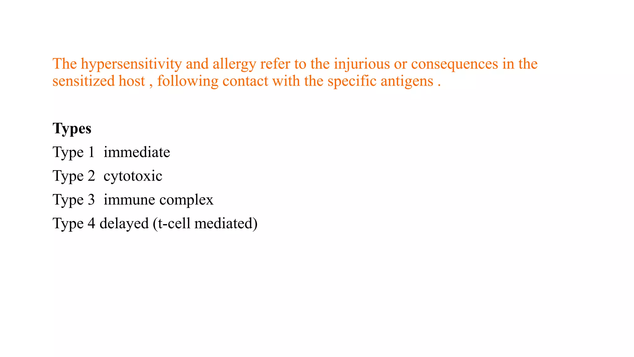
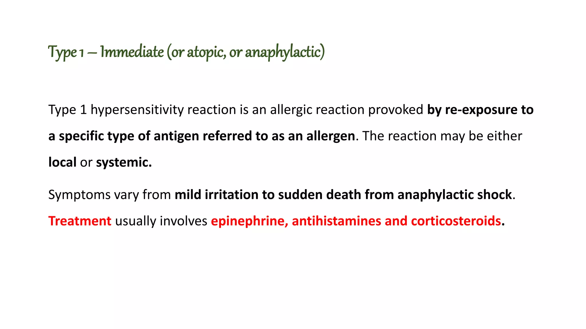
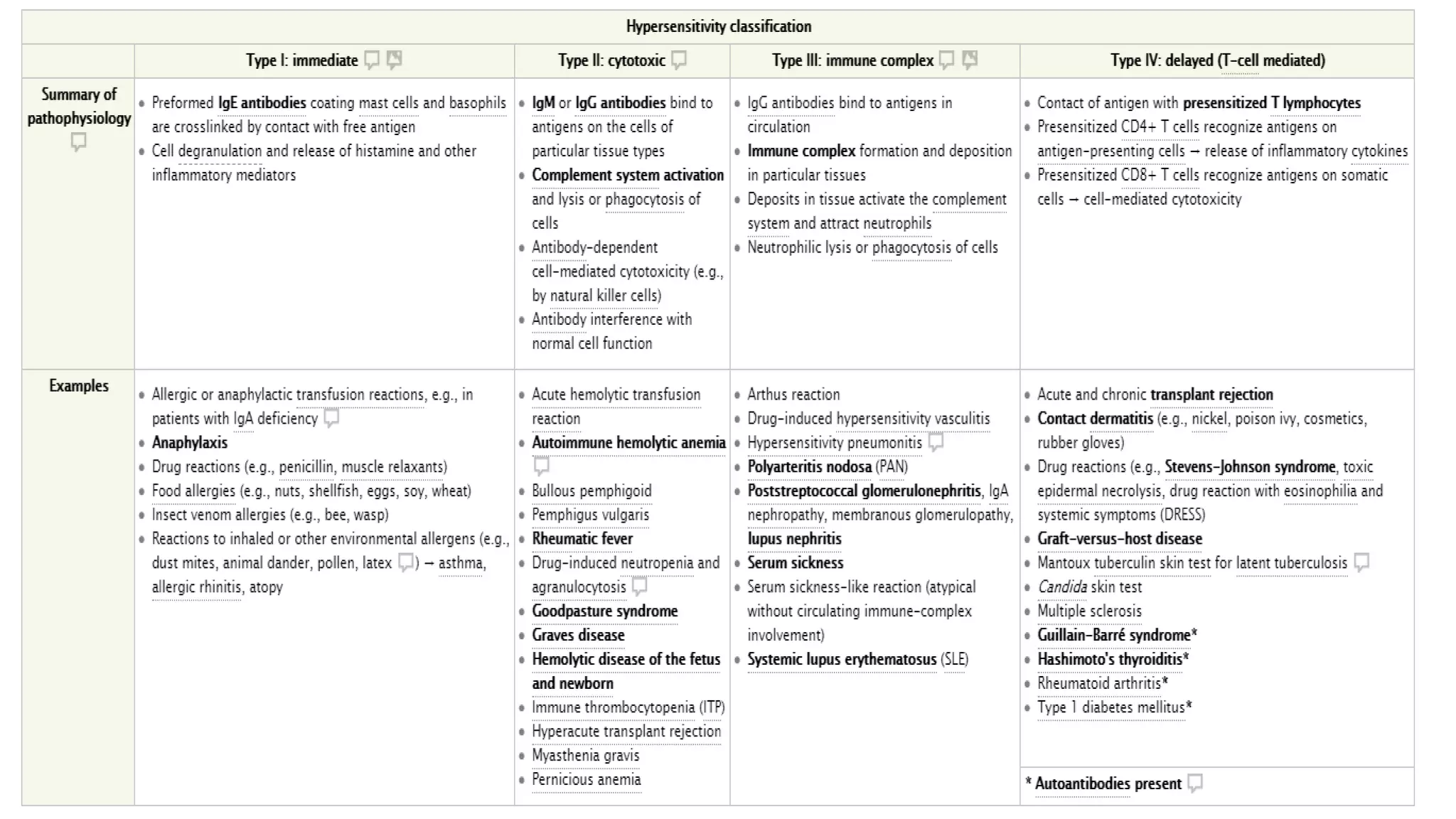
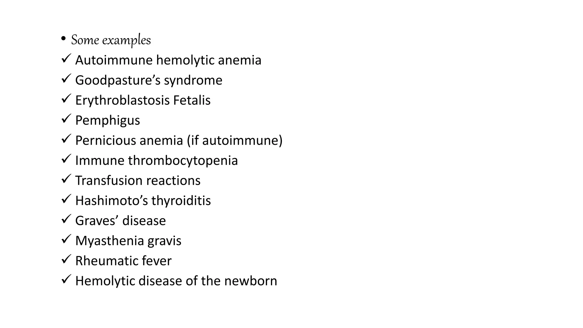
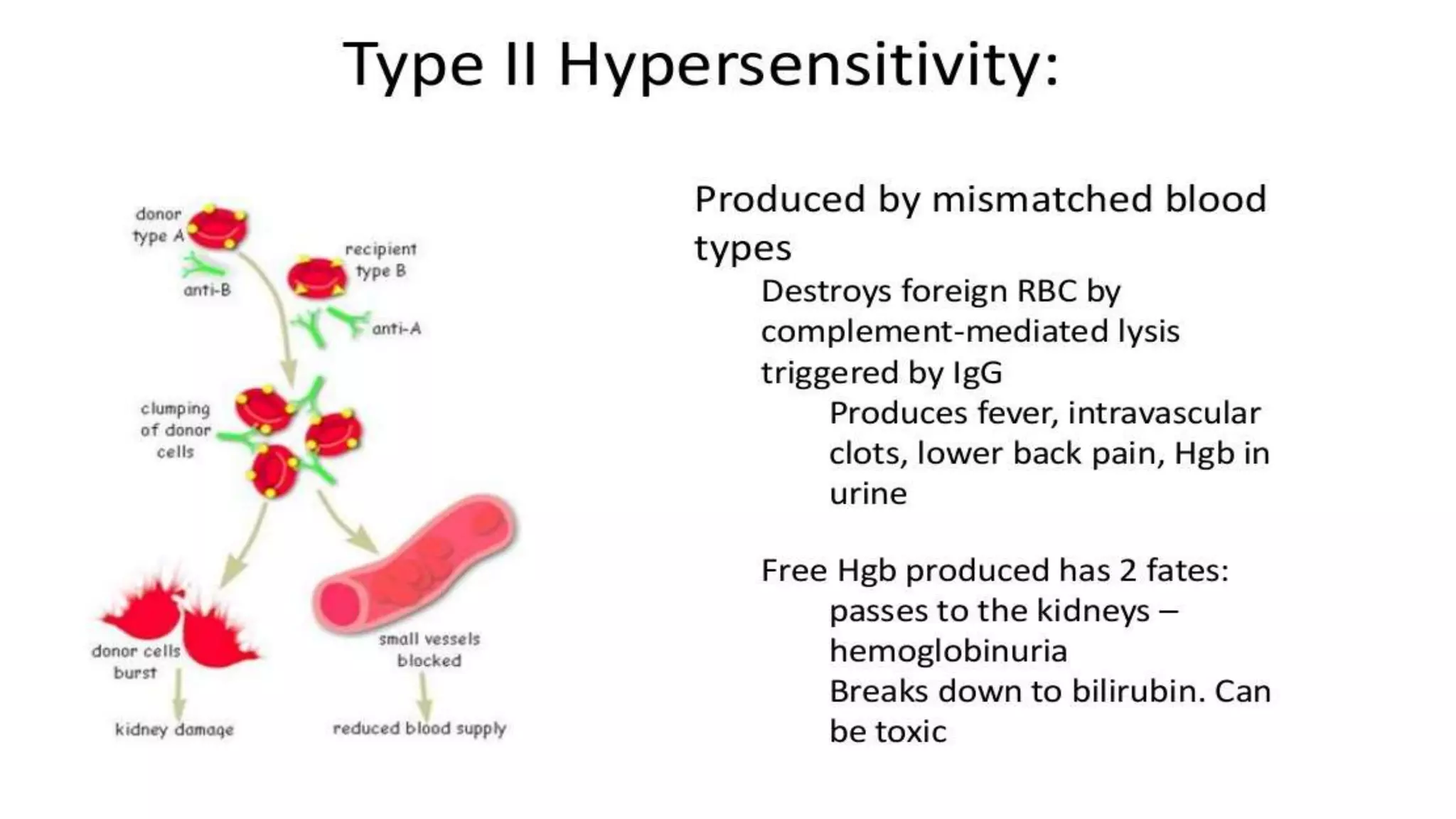
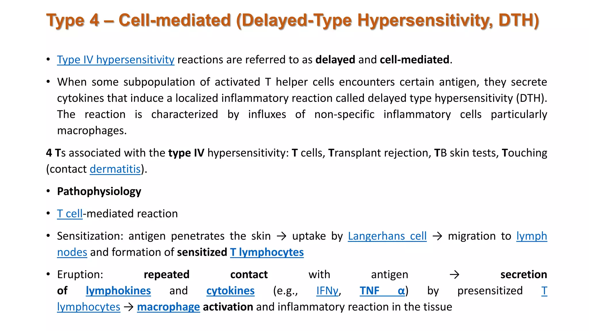
Allergies/hypersensitivity
reactionS
Mr. HEMANT KANHERE
M.PHARM 1ST YEAR
DEPARTMENT OF PHARMACOLOGY
SKBCOP, KAMPTEE
• Definitions
• Hypersensitivity reaction: a condition in which the normally
protective immune system has a harmful effect on the body
• Allergy: an abnormal immunological response to an otherwise harmless
environmental stimulus (e.g., Food, pollen, animal dander)
• Autoimmune disease: an abnormal immunological response directed against
an antigen that is actually part of the body itself
• Stages
• Sensitization: initial asymptomatic contact with an antigen
• Effect: harmful immune response following sensitization and subsequent
antigen contact
The hypersensitivity and allergy refer to the injurious or consequences in the
sensitized host , following contact with the specific antigens .
Types
Type 1 immediate
Type 2 cytotoxic
Type 3 immune complex
Type 4 delayed (t-cell mediated)
Type 1 – Immediate (or atopic, or anaphylactic)
Type 1 hypersensitivity reaction is an allergic reaction provoked by re-exposure to
a specific type of antigen referred to as an allergen. The reaction may be either
local or systemic.
Symptoms vary from mild irritation to sudden death from anaphylactic shock.
Treatment usually involves epinephrine, antihistamines and corticosteroids.
Exposure may be by ingestion, inhalation, injection, or direct contact. The
‘naïve’ b lymphocytes become primed and differentiate into an antibody–
secreting cell and this leads to class-switching of the antibody to the IgE class.
Clinicalfindings
Course
Immediate reaction: allergic reaction within minutes of contact with the antigen
Late-phase reaction: occurs hours after immediate reaction for a duration of 24–72 hours
Main symptoms: pruritus, edema, rash, rhinitis, bronchospasm, and abdominal cramping
Specific manifestations
• Allergic conjunctivitis
• Allergic rhinitis
• Allergic asthma
• Atopy: genetic predisposition to producing ige antibodies against certain
harmless environmental allergens (e.G., Pollen, mites, molds, certain foods)
• Associated conditions: asthma, atopic dermatitis, allergic rhinitis, allergic conjunctivitis, food
allergies
• Urticaria (hives): well-circumscribed, raised, pruritic, and erythematous plaques with a round, oval,
or serpiginous shape; up to several centimeters in diameter (wheals); caused by mast cell activation
in the superficial dermis
• Angioedema: due to mast cell activation in the dermis and/or subcutaneous tissue
• Anaphylaxis
• Diagnostics
• In vivo skin testing
• General principle: Small amounts of allergens (e.g., pollen) are introduced into the skin to test for
a local allergic reaction.
• Higher sensitivity may be achieved with more invasive testing. However, the more invasive
the test, the higher the risk of anaphylactic shock.
• Test results are usually available after 20 minutes.
• Evaluation: skin reddening and size of wheals
• Skin prick test
• Tiny amounts of various allergens are applied to the skin ; a lancet is then used to prick the
surface of the skin so that allergen extracts may enter.
• Positive result: wheal equal to or larger than histamine control (or greater than 3 mm)
• Scratch test: comparable to prick test; a scratch (about 1 cm) is made and
the allergen subsequently applied
• Intradermal test: intradermal injection of small amounts of the allergen on the back or arm.
• In vitro testingTryptase in serum (a relatively specific marker of mast
cell activation): if elevated → increased risk of severe reactions
 Allergen-specific IgE
Indicated in patients with known allergic triggers and clinical symptoms
Preferable to in vivo skin testing in patients in whom the risk
of anaphylaxis is high with skin testing
 Total IgE
Often elevated in patients with allergic conditions
Because normal levels of IgE do not exclude allergy, in vitro testing
should not be used as a definitive test for allergy diagnosis
Treatment
• Treatment of type I hypersensitivity reactions depends on the etiology of the reaction
Urticaria : avoid offending agent (if known), H1-receptor
blocker (e.g., cetirizine), glucocorticoids
• Drug reactions
• Mild reactions (mild urticaria/angioedema) may be treated by removing the offending
drug and monitoring ± antihistamines.
• Moderate reactions (more pronounced urticaria/angioedema) should be treated with
withdrawal of the offending drug and antihistamines ± glucocorticoids.
• Severe reactions require emergency resuscitation (see anaphylaxis).
• Emergency (self-) medication : Patients with known allergic reactions to food or insect
venom, for example, may be provided with antihistamines, corticosteroids,
and epinephrine auto-injectors for self-treatment (in patients at risk of anaphylaxis).
• Allergen immunotherapy (desensitization)
• Indication
• Documented IgE-mediated allergy (e.g., allergic rhinitis, allergic asthma, allergy to
wasp or bee venom)
• Significant symptoms and inadequate relief from symptomatic therapy and exposure
prophylaxis
• Significant symptoms despite symptomatic therapy and avoidance of the allergen
Type 2 – Antibody-dependent
• Type II hypersensitivity reactions are referred to as cytotoxic and play
a role in several autoimmune diseases.
• Clinical features, diagnostics, and treatment depend on the
underlying etiology
• Distribution of disease: often limited to a particular tissue type
• Diagnosis may involve autoantibody testing (see antibody diagnosis of
autoimmune diseases) and the Coombs test
Pathophysiology
• IgM and IgG bind to antigens on cells in the body mistakenly detected as
foreign and cause:
• Complement activation and Fc-mediated immune cell activation
• Cellular lysis or phagocytosis
• Opsonization → phagocytosis and/or complement activation
• Complement-mediated lysis
• Antibody-dependent cell-mediated cytotoxicity (NK cells or macrophages)
• Inhibition or activation of the downstream signaling pathways
Complement mediated lysis of cell
• Complement system is a system of lytic enzyme which are usually inactive in
blood.
• Enzymes of complement system are activated by antigen-antibody complex.
• When antibody binds to antigen (microorganism or RBC) they form Ag-ab
complex.
• Ag-ab complex can activate complement system by three different mechanism-
classical pathway, alternate pathway and lectin pathway.
• Activated complement proceeds in cascade mechanism.
• When complement is activated on the surface of cell (RBC) it causes lysis of cell.
Antibody dependent cell mediatedcytotoxicity (ADCC)
• Antibody binds with antigen by its Fab portion. However Fc
region of antibody has receptor on cytotoxic cells. So, antibody
cross link target cell (microorganism or RBC) with cytotoxic cells
and promote killing.
• Most cytotoxic cells contain storage of hydrolytic and digestive
enzymes. These enzymes are released on the surface of target
cell , killing them.
• Here antibody itself does not kill or destroy cell but rather
mediate killing by presenting antigen to cytotoxic cell. Similarly
cytotoxic cell depends upon antibody to bind antigen. So this
mechanism is known as Antibody dependent cell mediated
cytotoxicity
Opsonization
• When antigen enters into host body, antibodies are produced.
• Antibody binds to antigen through Fab region. Fc region of antibody remains free.
• Phagocytic cells such as Neutrophils, macrophages and monocytes have receptors that can bind to Fc
region of antibody. The receptor is known as FcR.
• In this case antibody molecule directly cross links antigen (Microrganism or RBC or target cell) with
phagocytic cells. This cross-linkage activates phagocytic cells and increases the rate of phagocytosis.
• This increased rate of phagocytosis by binding of antibody to antigen is called Opsonization.
• Some examples
 Autoimmune hemolytic anemia
 Goodpasture’s syndrome
 Erythroblastosis Fetalis
 Pemphigus
 Pernicious anemia (if autoimmune)
 Immune thrombocytopenia
 Transfusion reactions
 Hashimoto’s thyroiditis
 Graves’ disease
 Myasthenia gravis
 Rheumatic fever
 Hemolytic disease of the newborn
Type 3 – Immune Complex
• Type III hypersensitivity reactions are referred to as immune complex reactions and
include many glomerulonephritides and vasculitides.
• Clinical features, diagnostics, and treatment depend on the underlying etiology
• Distribution of disease: systemic
Pathophysiology
• Antigen (e.g., the molecules of a drug in circulation) binds to IgG to form
an immune complex = antigen-antibody complex
• Immune complexes are deposited in tissue, especially blood vessels → initiation
of complement cascade → release of lysosomal enzymes from neutrophils → cell
death → inflammation → vasculitis
• Type III hypersensitivity reaction develops when immune complex activates C3a
and C5a components of complement system.
• C3a and C5a are lymphotoxin (anaphylotoxin) that causes localized mast cell
degranulation.
• Degranulation of mast cell releases histamine which increases vascular
permeability of blood capillaries. This facilitates deposition of immune complexes
on wall of blood vessel.
• C5a, C3a and C5b67 also acts as chemotatic factors for neutrophils, So it attracts
neutrophils at the site of immune complex deposition.
• C3b acts as opsonin by binding with immune complex. Neutrophil binds to C3b
coated immune complex by means of type I complement receptor which is
specific for C3b.
• The neutrophils attempt to phagocytose the immune complex but phagocytosis is
not possible because immune complexes are deposited on basement membrane,
so the neutrophil releases lytic enzymes to destroy immune complex.
• The lytic enzymes cause tissue damage surrounding of immune complex deposits,
resulting hypersensitivity reaction. Furthermore complement proteins can also
contribute to tissue destruction.
Types of Type III hypersensitivity reaction
1. Localized Type III hypersensitivity reaction:
• Acute Arthus reaction is an example of localized
Type III hypersensitivity reaction.
• When antigen is injected or enters intradermally or
subcutaneously, they bind with antibody to form
localized immune complexes which mediate acute
Arthus reaction within 4 to 8 hours.
• As the reaction develops, localized tissue damage
and vascular damage results in accumulation of
fluids (edema) and RBCs (erythema) at the site of
antigen entry.
• The severity of reaction can vary from mild swelling
and redness to tissue necrosis.
2. Generalized Type III hypersensitivity reaction:
• Serum sickness is an example of generalized Type III
hypersensitivity reaction.
• When large amount of antigen enter blood stream
and bind to antibody, circulating immune complexes
forms. If antigens are in significantly excess
compared to antibody, the immune complexes
formed are smaller and soluble which are not
phagocytosed by phagocytic cells leading to Type III
hypersensitivity reaction.
• The manifestation of serum sickness depends on the
quantity of immune complex as well as overall site
of deposition. The site may vary but accumulation of
complexes occurs at site of blood filtration.
• Generalized Type III hypersensitivity reaction at
different site results in different diseases such as
Glomerulonephritis (Kideny), vasculitis (arteries),
Arthritis (synovial joints).
Factors that causes deposition of immune complex and increase susceptibility
to Type III hypersensitivity reaction:
1. Persistent infection:
• In persistent infection such as Malaria, large number of immune complexes are
formed and deposited in tissues.
2. Complement deficiency:
• Complement removes immune complexes from blood, but when complement
system is deficient, large amount of immune complexes circulates in blood and
deposits in tissues.
3. Autoimmunity:
• In autoimmune disease, large amount of immune complexes are formed and
deposited in tissues.
4. Genetic defects:
• In certain genetic defects, small and soluble immune complexes are formed
that can not be phagocytosed.
Examples
• Immune complex glomerulonephritis
• Rheumatoid arthritis
• Serum sickness
• Subacute bacterial endocarditis
• Symptoms of malaria
• Systemic lupus erythematosus
• Arthus reaction
• Farmer’s Lung (Arthus-type reaction)
Type 4 – Cell-mediated (Delayed-Type Hypersensitivity, DTH)
• Type IV hypersensitivity reactions are referred to as delayed and cell-mediated.
• When some subpopulation of activated T helper cells encounters certain antigen, they secrete
cytokines that induce a localized inflammatory reaction called delayed type hypersensitivity (DTH).
The reaction is characterized by influxes of non-specific inflammatory cells particularly
macrophages.
4 Ts associated with the type IV hypersensitivity: T cells, Transplant rejection, TB skin tests, Touching
(contact dermatitis).
• Pathophysiology
• T cell-mediated reaction
• Sensitization: antigen penetrates the skin → uptake by Langerhans cell → migration to lymph
nodes and formation of sensitized T lymphocytes
• Eruption: repeated contact with antigen → secretion
of lymphokines and cytokines (e.g., IFNγ, TNF α) by presensitized T
lymphocytes → macrophage activation and inflammatory reaction in the tissue
DTH occurs in two phases:
• DTH begins with initial sensitization by primary
contact with antigen. At first antigen is
processed and presented by antigen presenting
cells (APCs) to CD4+T cell.
• CD4 +T cells are activated to form TH cells.
• During this TH cells are clonally expanded by
binding with MHC-II molecule carrying antigen
by appropriate APCs. Varieties of APCs have
been shown to be involved in activation of DTH
response including langerhans cell and
macrophages.
• CD4+ T cell are the primary cell activated
during sensitizing phase of DTH response.
However in some cases CD8 + cells are also
found to induce DTH response.
• A subsequent or second time exposure to antigen induces the
effector phage.
• In this phase, TH1 cell secretes varieties of cytokines that recruits
and activates macrophages and other non-specific inflammatory
cells to the site of antigen injection.
• Macrophages are the principle effector of DTH response. The
activated macrophages exhibit increased level of phagocytosis and
increased ability to kill the antigen (microorganisms) by various
cytotoxic lytic enzymes.
• Activated macrophages releases lytic enzymes that damage
surrounding tissues and intracellular bacteria.
• The influx and activation of macrophages in DTH is important in
host defense against intracellular bacteria and parasite, where
circulating antibodies cannot reach them.
• Increased phagocytic activity and build up lytic enzyme from
macrophages in the area of infection leads to non-specific
destruction of tissues and intracellular pathogens.
• Generally pathogens are killed rapidly with little tissue damage.
However in some case, especially if the antigen is not easily cleared,
a pro-long DTH response can itself become destructive to host as
intensive and chronic inflammation develops into a visible
granulomatous reaction.
• A granuloma develops when continuous activation of macrophages
induces the epitheloid shape and some time fusion of macrophages
to form multinucleated giant cells. These giant cells displace normal
cells forming palpable nodules and release high concentration of
lytic enzymes which destroy surrounding tissues leading to necrosis.
Type IV drug reactions
• Local drug reaction following topical application of drug; see allergic contact
dermatitis above
• Maculopapular or morbilliform (measles-like) drug eruption
• Stevens-Johnson syndrome and toxic epidermal necrolysis
• DRESS syndrome (drug rash with eosinophilia and systemic symptoms syndrome; also
known as drug-induced hypersensitivity syndrome): delayed hypersensitivity reaction to
a drug (within 1–8 weeks following administration)
• Etiology
• Allopurinol
• Antiepileptic drugs (e.g., lamotrigine, phenytoin, carbamazepine)
• Antibiotics (e.g., sulfonamide)
• Clinical features
• Fever
• Pruritic morbilliform rash
• Facial edema
• Hepatomegaly
• Diffuse lymphadenopathy
• Possible multiorgan failure
• Laboratory tests
• Eosinophilia
• Thrombocytopenia
• Atypical lymphocytosis
• Treatment
• Drug withdrawal
• Symptomatic: Corticosteroids are often used, but their effect is disputed.
• Prognosis: fatal in ∼ 10% of cases
• Type IV is fourth and last (i.e., delayed).
Hypersensitivity Reactions & its types.

Hypersensitivity Reactions & its types.

  • 1.
    Allergies/hypersensitivity reactionS Mr. HEMANT KANHERE M.PHARM1ST YEAR DEPARTMENT OF PHARMACOLOGY SKBCOP, KAMPTEE
  • 2.
    • Definitions • Hypersensitivityreaction: a condition in which the normally protective immune system has a harmful effect on the body • Allergy: an abnormal immunological response to an otherwise harmless environmental stimulus (e.g., Food, pollen, animal dander) • Autoimmune disease: an abnormal immunological response directed against an antigen that is actually part of the body itself • Stages • Sensitization: initial asymptomatic contact with an antigen • Effect: harmful immune response following sensitization and subsequent antigen contact
  • 3.
    The hypersensitivity andallergy refer to the injurious or consequences in the sensitized host , following contact with the specific antigens . Types Type 1 immediate Type 2 cytotoxic Type 3 immune complex Type 4 delayed (t-cell mediated)
  • 4.
    Type 1 –Immediate (or atopic, or anaphylactic) Type 1 hypersensitivity reaction is an allergic reaction provoked by re-exposure to a specific type of antigen referred to as an allergen. The reaction may be either local or systemic. Symptoms vary from mild irritation to sudden death from anaphylactic shock. Treatment usually involves epinephrine, antihistamines and corticosteroids.
  • 5.
    Exposure may beby ingestion, inhalation, injection, or direct contact. The ‘naïve’ b lymphocytes become primed and differentiate into an antibody– secreting cell and this leads to class-switching of the antibody to the IgE class.
  • 8.
    Clinicalfindings Course Immediate reaction: allergicreaction within minutes of contact with the antigen Late-phase reaction: occurs hours after immediate reaction for a duration of 24–72 hours Main symptoms: pruritus, edema, rash, rhinitis, bronchospasm, and abdominal cramping Specific manifestations • Allergic conjunctivitis • Allergic rhinitis • Allergic asthma • Atopy: genetic predisposition to producing ige antibodies against certain harmless environmental allergens (e.G., Pollen, mites, molds, certain foods) • Associated conditions: asthma, atopic dermatitis, allergic rhinitis, allergic conjunctivitis, food allergies • Urticaria (hives): well-circumscribed, raised, pruritic, and erythematous plaques with a round, oval, or serpiginous shape; up to several centimeters in diameter (wheals); caused by mast cell activation in the superficial dermis • Angioedema: due to mast cell activation in the dermis and/or subcutaneous tissue • Anaphylaxis
  • 10.
    • Diagnostics • Invivo skin testing • General principle: Small amounts of allergens (e.g., pollen) are introduced into the skin to test for a local allergic reaction. • Higher sensitivity may be achieved with more invasive testing. However, the more invasive the test, the higher the risk of anaphylactic shock. • Test results are usually available after 20 minutes. • Evaluation: skin reddening and size of wheals • Skin prick test • Tiny amounts of various allergens are applied to the skin ; a lancet is then used to prick the surface of the skin so that allergen extracts may enter. • Positive result: wheal equal to or larger than histamine control (or greater than 3 mm) • Scratch test: comparable to prick test; a scratch (about 1 cm) is made and the allergen subsequently applied • Intradermal test: intradermal injection of small amounts of the allergen on the back or arm.
  • 11.
    • In vitrotestingTryptase in serum (a relatively specific marker of mast cell activation): if elevated → increased risk of severe reactions  Allergen-specific IgE Indicated in patients with known allergic triggers and clinical symptoms Preferable to in vivo skin testing in patients in whom the risk of anaphylaxis is high with skin testing  Total IgE Often elevated in patients with allergic conditions Because normal levels of IgE do not exclude allergy, in vitro testing should not be used as a definitive test for allergy diagnosis
  • 12.
    Treatment • Treatment oftype I hypersensitivity reactions depends on the etiology of the reaction Urticaria : avoid offending agent (if known), H1-receptor blocker (e.g., cetirizine), glucocorticoids • Drug reactions • Mild reactions (mild urticaria/angioedema) may be treated by removing the offending drug and monitoring ± antihistamines. • Moderate reactions (more pronounced urticaria/angioedema) should be treated with withdrawal of the offending drug and antihistamines ± glucocorticoids. • Severe reactions require emergency resuscitation (see anaphylaxis). • Emergency (self-) medication : Patients with known allergic reactions to food or insect venom, for example, may be provided with antihistamines, corticosteroids, and epinephrine auto-injectors for self-treatment (in patients at risk of anaphylaxis). • Allergen immunotherapy (desensitization) • Indication • Documented IgE-mediated allergy (e.g., allergic rhinitis, allergic asthma, allergy to wasp or bee venom) • Significant symptoms and inadequate relief from symptomatic therapy and exposure prophylaxis • Significant symptoms despite symptomatic therapy and avoidance of the allergen
  • 13.
    Type 2 –Antibody-dependent • Type II hypersensitivity reactions are referred to as cytotoxic and play a role in several autoimmune diseases. • Clinical features, diagnostics, and treatment depend on the underlying etiology • Distribution of disease: often limited to a particular tissue type • Diagnosis may involve autoantibody testing (see antibody diagnosis of autoimmune diseases) and the Coombs test
  • 14.
    Pathophysiology • IgM andIgG bind to antigens on cells in the body mistakenly detected as foreign and cause: • Complement activation and Fc-mediated immune cell activation • Cellular lysis or phagocytosis • Opsonization → phagocytosis and/or complement activation • Complement-mediated lysis • Antibody-dependent cell-mediated cytotoxicity (NK cells or macrophages) • Inhibition or activation of the downstream signaling pathways
  • 16.
    Complement mediated lysisof cell • Complement system is a system of lytic enzyme which are usually inactive in blood. • Enzymes of complement system are activated by antigen-antibody complex. • When antibody binds to antigen (microorganism or RBC) they form Ag-ab complex. • Ag-ab complex can activate complement system by three different mechanism- classical pathway, alternate pathway and lectin pathway. • Activated complement proceeds in cascade mechanism. • When complement is activated on the surface of cell (RBC) it causes lysis of cell.
  • 17.
    Antibody dependent cellmediatedcytotoxicity (ADCC) • Antibody binds with antigen by its Fab portion. However Fc region of antibody has receptor on cytotoxic cells. So, antibody cross link target cell (microorganism or RBC) with cytotoxic cells and promote killing. • Most cytotoxic cells contain storage of hydrolytic and digestive enzymes. These enzymes are released on the surface of target cell , killing them. • Here antibody itself does not kill or destroy cell but rather mediate killing by presenting antigen to cytotoxic cell. Similarly cytotoxic cell depends upon antibody to bind antigen. So this mechanism is known as Antibody dependent cell mediated cytotoxicity
  • 18.
    Opsonization • When antigenenters into host body, antibodies are produced. • Antibody binds to antigen through Fab region. Fc region of antibody remains free. • Phagocytic cells such as Neutrophils, macrophages and monocytes have receptors that can bind to Fc region of antibody. The receptor is known as FcR. • In this case antibody molecule directly cross links antigen (Microrganism or RBC or target cell) with phagocytic cells. This cross-linkage activates phagocytic cells and increases the rate of phagocytosis. • This increased rate of phagocytosis by binding of antibody to antigen is called Opsonization.
  • 19.
    • Some examples Autoimmune hemolytic anemia  Goodpasture’s syndrome  Erythroblastosis Fetalis  Pemphigus  Pernicious anemia (if autoimmune)  Immune thrombocytopenia  Transfusion reactions  Hashimoto’s thyroiditis  Graves’ disease  Myasthenia gravis  Rheumatic fever  Hemolytic disease of the newborn
  • 22.
    Type 3 –Immune Complex • Type III hypersensitivity reactions are referred to as immune complex reactions and include many glomerulonephritides and vasculitides. • Clinical features, diagnostics, and treatment depend on the underlying etiology • Distribution of disease: systemic Pathophysiology • Antigen (e.g., the molecules of a drug in circulation) binds to IgG to form an immune complex = antigen-antibody complex • Immune complexes are deposited in tissue, especially blood vessels → initiation of complement cascade → release of lysosomal enzymes from neutrophils → cell death → inflammation → vasculitis
  • 24.
    • Type IIIhypersensitivity reaction develops when immune complex activates C3a and C5a components of complement system. • C3a and C5a are lymphotoxin (anaphylotoxin) that causes localized mast cell degranulation. • Degranulation of mast cell releases histamine which increases vascular permeability of blood capillaries. This facilitates deposition of immune complexes on wall of blood vessel. • C5a, C3a and C5b67 also acts as chemotatic factors for neutrophils, So it attracts neutrophils at the site of immune complex deposition. • C3b acts as opsonin by binding with immune complex. Neutrophil binds to C3b coated immune complex by means of type I complement receptor which is specific for C3b. • The neutrophils attempt to phagocytose the immune complex but phagocytosis is not possible because immune complexes are deposited on basement membrane, so the neutrophil releases lytic enzymes to destroy immune complex. • The lytic enzymes cause tissue damage surrounding of immune complex deposits, resulting hypersensitivity reaction. Furthermore complement proteins can also contribute to tissue destruction.
  • 25.
    Types of TypeIII hypersensitivity reaction 1. Localized Type III hypersensitivity reaction: • Acute Arthus reaction is an example of localized Type III hypersensitivity reaction. • When antigen is injected or enters intradermally or subcutaneously, they bind with antibody to form localized immune complexes which mediate acute Arthus reaction within 4 to 8 hours. • As the reaction develops, localized tissue damage and vascular damage results in accumulation of fluids (edema) and RBCs (erythema) at the site of antigen entry. • The severity of reaction can vary from mild swelling and redness to tissue necrosis. 2. Generalized Type III hypersensitivity reaction: • Serum sickness is an example of generalized Type III hypersensitivity reaction. • When large amount of antigen enter blood stream and bind to antibody, circulating immune complexes forms. If antigens are in significantly excess compared to antibody, the immune complexes formed are smaller and soluble which are not phagocytosed by phagocytic cells leading to Type III hypersensitivity reaction. • The manifestation of serum sickness depends on the quantity of immune complex as well as overall site of deposition. The site may vary but accumulation of complexes occurs at site of blood filtration. • Generalized Type III hypersensitivity reaction at different site results in different diseases such as Glomerulonephritis (Kideny), vasculitis (arteries), Arthritis (synovial joints).
  • 26.
    Factors that causesdeposition of immune complex and increase susceptibility to Type III hypersensitivity reaction: 1. Persistent infection: • In persistent infection such as Malaria, large number of immune complexes are formed and deposited in tissues. 2. Complement deficiency: • Complement removes immune complexes from blood, but when complement system is deficient, large amount of immune complexes circulates in blood and deposits in tissues. 3. Autoimmunity: • In autoimmune disease, large amount of immune complexes are formed and deposited in tissues. 4. Genetic defects: • In certain genetic defects, small and soluble immune complexes are formed that can not be phagocytosed.
  • 27.
    Examples • Immune complexglomerulonephritis • Rheumatoid arthritis • Serum sickness • Subacute bacterial endocarditis • Symptoms of malaria • Systemic lupus erythematosus • Arthus reaction • Farmer’s Lung (Arthus-type reaction)
  • 28.
    Type 4 –Cell-mediated (Delayed-Type Hypersensitivity, DTH) • Type IV hypersensitivity reactions are referred to as delayed and cell-mediated. • When some subpopulation of activated T helper cells encounters certain antigen, they secrete cytokines that induce a localized inflammatory reaction called delayed type hypersensitivity (DTH). The reaction is characterized by influxes of non-specific inflammatory cells particularly macrophages. 4 Ts associated with the type IV hypersensitivity: T cells, Transplant rejection, TB skin tests, Touching (contact dermatitis). • Pathophysiology • T cell-mediated reaction • Sensitization: antigen penetrates the skin → uptake by Langerhans cell → migration to lymph nodes and formation of sensitized T lymphocytes • Eruption: repeated contact with antigen → secretion of lymphokines and cytokines (e.g., IFNγ, TNF α) by presensitized T lymphocytes → macrophage activation and inflammatory reaction in the tissue
  • 29.
    DTH occurs intwo phases: • DTH begins with initial sensitization by primary contact with antigen. At first antigen is processed and presented by antigen presenting cells (APCs) to CD4+T cell. • CD4 +T cells are activated to form TH cells. • During this TH cells are clonally expanded by binding with MHC-II molecule carrying antigen by appropriate APCs. Varieties of APCs have been shown to be involved in activation of DTH response including langerhans cell and macrophages. • CD4+ T cell are the primary cell activated during sensitizing phase of DTH response. However in some cases CD8 + cells are also found to induce DTH response.
  • 30.
    • A subsequentor second time exposure to antigen induces the effector phage. • In this phase, TH1 cell secretes varieties of cytokines that recruits and activates macrophages and other non-specific inflammatory cells to the site of antigen injection. • Macrophages are the principle effector of DTH response. The activated macrophages exhibit increased level of phagocytosis and increased ability to kill the antigen (microorganisms) by various cytotoxic lytic enzymes. • Activated macrophages releases lytic enzymes that damage surrounding tissues and intracellular bacteria. • The influx and activation of macrophages in DTH is important in host defense against intracellular bacteria and parasite, where circulating antibodies cannot reach them. • Increased phagocytic activity and build up lytic enzyme from macrophages in the area of infection leads to non-specific destruction of tissues and intracellular pathogens. • Generally pathogens are killed rapidly with little tissue damage. However in some case, especially if the antigen is not easily cleared, a pro-long DTH response can itself become destructive to host as intensive and chronic inflammation develops into a visible granulomatous reaction. • A granuloma develops when continuous activation of macrophages induces the epitheloid shape and some time fusion of macrophages to form multinucleated giant cells. These giant cells displace normal cells forming palpable nodules and release high concentration of lytic enzymes which destroy surrounding tissues leading to necrosis.
  • 31.
    Type IV drugreactions • Local drug reaction following topical application of drug; see allergic contact dermatitis above • Maculopapular or morbilliform (measles-like) drug eruption • Stevens-Johnson syndrome and toxic epidermal necrolysis • DRESS syndrome (drug rash with eosinophilia and systemic symptoms syndrome; also known as drug-induced hypersensitivity syndrome): delayed hypersensitivity reaction to a drug (within 1–8 weeks following administration) • Etiology • Allopurinol • Antiepileptic drugs (e.g., lamotrigine, phenytoin, carbamazepine) • Antibiotics (e.g., sulfonamide) • Clinical features • Fever • Pruritic morbilliform rash • Facial edema • Hepatomegaly • Diffuse lymphadenopathy • Possible multiorgan failure
  • 32.
    • Laboratory tests •Eosinophilia • Thrombocytopenia • Atypical lymphocytosis • Treatment • Drug withdrawal • Symptomatic: Corticosteroids are often used, but their effect is disputed. • Prognosis: fatal in ∼ 10% of cases • Type IV is fourth and last (i.e., delayed).