INTRODUCTIO
N
Hypersensitivity isthe term used when an immune
response results in exaggerated or inappropriate reactions
harmful to the host.
The term allergy is often equated with hypersensitivity but
more accurately should be limited to the IgE–mediated
reaction.
The clinical manifestations of these reactions are typical in a
given individual and occur on contact with the specific
antigen to which the individual is hypersensitive.
The first contact of the individual with the antigen
sensitizes (i.e., induces the antibody), and the subsequent
contacts elicit the allergic response.
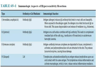
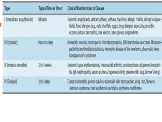
Hypersensitivity reactions can be subdivided into four main
types. Types I, II, and III are antibody-mediated, whereas
type IV is cell- mediated.
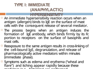
TYPE I: IMMEDIATE
(ANAPHYLACTIC)
HYPERSENSITIVITY
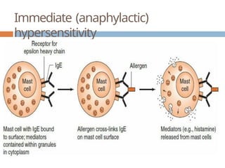
An immediate hypersensitivity reaction occurs when an
antigen (allergen) binds to IgE on the surface of mast
cells with the consequent release of several mediator.
The process begins when an antigen induces the
formation of IgE antibody, which binds firmly by its Fc
portion to receptors on the surface of basophils and
mast cells.
Reexposure to the same antigen results in cross-linking of
the cell-bound IgE, degranulation, and release of
pharmacologically active mediators within minutes
(immediate phase).
Symptoms such as edema and erythema (“wheal and
flare”) and itching appear rapidly because these
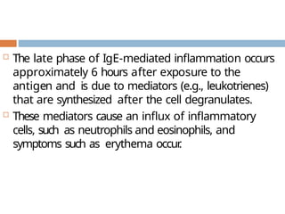
The latephase of IgE-mediated inflammation occurs
approximately 6 hours after exposure to the
antigen and is due to mediators (e.g., leukotrienes)
that are synthesized after the cell degranulates.
These mediators cause an influx of inflammatory
cells, such as neutrophils and eosinophils, and
symptoms such as erythema occur
.
8.
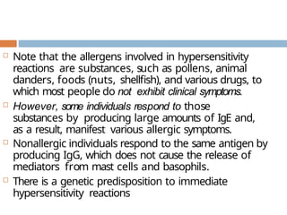
Note thatthe allergens involved in hypersensitivity
reactions are substances, such as pollens, animal
danders, foods (nuts, shellfish), and various drugs, to
which most people do not exhibit clinical symptoms.
However, some individuals respond to those
substances by producing large amounts of IgE and,
as a result, manifest various allergic symptoms.
Nonallergic individuals respond to the same antigen by
producing IgG, which does not cause the release of
mediators from mast cells and basophils.
There is a genetic predisposition to immediate
hypersensitivity reactions
9.
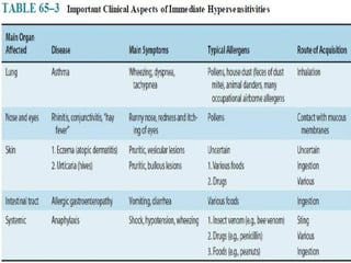
The clinicalmanifestations of type I hypersensitivity can
appear in various forms (e.g., urticaria [also known as
hives], eczema, rhinitis and conjunctivitis [also known as
hay fever], and asthma).
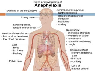
The most severe form of type I hypersensitivity is
systemic anaphylaxis, in which severe
bronchoconstriction and hypotension (shock) can be
life-threatening.
The most common causes of anaphylaxis are foods such
as peanuts and shellfish, bee venom, and drugs such as
penicillin
Major manifestations of anaphylaxis occur when large
amounts of mediators are suddenly released as a result
11.
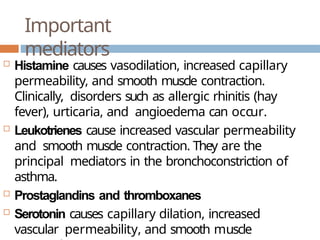
Important
mediators
Histamine causesvasodilation, increased capillary
permeability, and smooth muscle contraction.
Clinically, disorders such as allergic rhinitis (hay
fever), urticaria, and angioedema can occur.
Leukotrienes cause increased vascular permeability
and smooth muscle contraction. They are the
principal mediators in the bronchoconstriction of
asthma.
Prostaglandins and thromboxanes
Serotonin causes capillary dilation, increased
vascular permeability, and smooth muscle
13.
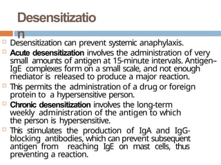
Desensitizatio
n
Desensitization canprevent systemic anaphylaxis.
Acute desensitization involves the administration of very
small amounts of antigen at 15-minute intervals. Antigen–
IgE complexes form on a small scale, and not enough
mediator is released to produce a major reaction.
This permits the administration of a drug or foreign
protein to a hypersensitive person.
Chronic desensitization involves the long-term
weekly administration of the antigen to which
the person is hypersensitive.
This stimulates the production of IgA and IgG-
blocking antibodies, which can prevent subsequent
antigen from reaching IgE on mast cells, thus
preventing a reaction.
14.
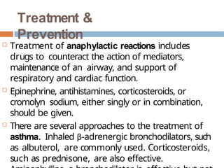
Treatment &
Prevention
Treatmentof anaphylactic reactions includes
drugs to counteract the action of mediators,
maintenance of an airway, and support of
respiratory and cardiac function.
Epinephrine, antihistamines, corticosteroids, or
cromolyn sodium, either singly or in combination,
should be given.
There are several approaches to the treatment of
asthma. Inhaled β-adrenergic bronchodilators, such
as albuterol, are commonly used. Corticosteroids,
such as prednisone, are also effective.
15.
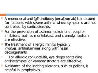
A monoclonalanti-IgE antibody (omalizumab) is indicated
for patients with severe asthma whose symptoms are not
controlled by corticosteroids.
For the prevention of asthma, leukotriene receptor
inhibitors, such as montelukast, and cromolyn sodium
are effective.
The treatment of allergic rhinitis typically
involves antihistamines along with nasal
decongestants.
For allergic conjunctivitis, eye drops containing
antihistamines or vasoconstrictors are effective.
Avoidance of the inciting allergens, such as pollens, is
helpful in prophylaxis.
16.
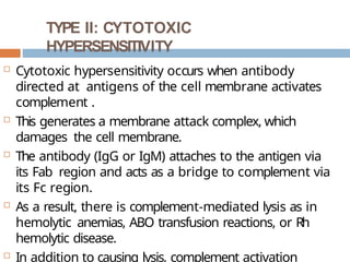
TYPE II: CYTOTOXIC
HYPERSENSITIVITY
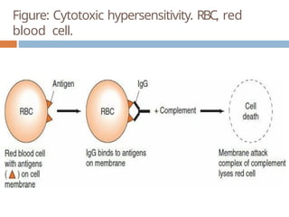
Cytotoxic hypersensitivity occurs when antibody
directed at antigens of the cell membrane activates
complement .
This generates a membrane attack complex, which
damages the cell membrane.
The antibody (IgG or IgM) attaches to the antigen via
its Fab region and acts as a bridge to complement via
its Fc region.
As a result, there is complement-mediated lysis as in
hemolytic anemias, ABO transfusion reactions, or Rh
hemolytic disease.
In addition to causing lysis, complement activation
Drugs (e.g.,penicillins, phenacetin, quinidine) can
attach to surface proteins on red blood cells and
initiate antibody formation. Such autoimmune
antibodies (IgG) then interact with the red blood cell
surface and result in hemolysis.
Other drugs (e.g., quinine) can attach to platelets and
induce autoantibodies that lyse the platelets, producing
thrombocytopenia and, as a consequence, a
bleeding tendency.
19.
TYPE III: IMMUNECOMPLEX
HYPERSENSITIVITY
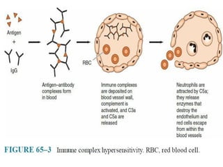
Immune complex hypersensitivity occurs when antigen–
antibody complexes induce an inflammatory response in
tissues.
Normally, immune complexes are promptly removed by the
reticuloendothelial system, but occasionally they persist and
are deposited in tissues, resulting in several disorders.
In persistent microbial or viral infections, immune complexes
may be deposited in organs (e.g., the kidneys), resulting in
damage.
In autoimmune disorders, “self” antigens may elicit antibodies
that bind to organ antigens or deposit in organs as complexes,
especially in joints (arthritis), kidneys (nephritis), or blood
vessels (vasculitis).
Wherever immune complexes are deposited, they activate the
21.
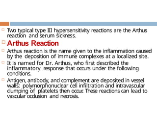
Two typicaltype III hypersensitivity reactions are the Arthus
reaction and serum sickness.
Arthus Reaction
Arthus reaction is the name given to the inflammation caused
by the deposition of immune complexes at a localized site.
It is named for Dr. Arthus, who first described the
inflammatory response that occurs under the following
conditions.
Antigen, antibody, and complement are deposited in vessel
walls; polymorphonuclear cell infiltration and intravascular
clumping of platelets then occur
. These reactions can lead to
vascular occlusion and necrosis.
22.
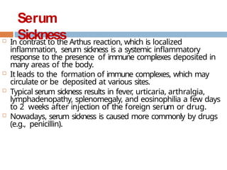
Serum
Sickness
In contrastto the Arthus reaction, which is localized
inflammation, serum sickness is a systemic inflammatory
response to the presence of immune complexes deposited in
many areas of the body.
It leads to the formation of immune complexes, which may
circulate or be deposited at various sites.
Typical serum sickness results in fever
, urticaria, arthralgia,
lymphadenopathy, splenomegaly, and eosinophilia a few days
to 2 weeks after injection of the foreign serum or drug.
Nowadays, serum sickness is caused more commonly by drugs
(e.g., penicillin).
23.
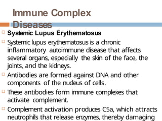
Immune Complex
Diseases
SystemicLupus Erythematosus
Systemic lupus erythematosus is a chronic
inflammatory autoimmune disease that affects
several organs, especially the skin of the face, the
joints, and the kidneys.
Antibodies are formed against DNA and other
components of the nucleus of cells.
These antibodies form immune complexes that
activate complement.
Complement activation produces C5a, which attracts
neutrophils that release enzymes, thereby damaging
24.
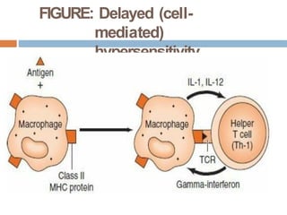
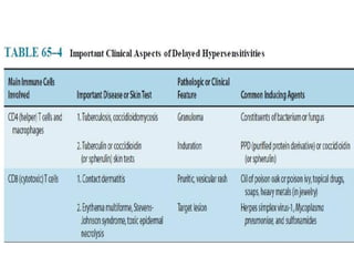
TYPE IV: DELAYED(CELL-MEDIATED)
HYPERSENSITIVITY
Delayed hypersensitivity is a function of T
lymphocytes, not antibody.
The response is “delayed” (i.e., it starts hours [or
days] after contact with the antigen and often
lasts for days).
In certain contact hypersensitivities, such as poison
oak, the pruritic, vesicular skin rash is caused by CD8-
positive cytotoxic T cells that attack skin cells that
display the plant oil as a foreign antigen.
In the tuberculin skin test, the indurated skin rash is
caused by CD4-positive helper T cells and
Clinically Important
Delayed Hypersensitivity
Reactions
Contact Hypersensitivity
This manifestation of cell-mediated hypersensitivity occurs after
sensitization with simple chemicals (e.g., nickel, formaldehyde), plant
materials (e.g., poison ivy, poison oak), topically applied drugs(e.g.,
sulfonamides, neomycin), some cosmetics, soaps, and other substances.
Neomycin is a very common cause.
In all cases, the small molecules acting as haptens enter the skin,
attach to body proteins, and become complete antigens.
Cell-mediated hypersensitivity is induced, particularly in the skin.
Upon a later skin contact with the offending agent, the sensitized
person develops contact dermatitis characterized by erythema,
itching, vesicles, eczema, or necrosis of skin within 12 to 48 hours
caused by the attack of cytotoxic T cells.
28.
Drug
allergy
A drugallergy is an allergy to a drug, most
commonly a medication.
Drugs often contain many different substances,
including dyes, which could cause allergic
reactions.
This can cause an allergic reaction on the first
administration of a drug. For example, a person
who developed an allergy to a red dye will be
allergic to any new drug which contains that red
dye.
When a medication causes an allergic reaction, it is
29.
Mechanis
m
Drug allergiesare attributed to "drug
hypersensitivity," initiated by exposure to a drug
at a dose normally tolerated by non-
hypersensitive persons.
There are two mechanisms for a drug allergy to
occur:
IgE or non-IgE mediated.
In IgE-mediated reactions, also know as
Immunoglobulin E mediated reactions, drug
allergens bind to IgE antibodies, which are attached
to mast cells and basophils, resulting in IgE cross-
30.
Drug
Hypersensitivity
Drugs, particularlyantimicrobial agents such as
penicillin, are now among the most common causes of
hypersensitivity reactions.
Usually it is not the intact drug that induces
antibody formation.
Rather
, a metabolic product of the drug, which acts
as a hapten and binds to a body protein, does
so.
The resulting antibody can react with the hapten or the
intact drug to give rise to type I hypersensitivity.
When reexposed to the drug, the person may exhibit a
drug rash, fever
, or local or systemic anaphylaxis of
variable severity.
31.
In non–IgE-mediated drug allergy, the mechanisms
include cytotoxic/cytolytic reactions involving the
interaction of IgG or IgM antibodies and
complement with a drug allergen associated with
cell membranes (e.g., immune hemolytic anemia,
thrombocytopenia), drug immune complex reactions
and T-cell–mediated reactions.
Drug allergy, occurs in 1% to 2% of all admissions
and 3% to 5% of hospitalized patients,
respectively but the true incidence of drug
allergy in the community, and among children
32.
Symptom
s
Identifying adrug allergy can sometimes be the hardest
part. Sometimes drug allergies are confused with
Nonallergic drug reactions because they both cause
somewhat similar reactions.
Symptoms of a drug allergy can include, but are not
limited to, the following list.
Hives
Itching
Rash
Fever
Facial swelling
Shortness of breath
Anaphylaxis, a life threatening drug reaction (produces
most of these symptoms as well as low blood pressure)

![ The clinical manifestations of type I hypersensitivity can
appear in various forms (e.g., urticaria [also known as
hives], eczema, rhinitis and conjunctivitis [also known as
hay fever], and asthma).
The most severe form of type I hypersensitivity is
systemic anaphylaxis, in which severe
bronchoconstriction and hypotension (shock) can be
life-threatening.
The most common causes of anaphylaxis are foods such
as peanuts and shellfish, bee venom, and drugs such as
penicillin
Major manifestations of anaphylaxis occur when large
amounts of mediators are suddenly released as a result](https://image.slidesharecdn.com/hypersensitivityppt-250309074951-a119f1a9/85/Hypersensitivity-ppt-Microbiology-for-Pharm-D-pptx-9-320.jpg)
![TYPE IV: DELAYED (CELL-MEDIATED)
HYPERSENSITIVITY
Delayed hypersensitivity is a function of T
lymphocytes, not antibody.
The response is “delayed” (i.e., it starts hours [or
days] after contact with the antigen and often
lasts for days).
In certain contact hypersensitivities, such as poison
oak, the pruritic, vesicular skin rash is caused by CD8-
positive cytotoxic T cells that attack skin cells that
display the plant oil as a foreign antigen.
In the tuberculin skin test, the indurated skin rash is
caused by CD4-positive helper T cells and](https://image.slidesharecdn.com/hypersensitivityppt-250309074951-a119f1a9/85/Hypersensitivity-ppt-Microbiology-for-Pharm-D-pptx-24-320.jpg)