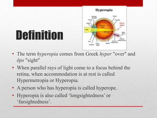
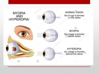
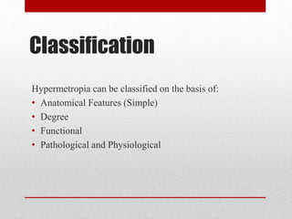
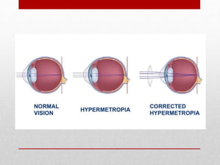
Retinoscopy is the primary objective method for determining a patient's refractive error. It involves using a retinoscope to illuminate the retina and observe the movement of the reflected light. For myopic patients, the light moves in the opposite direction of the retinoscope's movement, while for hyperopic patients it moves in the same direction. The goal is to find the neutralization point where no movement is seen, indicating the proper refractive correction. Factors like the working distance, type of mirror used, and patient's fixation can impact results. Retinoscopy is useful for initial refractive estimates and screening for ocular conditions.
Cycloplegic refraction involves temporarily paralyzing the ciliary muscle with eye drops in order to determine a person's full refractive error. This is important for children who accommodate too much. Common cycloplegic agents include atropine, homatropine, and cyclopentolate. Cyclopentolate is often the drug of choice due to its faster onset and shorter duration. A cycloplegic refraction allows an accurate assessment of refractive error, especially in children and other patients where accommodation can affect results.
This document discusses myopia, including its optics, classification, treatment, and prognosis. It defines myopia as a refractive error where parallel light rays focus in front of the retina. Myopia is classified as axial, curvatural, index, or acquired. Treatment options include optical correction with concave lenses, surgery, general measures like visual hygiene, and low vision aids for high myopia. Pathological myopia is a form characterized by a rapidly progressive refractive error and increased risks of retinal detachment and other complications.
- Aphakia is the absence of the crystalline lens from the eye. It can be congenital or caused by surgery or trauma.
- In aphakia, the eye becomes highly hyperopic, the anterior focal point moves forward, and the retinal image is magnified. This decreases visual acuity and field of view.
- Aphakia is treated with spectacles, contact lenses, or intraocular lenses. Spectacles cause issues like increased image size, ring scotomas, and reduced field of view. Contact lenses and IOLs provide better image quality but have risks of complications.
Hypermetropia, also known as hyperopia or longsightedness, is a refractive error where the eye focuses images behind the retina. It has various causes including a short axial length of the eye or flattened cornea. Symptoms include blurry near vision and asthenopia. Treatment involves prescribing convex lenses to correct refractive errors or refractive surgery for more severe cases. Early treatment of hypermetropia in children is important to prevent amblyopia and strabismus.
a detailed informative compilation on everything related to hypermetropia or hyperopia required in ophthalmic or optometric clinical practice and education
This document summarizes key concepts related to ametropia (conditions where the eye fails to focus light properly on the retina). It defines and compares different types of ametropia including myopia, hyperopia, astigmatism, and anisometropia. It also discusses optical correction of ametropia using lenses and the importance of considering lens position and back vertex distance when prescribing high-powered lenses. Key points covered include the differences between axial and refractive ametropia, types of hyperopia and astigmatism, and formulas for calculating effective lens power based on movement relative to the eye.
Correction of Ametropia is very basic topic in Optometry background. Hope the SlideShare may help you. This PPT will help Bachelor students (B.optoms).
The document provides information about the Jackson Crossed-Cylinder (JCC) technique for determining astigmatism during eye exams. It discusses the optics and proper use of the JCC. It describes the historical origins of the JCC, how it works, and the step-by-step procedure for using it to refine the axis and power of astigmatic corrections. Common sources of error are also outlined. The JCC is presented as an important tool for optometrists to accurately measure and correct astigmatism in clinical practice.
The document summarizes the Amsler grid, a diagnostic tool used since 1945 to screen for and monitor macular diseases. It consists of a grid with a central dot that patients look at to detect any distortions, gaps, or blurred areas in their central vision. Various versions are available, including ones with different colors, patterns of lines, or dot sizes to test specific parts of the visual field and detect different types of visual abnormalities that could indicate conditions like macular degeneration or glaucoma. The procedure involves having patients view the grid with each eye separately at 16 inches and report any anomalies in the lines of the grid.
This document discusses pediatric refraction and various techniques used for refracting children. Pediatric refraction is different from adult refraction due to active accommodation in children. Cycloplegic refraction is preferable to paralyze accommodation. Different techniques are used based on the age of the child, including near retinoscopy, dynamic retinoscopy, and book retinoscopy. Cycloplegics help obtain an accurate refraction by paralyzing accommodation.
This document discusses various congenital anomalies that can occur in the eye. It covers anomalies of the eyelid, cornea, lens, uveal tract, retina, choroid, lacrimal apparatus, orbit, and optic nerve. For each structure, it provides examples of specific anomalies such as ptosis, coloboma, aniridia, persistent pupillary membrane, optic nerve hypoplasia, and more. It describes the signs, causes, and other clinical features of each congenital ocular anomaly.
The document discusses two methods for measuring lens power: trial lens hand neutralization which uses linear or rotational motion of trial lenses to estimate power, and lensometry which uses a lensometer device to precisely measure power by neutralizing lenses against a standard lens. It provides details on how each method is performed and the components involved in lensometry measurements.
This document discusses low vision aids and their use for people with low vision. It defines low vision as visual acuity between 6/18 and 3/60 in the better eye after correction, or a field of vision between 20 to 30 degrees. Common causes of low vision include macular degeneration, glaucoma, and diabetic retinopathy. Optical low vision aids like magnifying spectacles, hand magnifiers, and telescopes use magnification to improve vision. Non-optical aids include increased lighting, contrast enhancement, and electronic magnifiers. Proper evaluation and prescribing of low vision aids depends on the patient's needs, vision status, and motivation. The goal is to prescribe simple, portable devices to help low vision
This presentation gives information about different myopia progression theories, reasons of increasing rate of myopia and it's available corrective measures.
- Presbyopia is the age-related loss of accommodation due to reduced elasticity of the lens and ciliary muscles. It starts in the 40s and complete loss of accommodation occurs by 50-60 years.
- Theories of accommodation include the Helmholtz theory of ciliary muscle contraction relaxing the zonules to allow lens curvature change, and the Schachar theory of reduced perilenticular space limiting ciliary muscle effect.
- Risk factors include occupation, geography, gender, medical conditions, and drugs. Treatment options include glasses, contact lenses, and various surgical procedures like LASIK, multifocal IOLs, and scleral expansion bands.
This document discusses several common complications that can arise from contact lens wear, including dry eye, papillary conjunctivitis, corneal abrasion, corneal hypoxia/edema, neovascularization, keratitis, and corneal ulcer. Dry eye is the most common problem and results from a lack of sufficient lubrication and moisture on the eye surface. Papillary conjunctivitis appears as localized swelling or papillae on the tarsal conjunctiva. Corneal abrasion is a scratch or staining on the cornea surface caused by a poorly fitted lens or lens deposits. Prolonged lens wear can lead to corneal hypoxia/edema from oxygen deprivation. Neovascularization is the growth of new blood
This document summarizes the physiology of the cornea. It discusses the cornea's gross anatomy, functions, histology, metabolism, hydration, transparency, and wound healing. Key points include that the cornea is transparent and avascular, has five layers, and maintains its structure and hydration through a balance of swelling pressure, metabolic pumping, and intraocular pressure. It obtains nutrients from tears and the aqueous humor and remains transparent through the uniform arrangement and small size of its stromal fibers.
Anisometropia is a condition where the two eyes have unequal refractive power. It can be congenital due to unequal eyeball growth, or acquired such as after cataract surgery. There are different types including simple where one eye is normal and the other myopic or hyperopic, compound where both eyes are myopic or hyperopic but to different degrees, and mixed where one eye is myopic and the other hyperopic. Treatments include spectacles up to 4 diopters of difference, contact lenses for higher degrees, and refractive surgery for high unilateral refractive errors.
This document discusses low vision aids and their use for people with visual impairments. It defines low vision according to the WHO and describes common causes of visual dysfunction like macular degeneration and glaucoma. The goals of low vision rehabilitation are to maintain and improve visual function through clinical assessment and optometric intervention. Low vision aids can be optical devices like magnifying glasses, telescopes, or non-optical devices that alter lighting, contrast and size of objects. Common optical devices discussed include magnifying spectacles, hand magnifiers, stand magnifiers, and telescopes.
Dr. Monika Soni presented on the topic of tear film at the upgraded department of ophthalmology at MGMMC & MYH Indore. The presentation discussed the anatomy and physiology of tear film, including the three layers of the tear film, mechanisms of tear secretion and distribution, functions of the tear film, tests to evaluate tear film such as tear breakup time, Schirmer's test, and osmolarity. A variety of glands contribute secretions to form and maintain the tear film, which is essential for maintaining a clear cornea and proper vision.
This document discusses coloboma, which is an embryologic defect resulting in a notch or gap in ocular structures. It can affect the iris, choroid, optic disc, or macula. Coloboma is usually sporadic but sometimes associated with genetic syndromes. Complications include retinal detachment, cataract, glaucoma, and amblyopia. Diagnosis involves examination and imaging. Management depends on the location and severity but may include treatment of refractive error, retinal detachment surgery, or cataract surgery. Prognosis depends on the structures involved, with macular or optic nerve coloboma having worse visual outcomes.
Presbyopia is the loss of accommodation that occurs with aging. It results in a decreased ability to focus on near objects and is caused by lenticular and extralenticular changes within the eye. Symptoms typically begin around age 40 and accommodation is completely lost by ages 50-60, affecting 100% of the population. Treatment options include reading glasses, bifocal and multifocal contact lenses, refractive surgery such as LASIK, and intraocular lens implants. Newer treatments being researched include corneal inlays and injectable accommodating intraocular lenses.
The document discusses non-contact tonometry, which measures intraocular pressure without touching the eye. It describes how non-contact tonometers work by using a puff of air to momentarily flatten the cornea, and an opto-electronic system to detect the reflected light and measure pressure. The document recommends taking multiple readings within a 3 mm Hg range and averaging them to account for natural fluctuations in pressure. Some limitations of non-contact tonometry include corneal opacity, hazy media, high astigmatism, and low visual acuity.
Cycloplegic refraction,spectacles and prescribing spectacles in childrenSIDESH HENDAVITHARANA
This document discusses cycloplegic refraction and cycloplegic agents used for cycloplegic refraction. It provides indications for cycloplegic refraction such as in accommodative esotropia, children under 3 years, and suspected latent hypermetropia. Common cycloplegic agents discussed are cyclopentolate and tropicamide. Details are given on their effects, dosages, and recovery times. Guidelines for prescribing glasses in children emphasize correcting refractive error to prevent amblyopia and delayed visual development while allowing the emmetropization process.
This document defines and describes astigmatism. It begins by defining astigmatism as a condition where parallel rays of light entering the eye do not focus to a single point on the retina. It then discusses the optics, etiology, classification, types (including corneal, lenticular, total, regular, irregular, with-the-rule, against-the-rule, oblique), and what patients see with different types of astigmatism (simple, compound, mixed). It concludes by covering prevalence based on age, gender, ethnicity and incidence, as well as effects on visual acuity.
Binocular single vision refers to simultaneous vision with two eyes that occurs when an individual fixates on an object. There are three grades of binocular vision: simultaneous perception, fusion, and stereopsis. Fusion is the ability to see a composite picture from two similar images, while stereopsis provides the impression of depth by superimposing images from slightly different angles. Tests for binocular vision include those for simultaneous perception, fusion, and stereopsis using instruments like the synaptophore. Binocular vision develops through infancy and childhood as the visual axes become coordinated to direct each fovea at the object of regard.
This document summarizes various tests for binocular single vision. It describes three grades of binocular single vision - simultaneous perception, fusion, and stereopsis. It also discusses normal and abnormal retinal correspondence, diplopia, confusion, and suppression. Several tests are described that evaluate retinal correspondence, suppression, fusion, and stereopsis, including the Worth four-dot test, Bagolini striated glasses test, after image test, 4 prism base out test, and red filter test. The document provides details on administering and interpreting the results of these common binocular vision tests.
Hypermetropia, also known as hyperopia or longsightedness, is a refractive error where the eye focuses images behind the retina rather than directly on it. It can be caused by the eyeball being too short, the cornea being too flat, or changes in the lens with age. Hypermetropia is classified as simple, pathological, or functional. It is usually corrected using convex lenses, contact lenses, or refractive surgery depending on the degree of nearsightedness and any other factors. Early diagnosis and treatment of hypermetropia in children is important to prevent vision problems.
A View On Hypermetropia by Robin Singh (BMCO)Robin Singh
This document discusses hypermetropia (farsightedness) including its terminology, causes, signs, symptoms, and treatment options. Hypermetropia occurs when light rays focus behind the retina rather than on it. It can be caused by the eyeball being too short or the cornea or lens being flatter than normal. Symptoms range from none for mild cases to defective vision and eyestrain for more severe cases. Treatment involves prescribing convex lenses to correct the refractive error through glasses or contacts. Surgery may also be used in some cases.
Hypermetropia, also known as farsightedness or hyperopia, is a refractive error where the eye focuses light behind the retina. It occurs when the eyeball is too short or the cornea is too flat. Hypermetropia can be classified as physiological, pathological, or functional. It is commonly diagnosed using a retinoscope or autorefractor. Symptoms include blurry vision and eye strain. Treatment options include corrective lenses, refractive surgery such as LASIK, or intraocular lens implantation.
Hypermetropia, or long-sightedness, is a refractive error where light rays focus behind the retina at rest. It occurs when the eye has insufficient converging power. There are different types including total, latent, and manifest hypermetropia. Symptoms include asthenopia and loss of near vision. Signs include esophoria/esotropia, a positive angle kappa, and pseudopapilledema. It is graded as low, moderate, or high. Treatment involves prescribing convex lenses through refraction under cycloplegia. Surgical options include laser and conductive keratoplasty procedures.
Hypermetropia, or farsightedness, occurs when the eye is too short, causing light rays to focus behind the retina. There are several types of hypermetropia based on its cause, including axial hypermetropia due to a short eyeball, and index hypermetropia which occurs in older patients due to changes in the lens. Hypermetropia is classified by severity and can range from asymptomatic to causing blurred vision. Treatment involves prescribing convex lenses to optically shift the focal point of light rays to the retina.
Hypermetropia, also known as farsightedness or longsightedness, is a vision disorder where parallel rays of light focus behind the retina when the eye is at rest. There are several types of hypermetropia including congenital, developmental, and acquired. Accommodation can affect the manifestation of hypermetropia as either total, latent, or manifest hypermetropia. Symptoms include difficulty with close work and blurred vision. Treatment involves prescribing convex spherical lenses through refraction. Several homeopathic remedies may help restore ciliary muscle power or treat eye strain symptoms.
Hypermetropia, also known as farsightedness or long-sightedness, is a refractive error where the eye focuses light behind the retina instead of directly on it. There are several types of hypermetropia based on etiology, including axial, curvatural, index, and positional. Hypermetropia is classified based on degree as low, moderate, or high. It can be diagnosed through visual acuity screening and retinoscopy. Management includes optical correction with convex lenses via spectacles or contact lenses, as well as refractive surgery for more severe cases once the eye is fully developed. Untreated hypermetropia can lead to complications and negatively impact quality of life.
This document discusses different types of optical anomalies of the eye including hypermetropia and astigmatism. It defines hypermetropia as a refractive error where light focuses behind the retina. The causes of hypermetropia include axial shortening of the eyeball or increased curvature of the refractive surfaces. Astigmatism is defined as a refractive error where refraction varies in different meridians, causing blurred vision. Regular astigmatism can be with-the-rule, against-the-rule or oblique, while irregular astigmatism results from corneal scarring. Treatment options discussed include refractive correction with glasses or contacts as well as refractive surgery.
This document summarizes hyperopia (farsightedness), including its etiology, clinical types, latent and manifest presentations, symptoms, signs, and treatment options. The main points are:
Hyperopia is caused by an eyeball that is too short or a cornea that is too flat. It can be developmental, pathological, or functional in nature. Symptoms include tiredness, headaches, and blurred distance vision. Examination may reveal a small eye size. Treatment includes glasses, contact lenses, or refractive surgery to bring light to a focus on the retina.
Refractive errors occur when there is a mismatch between the eye's optical power and its axial length, causing light rays to focus in front or behind the retina. The most common refractive errors are myopia, hyperopia, and astigmatism. Diagnosis involves using instruments like autorefractors and retinoscopes to measure how light enters the eye. Optical corrections include spectacle lenses, contact lenses, and intraocular lenses, with the type chosen based on factors like comfort, durability, and amount of correction needed.
This document provides guidelines for prescribing glasses in children. It defines various refractive errors such as myopia, hyperopia, and astigmatism. It recommends fully correcting refractive errors over ±4 diopters as these can cause amblyopia. For lower refractive errors, it recommends considering the child's age and visual needs. Anisometropia over 1.5 diopters should also be corrected. Special cases like accommodative esotropia may require bifocals. The goal of treatment is to provide a clear retinal image while maintaining proper accommodation and convergence.
Nearsightedness (myopia) is a common vision condition in which near objects appear clear, but objects farther away look blurry. It occurs when the shape of the eye — or the shape of certain parts of the eye — causes light rays to bend (refract) inaccurately. Light rays that should be focused on nerve tissues at the back of the eye (retina) are focused in front of the retina.
Nearsightedness usually develops during childhood and adolescence, and it usually becomes more stable between the ages of 20 and 40. Myopia tends to run in families.
A basic eye exam can confirm nearsightedness. You can compensate for the blurry vision with eyeglasses, contact lenses or refractive surgery.
This document defines various types of hyperopia (farsightedness) and discusses their prevalence, natural history, diagnosis, and management. It defines classifications of hyperopia including simple/physiological, pathological, and functional. It also discusses evaluating and managing hyperopia in young children, older children/younger adults, and those developing presbyopia. Key points covered include the need to monitor and treat moderate-high hyperopia to prevent amblyopia and strabismus in young children, and addressing both distance and near vision with age.
Hyperopia, also known as long-sightedness, is a refractive error where parallel rays of light focus behind the retina when the eye is at rest. It results from the eyeball being too short or the cornea being too flat. The main types are simple, pathological, and functional hyperopia. Symptoms include blurred near vision and asthenopia. Treatment involves prescribing convex lenses, contact lenses, or refractive surgery depending on the degree of hyperopia and presence of symptoms. Proper visual hygiene and periodic eye exams are also important to manage hyperopia.
This document provides an overview of common refractive errors including emmetropia, ametropia, hypermetropia, myopia, and astigmatism. It discusses the definitions, classifications, symptoms, signs, complications, and management of each condition. Key points include that hypermetropia results in light focusing behind the retina, myopia in front of the retina, and astigmatism in focal lines rather than a point. Management involves optical correction using lenses to focus light appropriately on the retina. Complications can include retinal detachment with high myopia.
This document provides an overview of myopia including its etiology, mechanisms, clinical types, signs and symptoms, complications, diagnosis, and correction. It discusses the optics of myopia and how parallel light is focused in front of the retina. The main types of myopia covered are simple, pathological, and acquired myopia. Diagnosis involves refraction procedures and various tests. Correction options discussed include spectacles, contact lenses, refractive surgeries like LASIK and PRK, and prevention methods.
This document discusses different types of refractive errors including emmetropia, ametropia, myopia, hypermetropia, and astigmatism. It provides details on:
- The definition and normal state of emmetropia
- Causes, symptoms, diagnosis and treatment options for myopia and hypermetropia such as prescription lenses, contact lenses, and refractive surgery
- Types of myopia including simple, pathological, and congenital myopia
- Causes of refractive errors like abnormal eyeball length, corneal or lens curvature, or refractive index
- Potential complications of high degrees of myopia like macular degeneration and retinal detachment
Hypermetropia
BY
RAIN HEALTH CARE
EYE & LIFESTYLE DISEASE CONSULTATION & MANAGEMENT CENTER
WHAT IS HYPERMETROPIA
TYPES OF HYPERMETROPIA
ETILOGY OF HYPERMETROPIA
CLINICAL FEATURES OF HYPERMETROPIA
HYPERMETROPIA PPT
Executive Directors Chat Leveraging AI for Diversity, Equity, and InclusionTechSoup
Let’s explore the intersection of technology and equity in the final session of our DEI series. Discover how AI tools, like ChatGPT, can be used to support and enhance your nonprofit's DEI initiatives. Participants will gain insights into practical AI applications and get tips for leveraging technology to advance their DEI goals.
This presentation includes basic of PCOS their pathology and treatment and also Ayurveda correlation of PCOS and Ayurvedic line of treatment mentioned in classics.
The simplified electron and muon model, Oscillating Spacetime: The Foundation...RitikBhardwaj56
Discover the Simplified Electron and Muon Model: A New Wave-Based Approach to Understanding Particles delves into a groundbreaking theory that presents electrons and muons as rotating soliton waves within oscillating spacetime. Geared towards students, researchers, and science buffs, this book breaks down complex ideas into simple explanations. It covers topics such as electron waves, temporal dynamics, and the implications of this model on particle physics. With clear illustrations and easy-to-follow explanations, readers will gain a new outlook on the universe's fundamental nature.
A workshop hosted by the South African Journal of Science aimed at postgraduate students and early career researchers with little or no experience in writing and publishing journal articles.
How to Fix the Import Error in the Odoo 17Celine George
An import error occurs when a program fails to import a module or library, disrupting its execution. In languages like Python, this issue arises when the specified module cannot be found or accessed, hindering the program's functionality. Resolving import errors is crucial for maintaining smooth software operation and uninterrupted development processes.
Macroeconomics- Movie Location
This will be used as part of your Personal Professional Portfolio once graded.
Objective:
Prepare a presentation or a paper using research, basic comparative analysis, data organization and application of economic information. You will make an informed assessment of an economic climate outside of the United States to accomplish an entertainment industry objective.
Physiology and chemistry of skin and pigmentation, hairs, scalp, lips and nail, Cleansing cream, Lotions, Face powders, Face packs, Lipsticks, Bath products, soaps and baby product,
Preparation and standardization of the following : Tonic, Bleaches, Dentifrices and Mouth washes & Tooth Pastes, Cosmetics for Nails.
বাংলাদেশের অর্থনৈতিক সমীক্ষা ২০২৪ [Bangladesh Economic Review 2024 Bangla.pdf] কম্পিউটার , ট্যাব ও স্মার্ট ফোন ভার্সন সহ সম্পূর্ণ বাংলা ই-বুক বা pdf বই " সুচিপত্র ...বুকমার্ক মেনু 🔖 ও হাইপার লিংক মেনু 📝👆 যুক্ত ..
আমাদের সবার জন্য খুব খুব গুরুত্বপূর্ণ একটি বই ..বিসিএস, ব্যাংক, ইউনিভার্সিটি ভর্তি ও যে কোন প্রতিযোগিতা মূলক পরীক্ষার জন্য এর খুব ইম্পরট্যান্ট একটি বিষয় ...তাছাড়া বাংলাদেশের সাম্প্রতিক যে কোন ডাটা বা তথ্য এই বইতে পাবেন ...
তাই একজন নাগরিক হিসাবে এই তথ্য গুলো আপনার জানা প্রয়োজন ...।
বিসিএস ও ব্যাংক এর লিখিত পরীক্ষা ...+এছাড়া মাধ্যমিক ও উচ্চমাধ্যমিকের স্টুডেন্টদের জন্য অনেক কাজে আসবে ...
Thinking of getting a dog? Be aware that breeds like Pit Bulls, Rottweilers, and German Shepherds can be loyal and dangerous. Proper training and socialization are crucial to preventing aggressive behaviors. Ensure safety by understanding their needs and always supervising interactions. Stay safe, and enjoy your furry friends!
2. Definition
• The term hyperopia comes from Greek hyper "over" and
ōps "sight"
• When parallel rays of light come to a focus behind the
retina, when accommodation is at rest is called
Hypermetropia or Hyperopia.
• A person who has hyperopia is called hyperope.
• Hyperopia is also called ‘longsightedness’ or
‘farsightedness’.
3.
4. Hypermetropia can be classified on the basis of:
• Anatomical Features (Simple)
• Degree
• Functional
• Pathological and Physiological
Classification
5. Classification by anatomical features:
• Axial Hypermetropia: If antero-posterior diameter of the eye
is less than the normal causes hypermetropia (Normal axial
length is 24mm). 1mm shortening will cause +3D
hypermetropia
• Curvature Hypermetropia: Normal radius of curvature of
cornea is 7.8mm anteriorly and 6.5mm posteriorly. Normal
radius of curvature of lens is 10mm anteriorly and 6mm
posteriorly. If the curvature of cornea and lens is less than the
normal causes hypermetropia. 1mm flattening of curvature of
cornea will cause +6D hypermetropia.
6. • Index Hypermetropia: If refractive index of cornea and lens
is less than the normal causes hypermetropia. Normal
refractive index of cornea is 1.377 and lens is 1.41 at centre
(Nucleus) & 1.386 at periphery (Cortex). This condition may
occur in diabetes under treatment.
• Displacement of lens: If the lens is displaced back ward, it
causes hypermetropia.
• Absence of lens: Absence of crystalline lens causes
hypermetropia. Usually lens is absence of the eye is called
Aphakia. It may be surgical removal or posterior dislocation.
7. Classification by degree of hypermetropia:
Low: +0.25 to +3D
• May have good distance vision and near vision, but may
have eyestrain and headaches with prolonged near work.
Medium: +3.25 to +5D
• Near vision blurred, but good distance vision. M ay have
eyestrain and headaches.
High: >+5D
• Both distance and near vision blurred( near vision is
worse than distance vision.)
8. Classification by the action of accommodation:
Also called Functional Hyperopia caused due to paralysis of
accommodation.
It is of following types:
• Latent Hypermetropia : It is the amount of hypermetropia which is
corrected normally by the normal tone of ciliary muscle. It is more
in young children than in adults. It can be revealed only after
cycloplegic drops are put in the eye.
• Manifest Hypermetropia : The strongest convex lens with which
the patient can still maintain full distance vision 6/6 ,indicates
manifest hypermetropia. Is made up of two components -
Facultative Hypermetropia and Absolute Hypermetropia
9. • Different type of manifest hypermetropia:
• Absolute Hypermetropia: It cannot be overcome by the effort of
accommodation. If the patient can not normally see 6/6 without a lens
then the weakest convex lens that will allow him to read this line
,indicates absolute hypermetropia.
• Facultative Hypermetropia:It is that part of hypermetropia which can
be corrected by the effort of accommodation. Facultative
hypermetropia=Manifest hypermetropia -Absolute hypermetropia.
Total Hypermetropia: It can be find out by abolishing the tone of ciliary
muscle by cycloplegics like atropine.
Total hypermetropia=Latent hypermetropia+Manifest hypermetropia
. (Facultative + Absolute).
10. Pathological Hypermetropia
• This is due to some underlying pathology
• Acquired: Corneal trauma, chalazion, chemical thermal burn, developing
cataract, cyclopegic agents, albinism, aniridia, Lebers congenital amurosis.
A reduction in axial length due to space-occupying lesion within the eye
such as retinal detachment, CSR, orbital tumours, retinal tumour etc.
• Congenital: Micropthalmia, Nanaopthalmia, Cornea plana, lens plana,
sclero cornea, anterior chamber cleavage syndrome, limbal dermoids.
Physiological Hypermetropia
It is normal biological variation. It includes axial and curvatural
hypermetropia. It may be hereditary.
11. Epidemiology
• Age-Related
• Full term infants have mild hypermetropia
• Higher level of astigmatism are associated with moderate to high
hypermetropia
• No gender difference
• Most full-term infants are mildly hyperopic. By age 6-9 months
approximately 4-9% of infants are hyperopic and by age 12
months the prevalence is approximately 3.6%. Infants with
moderate to high hyperopia (greater than +3.50D) are up to 13
times more likely to develop strabismus by age 4 if left
uncorrected.
• Hyperopia is most common in the Hispanic population, next most
common in Native Americans, African Americans, and Pacific
Islanders, and least most common in Asians and Caucasians,
according to a multi-ethnic study of atherosclerosis.
12. Symptoms
• Blurred vision –more for near than for distance.
• Accommodative asthenopia, i.e., Tiredness of eyes
and Frontal or fronto-temporal headache.
• Eyestrain(sore, tired, red, dry, or watery eyes)
• Difficulty reading or performing near tasks
• Vision that seems worse at night or in dim light.
• Squinting of eyes
• Frequent blinking
• Decreased Binocularity
• Eye hand coordination can be decreased.
13. Signs
• Small size of eye ball.
• Small size of cornea.
• Anterior chamber is shallow and the angle is narrow.
• Visual acuity varies with the degree of hypermetropia and
the power of accommodation.
• Divergent squint or Convergent Squint
• Ophthalmoscopically-
a) Optic disc is smaller, hyperaemia with less defined
cup disc ratio.
b) It may show a characteristic appearance which may
resemble optic neuritis or papilloedema.
c) Tortuosity and abnormal branching of blood vessels.
14. Complications:
• Recurrent stye, blepharitis or chalazion may occur due to
repeated rubbing of the eyes to get clear vision.
• Accommodative Convergent squint may develop in
children due to excessive use of accommodation.
• Amblyopia.
• Angle closure glaucoma due to small eye with shallow
anterior chamber and narrow anterior chamber angle.
15. Diagnosis
• The gold standard for visual acuity testing is to use the
Snellen chart using manifest and cycloplegic refraction.
• Subjective refraction can be performed with a visual
acuity chart at far distance and near distance
• Objective refraction can be performed using an auto-
refraction machine or retinoscopy.
16. Treatment:
A)Optical:
1.Glasses: Convex lenses are prescribed after
cycloplegic refraction, particularly children below
10 years.
2.Contact lenses.
B)Surgical:
1.Keratophakia.
2. Epikeratophakia.
3.Keratomileusis.
4.Secondary IOL implantation in Aphakia.: