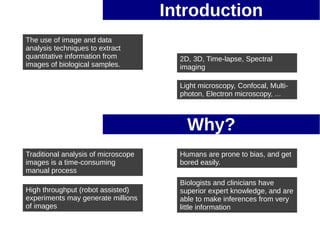
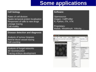
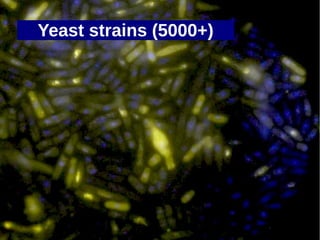
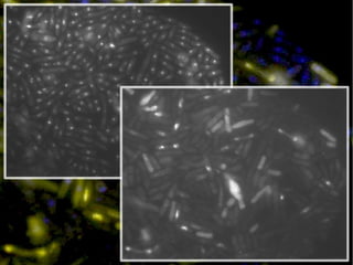
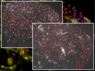
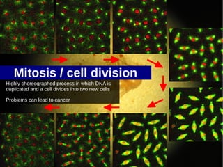
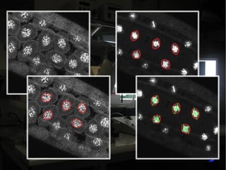
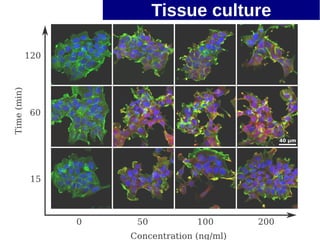
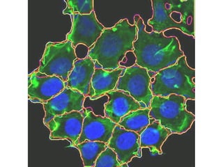
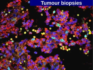
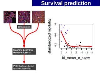
Quantitative Image Analysis
   in the Life Sciences


                 Simon Li
                 someone@pitpe.co.uk
                                   1
Introduction
The use of image and data
analysis techniques to extract
quantitative information from      2D, 3D, Time-lapse, Spectral
images of biological samples.      imaging

                                   Light microscopy, Confocal, Multi-
                                   photon, Electron microscopy, ...
Introduction
The use of image and data
analysis techniques to extract
quantitative information from          2D, 3D, Time-lapse, Spectral
images of biological samples.          imaging

                                       Light microscopy, Confocal, Multi-
                                       photon, Electron microscopy, ...


                                         Why?
Traditional analysis of microscope     Humans are prone to bias, and get
images is a time-consuming             bored easily.
manual process
                                       Biologists and clinicians have
High throughput (robot assisted)       superior expert knowledge, and are
experiments may generate millions      able to make inferences from very
of images                              little information
Some applications
Cell biology                           Software:

Rates of cell division                 Opensource:
Spatio-temporal protein localisation   ImageJ, CellProfiler
Responses of cells to new drugs        R, Python, ITK, VTK
Lineage tracing
Cell motility                          Proprietary:
                                       Matlab, MetaMorph, Volocity, ...
Disease detection and diagnosis

Analysis of tumour biopsies
Retinal blood vessel tracing
Cell counting

Analysis of fungal networks
3D reconstruction
Tracing embryo development
Yeast strains (5000+)
Mitosis / cell division
Highly choreographed process in which DNA is
duplicated and a cell divides into two new cells

Problems can lead to cancer
Tissue culture
Tumour biopsies
Survival prediction


    100 patients


Machine Learning:
Random forests


Potentially predictive
features identified
Simon Li
someone@pitpe.co.uk

Hydrahack 1.5: Bioimaging intro (higher contrast)

  • 1.
    Quantitative Image Analysis in the Life Sciences Simon Li someone@pitpe.co.uk 1
  • 2.
    Introduction The use ofimage and data analysis techniques to extract quantitative information from 2D, 3D, Time-lapse, Spectral images of biological samples. imaging Light microscopy, Confocal, Multi- photon, Electron microscopy, ...
  • 3.
    Introduction The use ofimage and data analysis techniques to extract quantitative information from 2D, 3D, Time-lapse, Spectral images of biological samples. imaging Light microscopy, Confocal, Multi- photon, Electron microscopy, ... Why? Traditional analysis of microscope Humans are prone to bias, and get images is a time-consuming bored easily. manual process Biologists and clinicians have High throughput (robot assisted) superior expert knowledge, and are experiments may generate millions able to make inferences from very of images little information
  • 4.
    Some applications Cell biology Software: Rates of cell division Opensource: Spatio-temporal protein localisation ImageJ, CellProfiler Responses of cells to new drugs R, Python, ITK, VTK Lineage tracing Cell motility Proprietary: Matlab, MetaMorph, Volocity, ... Disease detection and diagnosis Analysis of tumour biopsies Retinal blood vessel tracing Cell counting Analysis of fungal networks 3D reconstruction Tracing embryo development
  • 5.
  • 8.
    Mitosis / celldivision Highly choreographed process in which DNA is duplicated and a cell divides into two new cells Problems can lead to cancer
  • 10.
  • 12.
  • 13.
    Survival prediction 100 patients Machine Learning: Random forests Potentially predictive features identified
  • 14.