This power point presentation may help you identify the parts of the human eye, feel free to save and share it! PLEASE DO LIKE MY POWERPOINT PRESENTATION THANKIE! (All rights are reserved).
Note the structures through which light passes to reach the retina. W.pdfseamusschwaabl99557
Note the structures through which light passes to reach the retina. Write these structures in
order.
Solution
The order of light passage from outside till to retina:
Initially light enters cornea & sclera layers ---> pupil ---> circular Iris that surrounds pupil using
pupillary sphincter muscle --> light enters lens---> retinal layers ---> rods & cones
Explanation:
Neural layer is the inner layer; it is in contact with the vitreous humour. The neural layer
contains three distinct populations of cells namely,
From the outer (pigmented epithelial layer) to the inner (towards the vitreous humour), these
cells are arranged in the order of, photoreceptors, bipolar cells, ganglion cells.
Photoreceptor cells (rods and cones) are communicated with the nerve cells called bipolar cells.
These bipolar cells synapse with the ganglion cells, which transmit the impulses to the brain via
axons in the optic nerve.
Retina is the innermost nervous coat of eyeball, and it contains some special photo receptor
structures called “rods and cones.” These rods and cones receive the light and send impulses to
the optic nerve.
The inner nuclear layer of the retina contains oval shaped bipolar cells. The dendrites of bipolar
cells synapse with the photoreceptor cells, and axons of bipolar cells synapse with the ganglionic
cells.
Ganglion cells are multipolar cells present in the ganglion cell layer of retina; their axons are
present in the inner surface of the retina, forming the optic nerve. The dendrites of ganglion cells
synapse with the axons of bipolar cells. These ganglion cells transmit the impulses to the brain
via axons in the optic nerve..
THIS POWER POINT PRESENTATION IS TO GIVE READERS AN OVERVIEW ON THE ANATOMY AND PHYSIOLOGY OF THE EYE: STRUCTURES, FUNCTIONS OF EACH PART OF THE EYE, AS WELL AS THE PHYSIOLOGY ON HOW THE IMAGE IS CAPTURED IN THE EYE AND TRANSLATED BY THE BRAIN IN ORDER TO HAVE THE MEANINGFUL VIEW OF THE IMAGE.
Eye Anatomy and Physiology in b.pharm 1 semester and 2 semester of pharmacy education.
This slide help to more to make notes and easily read out this subject.
The visual pathway/visual system is the part of central nervous system which gives organisms the ability to process visual detail , as well as enabling the formation of several non-image photo response functions.
It detects interprets information from visible light to build a representation of the surrounding environment .
The visual system carries out a number of complex tasks , including the reception of light and the formation of monocular representations; the buildup of a nuclear binocular perception from a pair of two dimensional projections ; the identification and categorization of visual objects ; assessing distances to and between objects and guiding body movements in relation to the objects seen.
In anatomy, special senses are the senses that have organs specifically devoted to them such as vision, gustation, olfaction, audition, and equilibrioception. These senses have specialized organs that detect and process stimuli and send signals to the brain which lead to the perception of that stimulus.
The eye is composed of a series of lenses and spaces that give focus to images, just as a camera does. It is composed of the vitreous humor, aqueous humor, the crystalline lens, and the cornea, and each of these has its own refraction index (the average being 1.34, because of the content of these tissues).Functions
Pupil. Opens and closes in order to regulate and control the amount of light.
Iris. Controls light level similar to the aperture of a camera.
Sclera. Protects the outer coat.
Cornea. A thin membrane which provides 67% of the eye's focusing power.
Crystalline lens. ...
Conjunctive. ...
Aqueous humour.
Vitreous humour.
Acetabularia Information For Class 9 .docxvaibhavrinwa19
Acetabularia acetabulum is a single-celled green alga that in its vegetative state is morphologically differentiated into a basal rhizoid and an axially elongated stalk, which bears whorls of branching hairs. The single diploid nucleus resides in the rhizoid.
Francesca Gottschalk - How can education support child empowerment.pptxEduSkills OECD
Francesca Gottschalk from the OECD’s Centre for Educational Research and Innovation presents at the Ask an Expert Webinar: How can education support child empowerment?
The French Revolution, which began in 1789, was a period of radical social and political upheaval in France. It marked the decline of absolute monarchies, the rise of secular and democratic republics, and the eventual rise of Napoleon Bonaparte. This revolutionary period is crucial in understanding the transition from feudalism to modernity in Europe.
For more information, visit-www.vavaclasses.com
June 3, 2024 Anti-Semitism Letter Sent to MIT President Kornbluth and MIT Cor...Levi Shapiro
Letter from the Congress of the United States regarding Anti-Semitism sent June 3rd to MIT President Sally Kornbluth, MIT Corp Chair, Mark Gorenberg
Dear Dr. Kornbluth and Mr. Gorenberg,
The US House of Representatives is deeply concerned by ongoing and pervasive acts of antisemitic
harassment and intimidation at the Massachusetts Institute of Technology (MIT). Failing to act decisively to ensure a safe learning environment for all students would be a grave dereliction of your responsibilities as President of MIT and Chair of the MIT Corporation.
This Congress will not stand idly by and allow an environment hostile to Jewish students to persist. The House believes that your institution is in violation of Title VI of the Civil Rights Act, and the inability or
unwillingness to rectify this violation through action requires accountability.
Postsecondary education is a unique opportunity for students to learn and have their ideas and beliefs challenged. However, universities receiving hundreds of millions of federal funds annually have denied
students that opportunity and have been hijacked to become venues for the promotion of terrorism, antisemitic harassment and intimidation, unlawful encampments, and in some cases, assaults and riots.
The House of Representatives will not countenance the use of federal funds to indoctrinate students into hateful, antisemitic, anti-American supporters of terrorism. Investigations into campus antisemitism by the Committee on Education and the Workforce and the Committee on Ways and Means have been expanded into a Congress-wide probe across all relevant jurisdictions to address this national crisis. The undersigned Committees will conduct oversight into the use of federal funds at MIT and its learning environment under authorities granted to each Committee.
• The Committee on Education and the Workforce has been investigating your institution since December 7, 2023. The Committee has broad jurisdiction over postsecondary education, including its compliance with Title VI of the Civil Rights Act, campus safety concerns over disruptions to the learning environment, and the awarding of federal student aid under the Higher Education Act.
• The Committee on Oversight and Accountability is investigating the sources of funding and other support flowing to groups espousing pro-Hamas propaganda and engaged in antisemitic harassment and intimidation of students. The Committee on Oversight and Accountability is the principal oversight committee of the US House of Representatives and has broad authority to investigate “any matter” at “any time” under House Rule X.
• The Committee on Ways and Means has been investigating several universities since November 15, 2023, when the Committee held a hearing entitled From Ivory Towers to Dark Corners: Investigating the Nexus Between Antisemitism, Tax-Exempt Universities, and Terror Financing. The Committee followed the hearing with letters to those institutions on January 10, 202
Introduction to AI for Nonprofits with Tapp NetworkTechSoup
Dive into the world of AI! Experts Jon Hill and Tareq Monaur will guide you through AI's role in enhancing nonprofit websites and basic marketing strategies, making it easy to understand and apply.
Unit 8 - Information and Communication Technology (Paper I).pdfThiyagu K
This slides describes the basic concepts of ICT, basics of Email, Emerging Technology and Digital Initiatives in Education. This presentations aligns with the UGC Paper I syllabus.
How to Make a Field invisible in Odoo 17Celine George
It is possible to hide or invisible some fields in odoo. Commonly using “invisible” attribute in the field definition to invisible the fields. This slide will show how to make a field invisible in odoo 17.
Model Attribute Check Company Auto PropertyCeline George
In Odoo, the multi-company feature allows you to manage multiple companies within a single Odoo database instance. Each company can have its own configurations while still sharing common resources such as products, customers, and suppliers.
Normal Labour/ Stages of Labour/ Mechanism of LabourWasim Ak
Normal labor is also termed spontaneous labor, defined as the natural physiological process through which the fetus, placenta, and membranes are expelled from the uterus through the birth canal at term (37 to 42 weeks
2. Objectives:
• You will Identify different parts of
the human eye.
• Explain the function of the eye.
• Enumerate the parts of the eye.
• Describe each function of the
human eye.
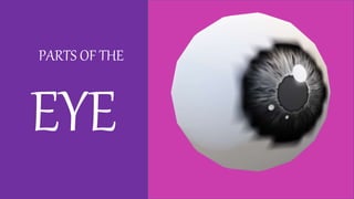
3. PARTS OF THE EYE
Vitreous Humor
Sclera
Iris
Optic nerve
Retina
Ciliary body
and muscle
5. Iris
The iris is the colored
part of the eye that
surrounds the pupil. It
regulates the amount
of light that enters the
eye.
6. Sclera
is the white outer coating of
the eye. It is tough, fibrous
tissue that extends from the
cornea (the clear front section
of the eye) to the optic nerve
at the back of the eye.
7. Optic
nerve
It's an extension of your
central nervous system, which
includes your brain and spine.
The optic nerve transmits
electrical impulses from your
eyes to your brain. Your brain
processes this sensory
information so that you can
see.
8. Retina
is a layer of photoreceptors
cells and glial cells within the
eye that captures incoming
photons and transmits them
along neuronal pathways as
both electrical and chemical
signals for the brain to
perceive a visual picture.
9. Ciliary body
and muscle
produces the fluid in the eye
called aqueous humor. It also
contains the ciliary muscle,
which changes the shape of
the lens when your eyes focus
on a near object. This process
is called accommodation.