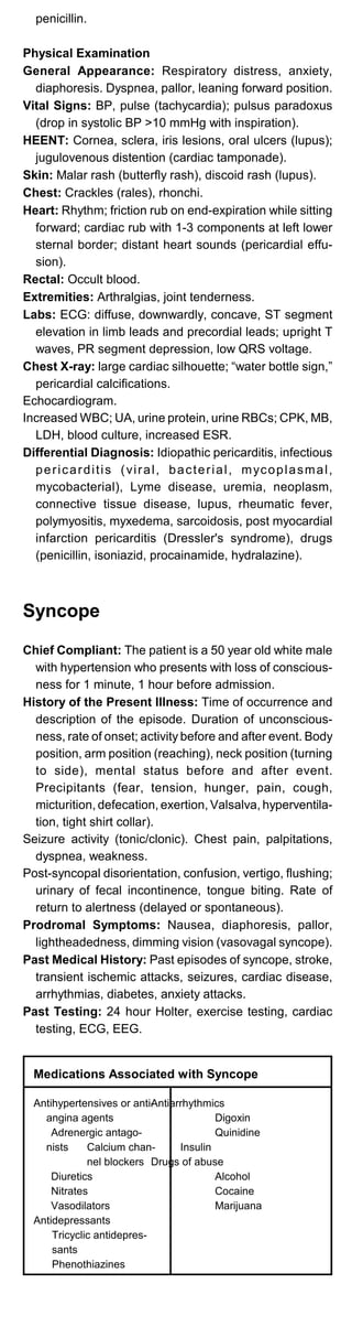
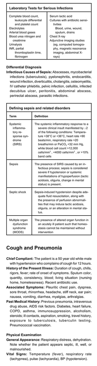
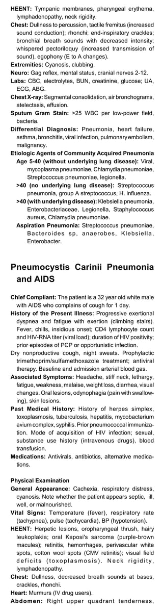
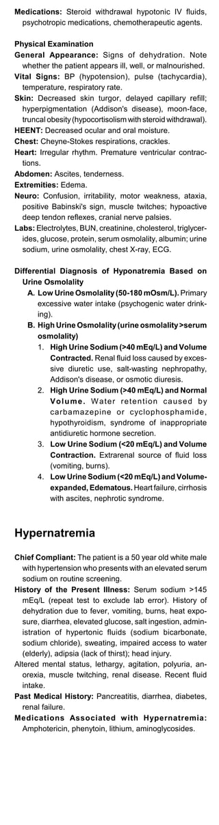
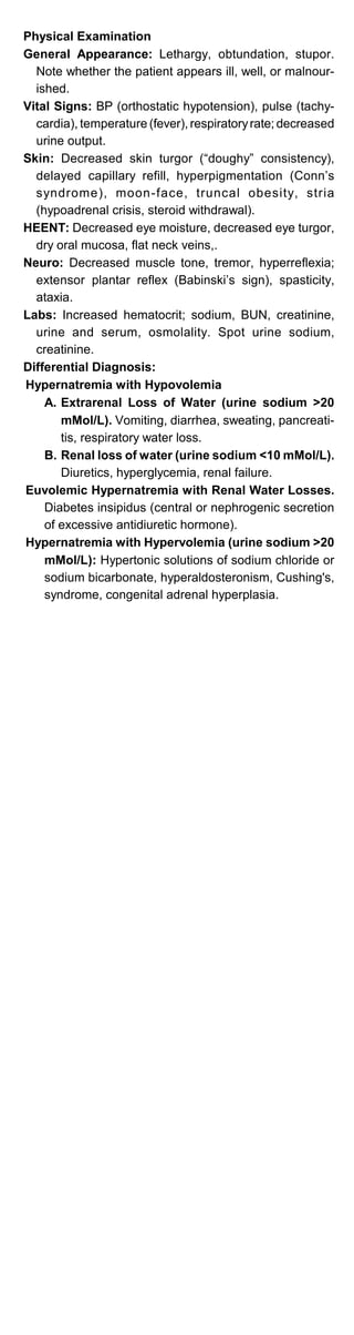
The document provides guidelines and templates for medical documentation including history and physical examinations, progress notes, procedure notes, discharge notes, prescription writing, and discharge summaries. It details the key information to include in each type of note such as patient identifying data, chief complaint, review of systems, physical exam findings, assessment, plan, and follow-up arrangements.

![calcifications.
Differential Diagnosis
Acute Infectious Diarrhea:Infectious diarrhea (salmonella,
shigella, E coli, Campylobacter, Bacillus cereus), enteric
viruses (rotavirus, Norwalk virus), traveler's diarrhea,
antibiotic-related diarrhea
Chronic Diarrhea:
Osmotic Diarrhea: Laxatives, lactulose, lactase defi
ciency(gastroenteritis, sprue), other disaccharidase
deficiencies, ingestion of mannitol, sorbitol, enteral
feeding.
Secretory Diarrhea: Bacterial enterotoxins, viral
infection; AIDS-associated disorders (mycobacterial,
HIV enteropathy), Zollinger-Ellison syndrome,
vasoactive intestinal peptide tumor, carcinoid
tumors, medullary thyroid cancer, colonic villus
adenoma.
Exudative Diarrhea: Bacterial infection, Clostridium
difficile, parasites, Crohn's disease, ulcerative
colitis, diverticulitis, intestinal ischemia, diverticulitis.
Diarrhea Secondary to Altered Intestinal Motility:
Diabetic gastroparesis, hyperthyroidism, laxatives,
cholinergics, irritable bowel syndrome, bacterial
overgrowth, constipation-related diarrhea.
Hematemesis and Upper Gastroin-
testinal Bleeding
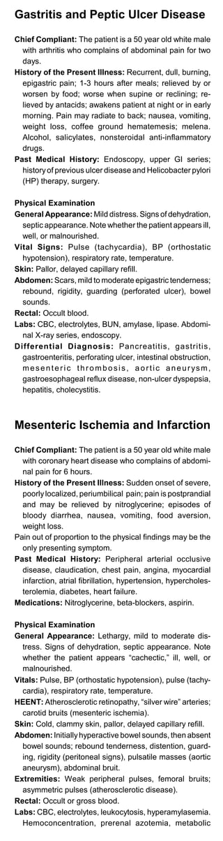
Chief Compliant: The patient is a 50 year old white male
with peptic ulcer disease who complains of emesis of
blood for 4 hours.
History of the Present Illness: Duration and frequency
of hematemesis (bright red blood, coffee ground
material), volume of blood, hematocrit. Forceful retching
prior to hematemesis (Mallory-Weiss tear).
Abdominal pain, melena, hematochezia (bright red blood
per rectum); history of peptic ulcer, esophagitis, prior
bleeding episodes. Nose bleeds, syncope,
lightheadedness, nausea.
Ingestion of alcohol. Weight loss, malaise, fatigue, an
orexia, early satiety, jaundice.
Nasogastric aspirate quantity and character; transfusions
given previously.
Past Medical History: Liver or renal disease, hepatic
encephalopathy, esophageal varices, aortic surgery.
Past Testing: X-ray studies, endoscopy. Past Treat-
ment: Endoscopic sclerotherapy, shunt surgery.
Medications: Aspirin, nonsteroidal anti-inflammatory
drugs, steroids, anticoagulants.
Family History: Liver disease or bleeding disorders.
Physical Examination
General Appearance: Pallor, diaphoresis, cold extremi
ties, confusion. Note whether the patient appears ill,
well, or malnourished.
Vital Signs: Supine and upright pulse and blood pressure
(orthostatic hypotension; resting tachycardia indicates
a 10% blood volume loss; postural hypotension indi
cates a 20-30% blood loss); oliguria (<20 mL of urine
per hour), temperature.
Skin: Delayed capillary refill, pallor, petechiae. Stigmata
of liver disease (jaundice, umbilical venous collaterals
[caput medusae], spider angiomas, parotid gland
hypertrophy). Hemorrhagic telangiectasia (Osler-
Weber-Rendu syndrome), abnormal pigmentation
(Peutz-Jeghers syndrome); purple-brown nodules
(Kaposi's sarcoma).
HEENT: Scleral pallor, oral telangiectasia, flat neck veins.
Chest: Gynecomastia (cirrhosis), breast masses (meta
static disease).
Heart: Systolic ejection murmur.
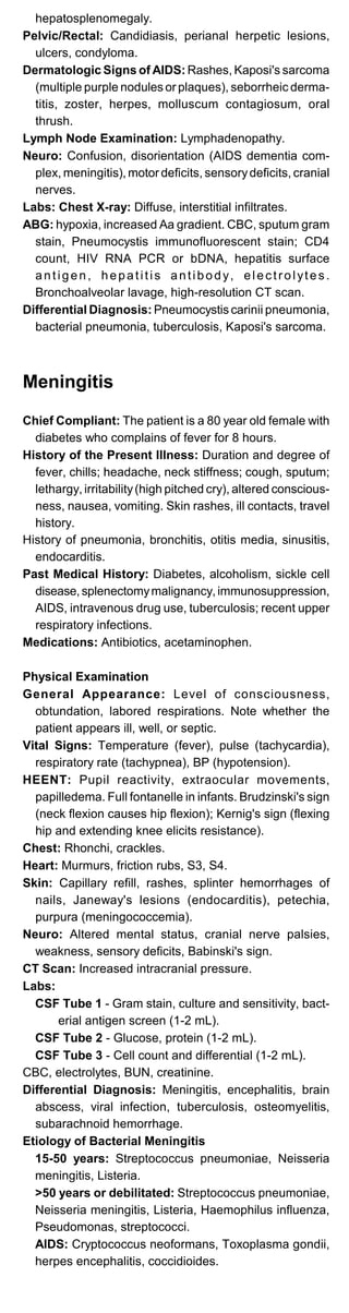
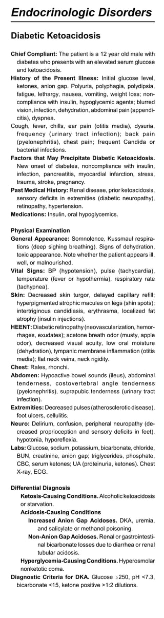
Abdomen: Scars, tenderness, rebound, masses,](https://image.slidesharecdn.com/historyandphysicalexam-160207003655/85/History-and-physical_exam-32-320.jpg)
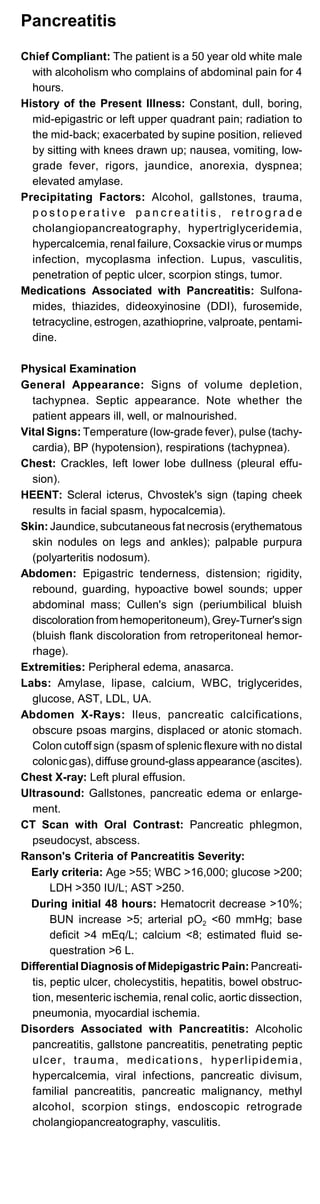
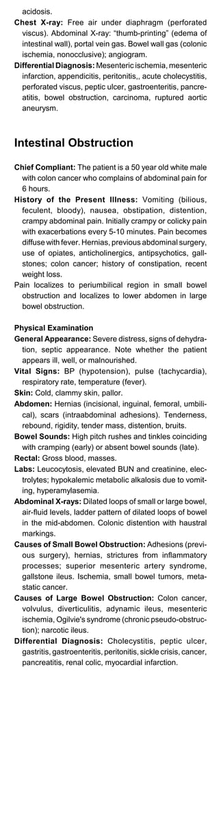

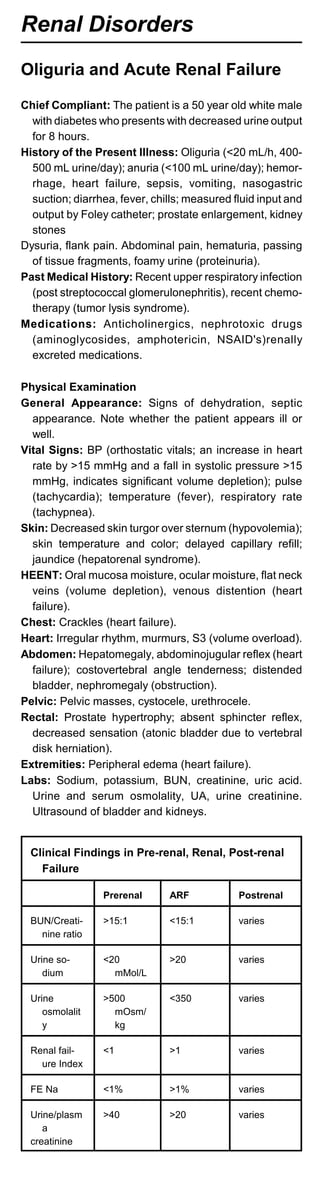
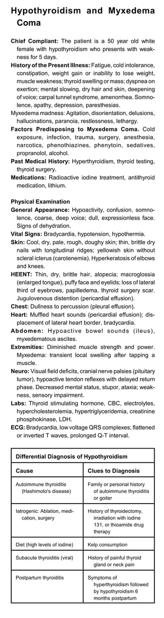
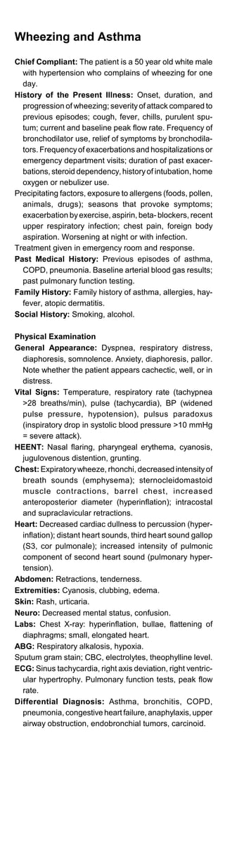
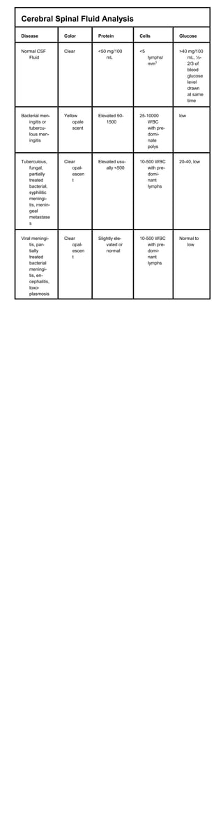
![Commonly Used Formulas
A-a gradient = [(PB-PH2O) FiO2 - PCO2/R] - PO2 arterial
= (713 x FiO2 - pCO2/0.8 ) -pO2 arterial
PB = 760 mmHg; PH2O = 47 mmHg ; R . 0.8
normal Aa gradient <10-15 mmHg (room air)
Arterial oxygen capacity =(Hgb(gm)/100 mL) x 1.36 mL
O2/gm Hgb
Arterial O2 content = 1.36(Hgb)(SaO2)+0.003(PaO2)= NL
20 vol%
O2 delivery = CO x arterial O2 content = NL 640-1000 mL
O2/min
Cardiac output = HR x stroke volume
Normal CO = 4-6 L/min
SVR = MAP - CVP x 80 = NL 800-1200 dyne/sec/cm2
COL/min
PVR = PA - PCWP x 80 = NL 45-120 dyne/sec/cm2
CO L/min
GFR mL/min = (140 - age) x wt in Kg
72 (males) x serum Cr (mg/dL)
85 (females) x serum Cr (mg/dL)
Normal creatinine clearance = 100-125 mL/min(males),
85-105(females)
Fractional excreted Na = U Na/ Serum Na x 100 = NL<1%
U Cr/ Serum Cr
Anion Gap = Na - (Cl + HCO3)
For each 100 mg/dL increase in glucose, Na+ decrease
by 1.6 mEq/L.
Ideal body weight males = 50 kg for first 5 feet of height +
2.3 kg for each additional inch.
Ideal body weight females = 45.5 kg for first 5 feet + 2.3
kg for each additional inch.
Basal energy expenditure (BEE):
Males=66 + (13.7 x actual weight Kg) + (5 x height
cm)-(6.8 x age)
Females= 655+(9.6 x actual weight Kg)+(1.7 x height
cm)-(4.7 x age)
Nitrogen Balance = Gm protein intake/6.25 - urine urea
nitrogen - (3-4
gm/d insensible loss)
Predicted Maximal Heart Rate = 220 - age
Normal ECG Intervals (sec)
PR 0.12-0.20
QRS 0.06-0.08
Heart rate/min Q-T
60 0.33-0.43
70 0.31-0.41
80 0.29-0.38
90 0.28-0.36
100 0.27-0.35](https://image.slidesharecdn.com/historyandphysicalexam-160207003655/85/History-and-physical_exam-68-320.jpg)