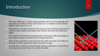
High Resolution
Electron Energy-
Loss
Spectroscopy
SHAHRUKH KHAN AP-037
M HAMZA ABBASI AP-065
1
High resolution electron energy loss spectroscopy
 High resolution electron energy loss spectroscopy (HREELS) is a tool
used in surface science.
 In contrast to other electron energy loss spectroscopies (EELS) HREELS
deals with small energy losses in the range of 10−3 eV to 1 eV.
2
 Measured physical quantities : the number of counts (intensity) as a
function of the energy loss of inelastic-ally backscattered low-energy
electrons.
 Information available : the vibrational modes, and the corresponding
energies, of species at the interface: substrate, physiosorbed adsorbate,
chemisorbed adsorbate, adsorbate–adsorbate interactions.
3
Introduction
 The main difficulty in surface characterization lies in the exceedingly low
population of surface atoms (1015 atoms cm−2) relative to that of bulk
species (1023 atoms cm−3).
 Experiments intended to examine the physical and chemical properties of
surfaces must employ techniques that interact only with the outermost
layers
 The majority of interfacial characterization techniques take advantage of
the unique surface sensitivity of low-energy electrons
 Such surface specificity arises because the mean free path of an electron
through a solid is dependent upon its kinetic energy.
 In particular, an electron whose kinetic energy is between 10 and 500 eV
can traverse no more than 2 nm within the solid
4
Examples of questions that can be
answered
 What molecular species are present on the surface?
 What is the mode of bonding between the adsorbate and the substrate?
 How does the surface chemical bond influence the vibrational properties
of the adsorbate?
 What is the surface coverage of the adsorbed species?
5
Region of Electrons
 A solid subjected to a beam of electrons of primary (incident) energy Ep
gives rise to backscattered (primary) and emitted (secondary) electrons.
The energy distribution of these electrons can be divided into four regions
based upon the origin of the scattered electrons. These regions
correspond to the following:
1. Secondary Electrons
2. Auger Electrons
3. In-Elastically Scattered Primary Electrons
4. Elastically Scattered Primary Electrons
6
Basis of HREELS
 Basis of high-resolution electron energy-loss spectroscopy (HREELS) are in-
Elastically Scattered Primary Electrons.
 Which are in-elastically scattered upon interactions with surface vibrational
states.
 Peaks associated with these electrons reside close to the elastic peak as
their energy losses are comparatively small
7
Loss Mechanism
 Almost all of the electrons incident at a solid surface undergo inelastic events that
cause them to be backscattered at energies lower than their initial or primary energy
𝐸 𝑝.
 If 𝐸𝑖 is the energy lost to the surface, peaks would appear in the spectrum at energies.
 ∆𝐸 = 𝐸 𝑝 − 𝐸𝑖
 Such peaks, commonly referred to as energy-loss peaks, originate from:
 1) Core level ionizations
 2) Valence-level excitations
 3) Vibrational excitations (0.50 eV)
8
Spectra Mechanism
 Two mechanisms give rise to high-resolution electron energy-loss (HREEL)
spectra.
 1) Dipole scattering
 2) Impact scattering
 3) Negative Ion resonance scattering
9
Dipole Scattering
 In dipole scattering, the incident electron may be treated similarly to an
electromagnetic (infrared) wave that interacts, at long range, with
oscillating dipoles created by the vibration of species at the surface.
 Electrons are scattered at long range (100 A) by oscillation of Coulombic
field of surface.
 Electron momentum perpendicular to surface is conserved.
10
Impact Scattering
 Electron is kinematic ally scattered at short range (few A) by atomic core.
 By potential of surface-modulated by nuclear vibrations
 Electron Scattered by wide range of angle.
 Electron momentum is not conserve-red in both parallel and perpendicular
direction.
11
Negative Ion Resonance Scattering
 Incident electron becomes transiently trapped in empty , high lying
molecular orbitals adsorbate.
 Vibrational feature intensity strongly depend on incident energy –
resonance.
12
HREELS Spectrum
 Zeros loss peak
Elastically Scattered Electrons
 Low loss region (<100ev)
Plasmon Resonance
 High loss region(>100ev)
Inner core Ionization
13
Instrumentation 14
Instrumentation
15
 Figure shows a schematic diagram of a first generation HREEL
spectrometer.
 In this model, the energy of incident electrons can be varied from 1 to 10
eV.
 To afford high-resolution, energy monochromat ion and analysis are done
with a cylindrical mirror analyzer.
 Owing to extremely low signals (10−10 A), continuous dynode electron
multiplier detectors are employed
Instrumentation 16
Instrumentation
 This model consists of an electron gun, a two-stage monochromator, a
single-stage energy analyzer, and a channel electron multiplier detector
 Both energy monochromation and energy analysis are carried out with
127◦ cylindrical deflection analyzers
17
Working Principle
 The incident electrons have an initial energy spread of 0.3 eV.
 The two- stage monochromator serves to narrow the energy spread to <1
MeV and generate a highly monoenergetic beam of low-energy electrons
(typically 1 to 10 eV).
 A zoom lens system focuses and accelerates the electron beam onto the
sample.
18
Working Principle
 The backscattered electrons are passed through a separate zoom lens for
focusing and deceleration before they are sorted out by a single-stage
energy analyzer into the detector
 The signal is fed to a preamplifier as pulses for electron counting
 The analyzer is designed to be rotatable from 0◦ to 78◦ for off-specular
(impact-scattering) measurements
19
Major advantages
 Extremely sensitive
 Limit of detection is 0.01% of a monolayer
 Broad spectral range; typically, 50 (6.2 MeV) to 4000 cm−1 (496 MeV)
 Impact-scattering mechanism not restricted by selection rules; in
combination with dipole scattering, all vibrational
 Modes may be discernible
 Nondestructive.
20
Major disadvantages
 Requiring an ultrahigh vacuum environment
 Backscattering may be inhibited by cationic species.
 The sample surface must be conductive and relatively smooth
21
Thank You.
22

High resolution electron energy loss spectroscopy

  • 1.
  • 2.
    High resolution electronenergy loss spectroscopy  High resolution electron energy loss spectroscopy (HREELS) is a tool used in surface science.  In contrast to other electron energy loss spectroscopies (EELS) HREELS deals with small energy losses in the range of 10−3 eV to 1 eV. 2
  • 3.
     Measured physicalquantities : the number of counts (intensity) as a function of the energy loss of inelastic-ally backscattered low-energy electrons.  Information available : the vibrational modes, and the corresponding energies, of species at the interface: substrate, physiosorbed adsorbate, chemisorbed adsorbate, adsorbate–adsorbate interactions. 3
  • 4.
    Introduction  The maindifficulty in surface characterization lies in the exceedingly low population of surface atoms (1015 atoms cm−2) relative to that of bulk species (1023 atoms cm−3).  Experiments intended to examine the physical and chemical properties of surfaces must employ techniques that interact only with the outermost layers  The majority of interfacial characterization techniques take advantage of the unique surface sensitivity of low-energy electrons  Such surface specificity arises because the mean free path of an electron through a solid is dependent upon its kinetic energy.  In particular, an electron whose kinetic energy is between 10 and 500 eV can traverse no more than 2 nm within the solid 4
  • 5.
    Examples of questionsthat can be answered  What molecular species are present on the surface?  What is the mode of bonding between the adsorbate and the substrate?  How does the surface chemical bond influence the vibrational properties of the adsorbate?  What is the surface coverage of the adsorbed species? 5
  • 6.
    Region of Electrons A solid subjected to a beam of electrons of primary (incident) energy Ep gives rise to backscattered (primary) and emitted (secondary) electrons. The energy distribution of these electrons can be divided into four regions based upon the origin of the scattered electrons. These regions correspond to the following: 1. Secondary Electrons 2. Auger Electrons 3. In-Elastically Scattered Primary Electrons 4. Elastically Scattered Primary Electrons 6
  • 7.
    Basis of HREELS Basis of high-resolution electron energy-loss spectroscopy (HREELS) are in- Elastically Scattered Primary Electrons.  Which are in-elastically scattered upon interactions with surface vibrational states.  Peaks associated with these electrons reside close to the elastic peak as their energy losses are comparatively small 7
  • 8.
    Loss Mechanism  Almostall of the electrons incident at a solid surface undergo inelastic events that cause them to be backscattered at energies lower than their initial or primary energy 𝐸 𝑝.  If 𝐸𝑖 is the energy lost to the surface, peaks would appear in the spectrum at energies.  ∆𝐸 = 𝐸 𝑝 − 𝐸𝑖  Such peaks, commonly referred to as energy-loss peaks, originate from:  1) Core level ionizations  2) Valence-level excitations  3) Vibrational excitations (0.50 eV) 8
  • 9.
    Spectra Mechanism  Twomechanisms give rise to high-resolution electron energy-loss (HREEL) spectra.  1) Dipole scattering  2) Impact scattering  3) Negative Ion resonance scattering 9
  • 10.
    Dipole Scattering  Indipole scattering, the incident electron may be treated similarly to an electromagnetic (infrared) wave that interacts, at long range, with oscillating dipoles created by the vibration of species at the surface.  Electrons are scattered at long range (100 A) by oscillation of Coulombic field of surface.  Electron momentum perpendicular to surface is conserved. 10
  • 11.
    Impact Scattering  Electronis kinematic ally scattered at short range (few A) by atomic core.  By potential of surface-modulated by nuclear vibrations  Electron Scattered by wide range of angle.  Electron momentum is not conserve-red in both parallel and perpendicular direction. 11
  • 12.
    Negative Ion ResonanceScattering  Incident electron becomes transiently trapped in empty , high lying molecular orbitals adsorbate.  Vibrational feature intensity strongly depend on incident energy – resonance. 12
  • 13.
    HREELS Spectrum  Zerosloss peak Elastically Scattered Electrons  Low loss region (<100ev) Plasmon Resonance  High loss region(>100ev) Inner core Ionization 13
  • 14.
  • 15.
    Instrumentation 15  Figure showsa schematic diagram of a first generation HREEL spectrometer.  In this model, the energy of incident electrons can be varied from 1 to 10 eV.  To afford high-resolution, energy monochromat ion and analysis are done with a cylindrical mirror analyzer.  Owing to extremely low signals (10−10 A), continuous dynode electron multiplier detectors are employed
  • 16.
  • 17.
    Instrumentation  This modelconsists of an electron gun, a two-stage monochromator, a single-stage energy analyzer, and a channel electron multiplier detector  Both energy monochromation and energy analysis are carried out with 127◦ cylindrical deflection analyzers 17
  • 18.
    Working Principle  Theincident electrons have an initial energy spread of 0.3 eV.  The two- stage monochromator serves to narrow the energy spread to <1 MeV and generate a highly monoenergetic beam of low-energy electrons (typically 1 to 10 eV).  A zoom lens system focuses and accelerates the electron beam onto the sample. 18
  • 19.
    Working Principle  Thebackscattered electrons are passed through a separate zoom lens for focusing and deceleration before they are sorted out by a single-stage energy analyzer into the detector  The signal is fed to a preamplifier as pulses for electron counting  The analyzer is designed to be rotatable from 0◦ to 78◦ for off-specular (impact-scattering) measurements 19
  • 20.
    Major advantages  Extremelysensitive  Limit of detection is 0.01% of a monolayer  Broad spectral range; typically, 50 (6.2 MeV) to 4000 cm−1 (496 MeV)  Impact-scattering mechanism not restricted by selection rules; in combination with dipole scattering, all vibrational  Modes may be discernible  Nondestructive. 20
  • 21.
    Major disadvantages  Requiringan ultrahigh vacuum environment  Backscattering may be inhibited by cationic species.  The sample surface must be conductive and relatively smooth 21
  • 22.