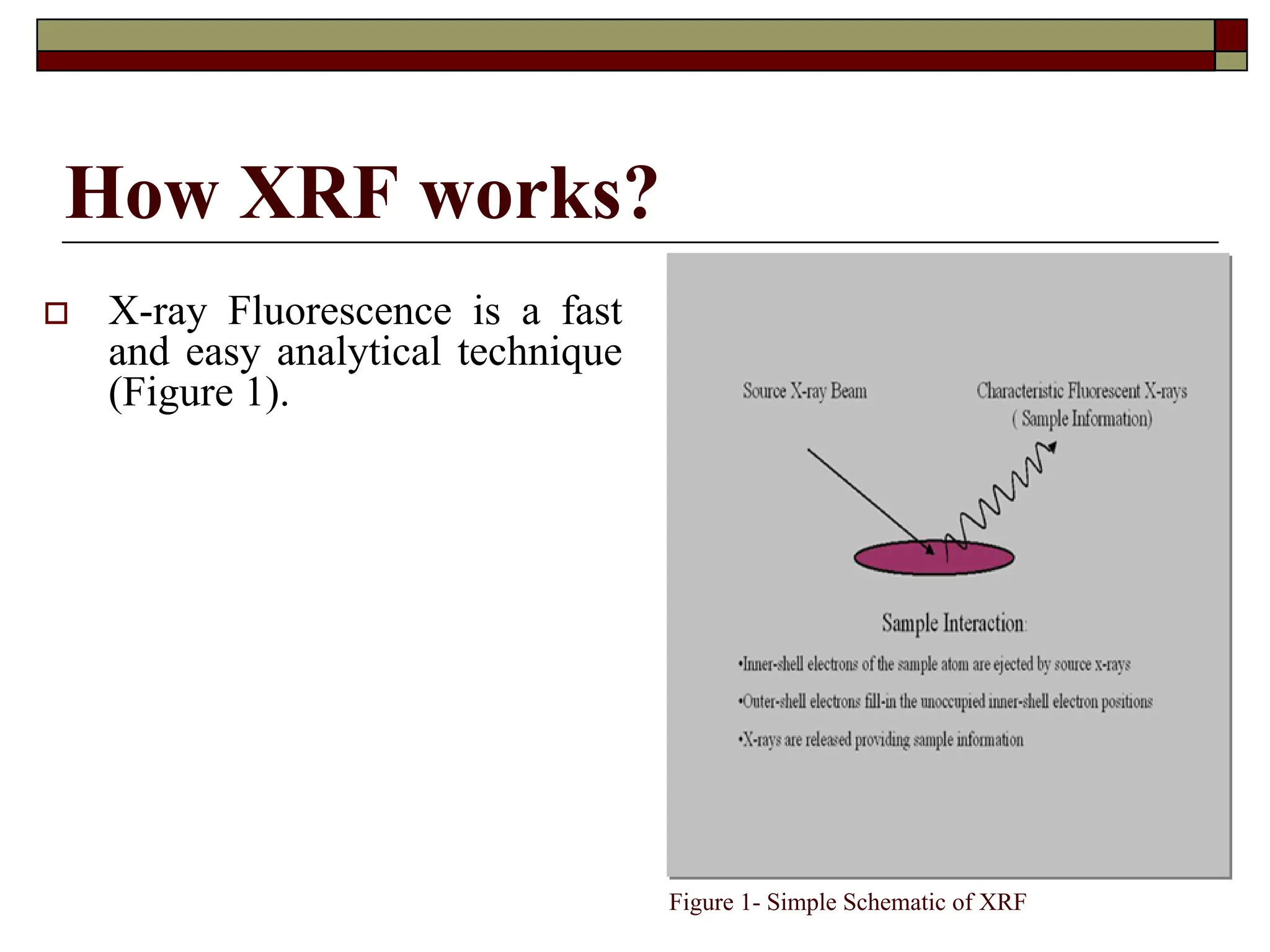
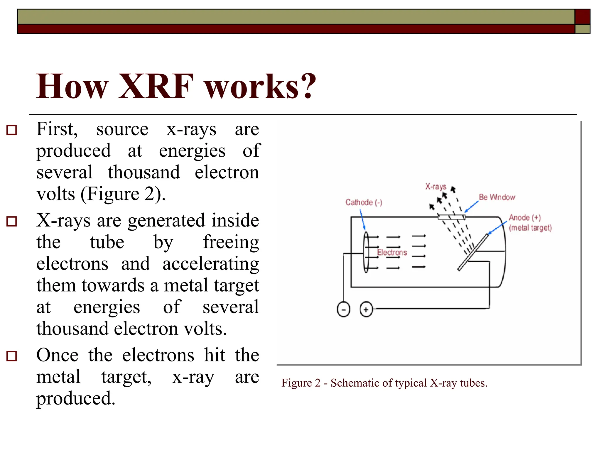
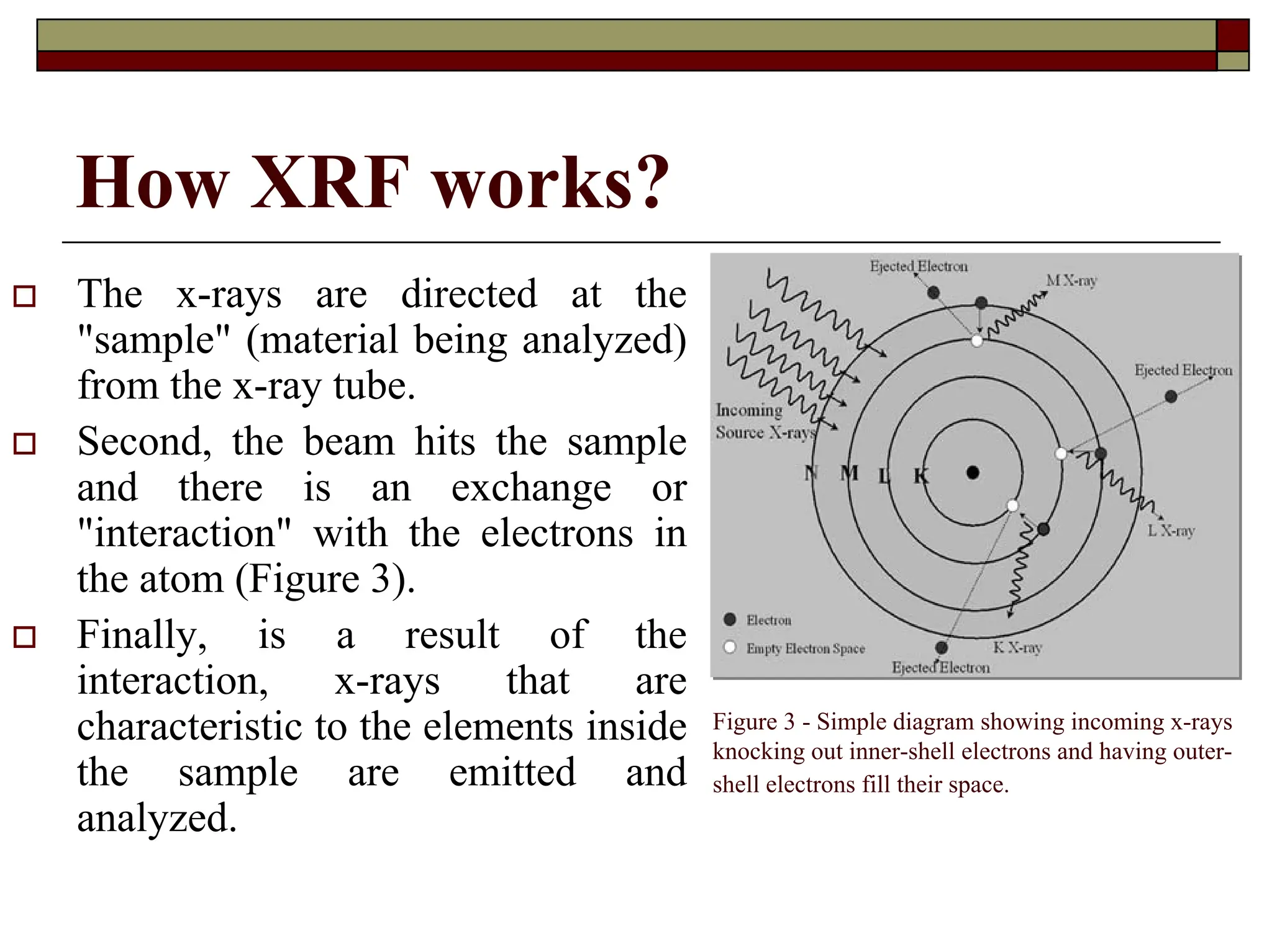
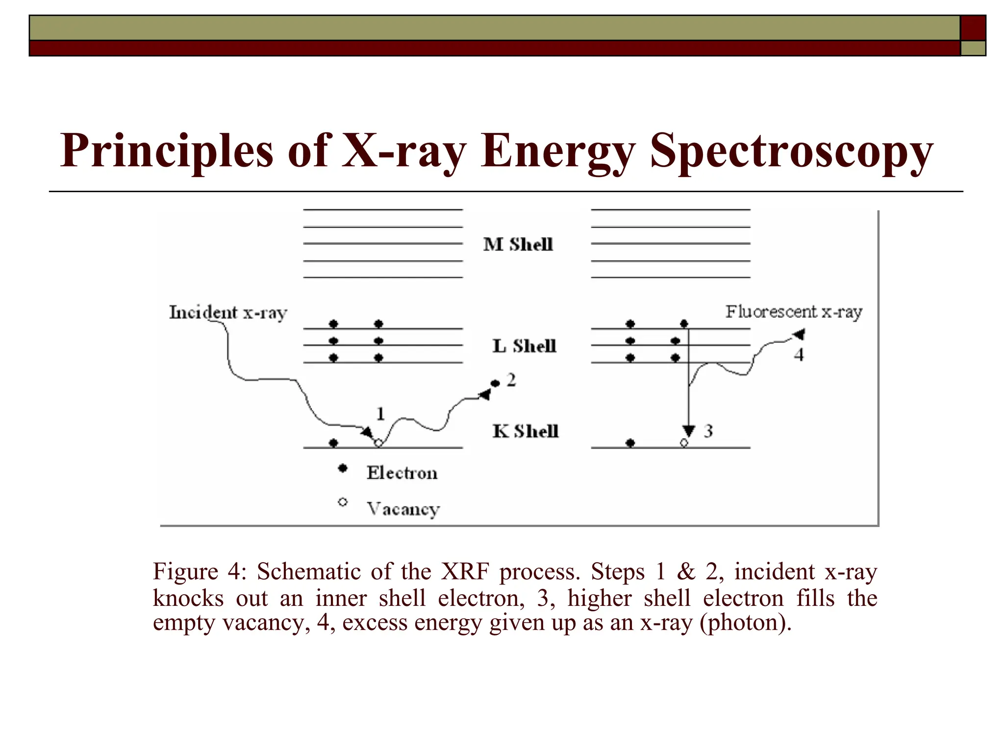
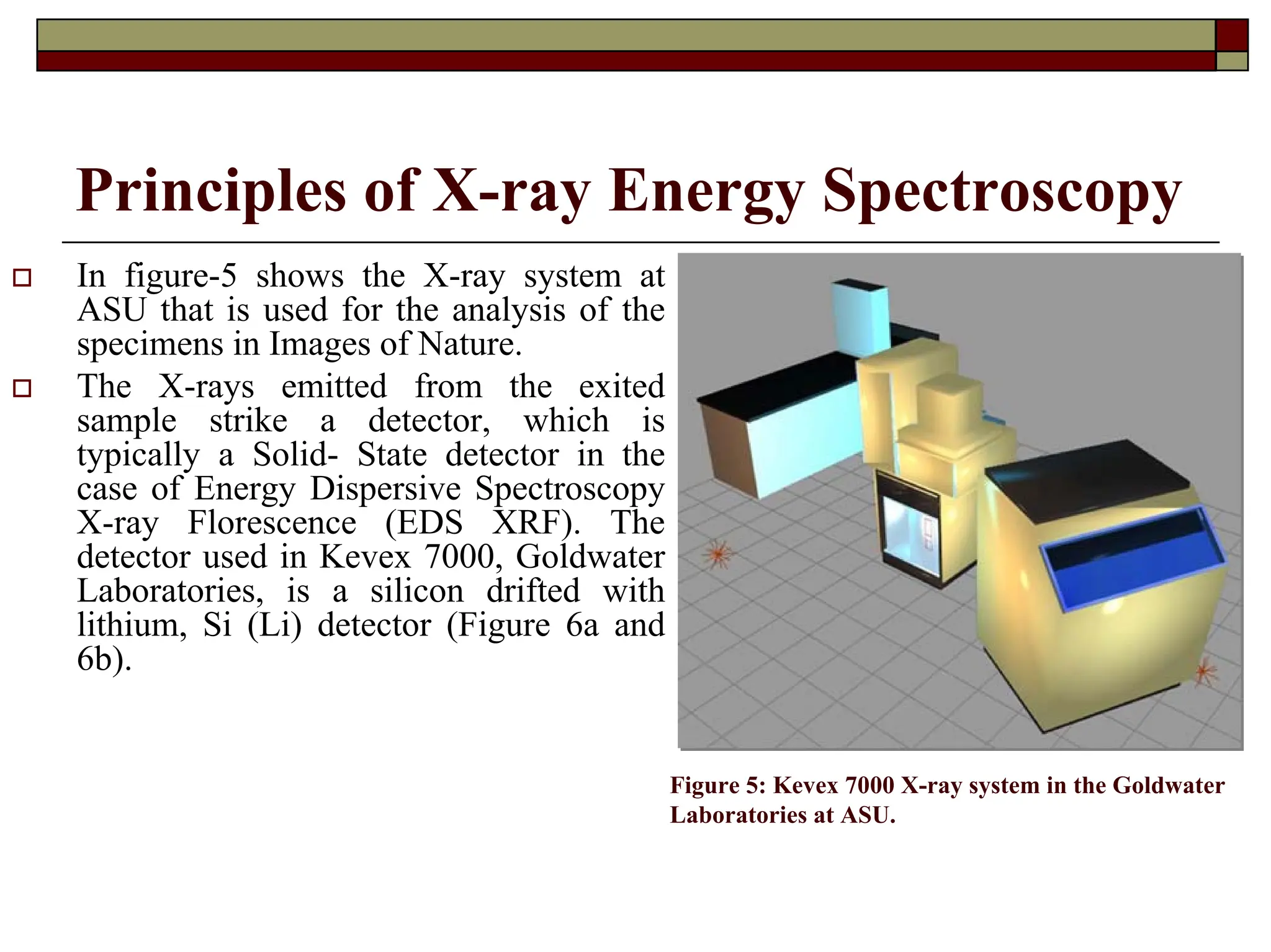
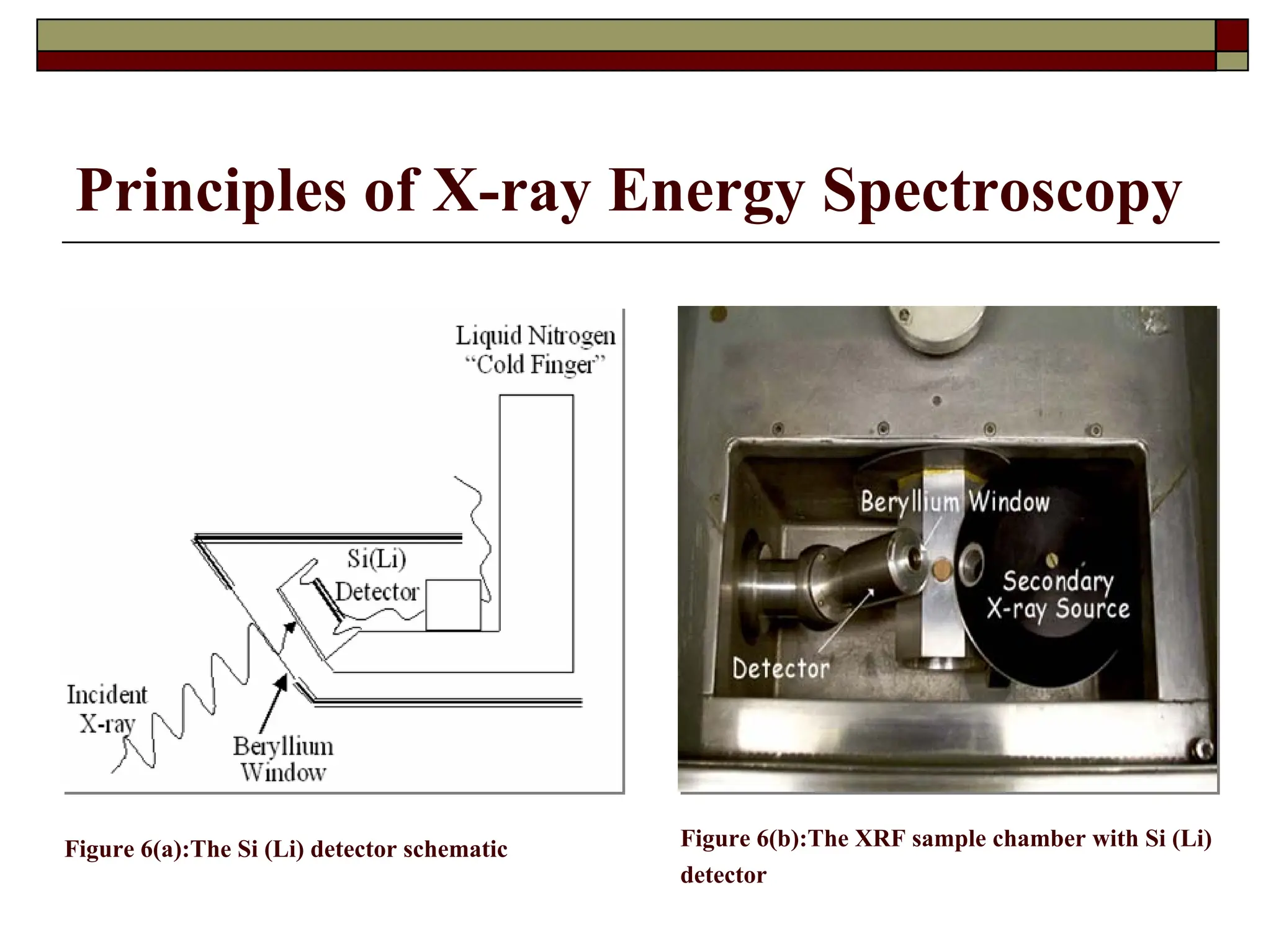
Basic Concepts of X
Basic Concepts of X-
-ray
ray
Fluorescence
Fluorescence
by Miguel Santiago,
Scientific Instrumentation Specialist
University of Puerto Rico
University of Puerto Rico –
– Mayag
Mayagü
üez
ez Campus
Campus
Department of Geology
X
X-
-ray / Microanalysis Laboratory
ray / Microanalysis Laboratory
F
F -
- 304
304
Introduction
† X-ray Fluorescence (XRF) is a nondestructive method for the elemental
analysis of solids and liquids.
† The sample is irradiated by an intense x-ray beam, which causes the emission
of fluorescent x-rays.
† The emitted x-rays can either be detected using energy dispersive or
wavelength dispersive detector.
† Either the energies or wavelengths of the emitted x-rays are used
to identify the elements present in the sample while the concentrations
(how much) of the elements are determined by the intensity of the x-rays.
† XRF is a bulk analysis technique with the depth of sample analyzed varying
from less than 1 mm to 1 cm depending on the energy of the emitted x-ray and
the sample composition.
† The elements commonly detected range from sodium to uranium.
† Lighter elements from boron to fluorine may also be detected.
How XRF works?
† X-ray Fluorescence is a fast
and easy analytical technique
(Figure 1).
Figure 1- Simple Schematic of XRF
How XRF works?
† First, source x-rays are
produced at energies of
several thousand electron
volts (Figure 2).
† X-rays are generated inside
the tube by freeing
electrons and accelerating
them towards a metal target
at energies of several
thousand electron volts.
† Once the electrons hit the
metal target, x-ray are
produced.
Figure 2 - Schematic of typical X-ray tubes.
How XRF works?
† The x-rays are directed at the
"sample" (material being analyzed)
from the x-ray tube.
† Second, the beam hits the sample
and there is an exchange or
"interaction" with the electrons in
the atom (Figure 3).
† Finally, is a result of the
interaction, x-rays that are
characteristic to the elements inside
the sample are emitted and
analyzed.
Figure 3 - Simple diagram showing incoming x-rays
knocking out inner-shell electrons and having outer-
shell electrons fill their space.
How XRF works?
† The outer-shell electrons are bound to the
atom with less energy than the inner-shell
electrons.
† When the outer-shell electron fills the inner-
shell vacancies (empty spaces) the energy
difference is accounted for by the energy of
the emitted x-ray and therefore energy is
conserved.
Principles of X-ray Energy Spectroscopy
† X-ray Energy Spectroscopy (XES) is a technique for
rapid, simultaneous multi-element analysis. When
excited by an appropriate source, a sample will emit x-
rays of energies that are characteristic for the elements
composing the sample.
† By measuring the energies of x-rays that are emitted
from an excited sample and counting the number of x-
rays of each energy, XRF allows us to identify which
elements are present in a sample, and also determine the
relative concentration of these elements within the
sample.
Principles of X-ray Energy Spectroscopy
† The sample can be excited by an x-ray source, by a
radioisotope source, or by an electron beam.
Specifically, in X-ray Florescence Spectroscopy
(XRF), generated x-rays are used as the primacy
source. The primary source "excites" the sample by
removing / "knocking out“ tightly bound electrons
from the inner-shell orbital of the excited atom in the
sample.
† Relaxation of the excited atom to the ground state is
accompanied by the emission of fluorescent x-rays.
Principles of X-ray Energy Spectroscopy
Figure 4: Schematic of the XRF process. Steps 1 & 2, incident x-ray
knocks out an inner shell electron, 3, higher shell electron fills the
empty vacancy, 4, excess energy given up as an x-ray (photon).
Principles of X-ray Energy Spectroscopy
† In figure-5 shows the X-ray system at
ASU that is used for the analysis of the
specimens in Images of Nature.
† The X-rays emitted from the exited
sample strike a detector, which is
typically a Solid- State detector in the
case of Energy Dispersive Spectroscopy
X-ray Florescence (EDS XRF). The
detector used in Kevex 7000, Goldwater
Laboratories, is a silicon drifted with
lithium, Si (Li) detector (Figure 6a and
6b).
Figure 5: Kevex 7000 X-ray system in the Goldwater
Laboratories at ASU.
Principles of X-ray Energy Spectroscopy
Figure 6(a):The Si (Li) detector schematic Figure 6(b):The XRF sample chamber with Si (Li)
detector
Principles of X-ray Energy Spectroscopy
† The elements that are present in a sample can be
identified by the location of their energy peaks along
the horizontal axis. Since, in theory, the number of
x-rays produced is proportional to the number of
atoms present in a sample, quantitative elemental
concentrations can be determined from the net
intensities of the energy peak.
Sample Information
† Most samples can be analyzed "as is" for most qualitative analysis and
quantitative analysis.
† Some samples may require sample preparation such as pelletizing or casting as a
fusion disk.
† Vacuum compatibility of the sample depends upon the element to be detected.
† The sample need not be conductive or insulating.
† For quantitative analysis the samples should be homogeneous with a flat smooth
surface.
† Quantitative and qualitative elemental information can be obtained from a bulk
infinite thickness sample.
† Qualitative elemental concentrations in solids/liquids are of the order of parts per
million weight percent (ppm wt%) range and qualitative elemental data from
boron to uranium can be acquired.
† The quality of these results depends on the calibration standards.
References
† The Kevex 7000 User's Manual, Kevex Corp. California, 1979.
† www.analytical.phylips.com/products/xrf/2830/
† Practical X-Ray Spectrometry by R. Jenkins and J.L. de Vries,
published by The Macmillan Press Ltd.
† Y. Yoneda and T. Horiuchi, Rev. Science Instruments 42:1069
(1971).
† H. Aiginger and P. Wobrauschek, Nuclear Instrument Methods
114: 157 (1974).
† Grieken, Rene E. and Andzrej Markowski. Handbook of X-ray
Spectrometry: Methods and Techniques. Marcel Dekker, Inc.,
New York (19993) P. 453.

Presentation on environmental science notes

  • 1.
    Basic Concepts ofX Basic Concepts of X- -ray ray Fluorescence Fluorescence by Miguel Santiago, Scientific Instrumentation Specialist University of Puerto Rico University of Puerto Rico – – Mayag Mayagü üez ez Campus Campus Department of Geology Department of Geology X X- -ray / Microanalysis Laboratory ray / Microanalysis Laboratory F F - - 304 304
  • 2.
    Introduction † X-ray Fluorescence(XRF) is a nondestructive method for the elemental analysis of solids and liquids. † The sample is irradiated by an intense x-ray beam, which causes the emission of fluorescent x-rays. † The emitted x-rays can either be detected using energy dispersive or wavelength dispersive detector. † Either the energies or wavelengths of the emitted x-rays are used to identify the elements present in the sample while the concentrations (how much) of the elements are determined by the intensity of the x-rays. † XRF is a bulk analysis technique with the depth of sample analyzed varying from less than 1 mm to 1 cm depending on the energy of the emitted x-ray and the sample composition. † The elements commonly detected range from sodium to uranium. † Lighter elements from boron to fluorine may also be detected.
  • 3.
    How XRF works? †X-ray Fluorescence is a fast and easy analytical technique (Figure 1). Figure 1- Simple Schematic of XRF
  • 4.
    How XRF works? †First, source x-rays are produced at energies of several thousand electron volts (Figure 2). † X-rays are generated inside the tube by freeing electrons and accelerating them towards a metal target at energies of several thousand electron volts. † Once the electrons hit the metal target, x-ray are produced. Figure 2 - Schematic of typical X-ray tubes.
  • 5.
    How XRF works? †The x-rays are directed at the "sample" (material being analyzed) from the x-ray tube. † Second, the beam hits the sample and there is an exchange or "interaction" with the electrons in the atom (Figure 3). † Finally, is a result of the interaction, x-rays that are characteristic to the elements inside the sample are emitted and analyzed. Figure 3 - Simple diagram showing incoming x-rays knocking out inner-shell electrons and having outer- shell electrons fill their space.
  • 6.
    How XRF works? †The outer-shell electrons are bound to the atom with less energy than the inner-shell electrons. † When the outer-shell electron fills the inner- shell vacancies (empty spaces) the energy difference is accounted for by the energy of the emitted x-ray and therefore energy is conserved.
  • 7.
    Principles of X-rayEnergy Spectroscopy † X-ray Energy Spectroscopy (XES) is a technique for rapid, simultaneous multi-element analysis. When excited by an appropriate source, a sample will emit x- rays of energies that are characteristic for the elements composing the sample. † By measuring the energies of x-rays that are emitted from an excited sample and counting the number of x- rays of each energy, XRF allows us to identify which elements are present in a sample, and also determine the relative concentration of these elements within the sample.
  • 8.
    Principles of X-rayEnergy Spectroscopy † The sample can be excited by an x-ray source, by a radioisotope source, or by an electron beam. Specifically, in X-ray Florescence Spectroscopy (XRF), generated x-rays are used as the primacy source. The primary source "excites" the sample by removing / "knocking out“ tightly bound electrons from the inner-shell orbital of the excited atom in the sample. † Relaxation of the excited atom to the ground state is accompanied by the emission of fluorescent x-rays.
  • 9.
    Principles of X-rayEnergy Spectroscopy Figure 4: Schematic of the XRF process. Steps 1 & 2, incident x-ray knocks out an inner shell electron, 3, higher shell electron fills the empty vacancy, 4, excess energy given up as an x-ray (photon).
  • 10.
    Principles of X-rayEnergy Spectroscopy † In figure-5 shows the X-ray system at ASU that is used for the analysis of the specimens in Images of Nature. † The X-rays emitted from the exited sample strike a detector, which is typically a Solid- State detector in the case of Energy Dispersive Spectroscopy X-ray Florescence (EDS XRF). The detector used in Kevex 7000, Goldwater Laboratories, is a silicon drifted with lithium, Si (Li) detector (Figure 6a and 6b). Figure 5: Kevex 7000 X-ray system in the Goldwater Laboratories at ASU.
  • 11.
    Principles of X-rayEnergy Spectroscopy Figure 6(a):The Si (Li) detector schematic Figure 6(b):The XRF sample chamber with Si (Li) detector
  • 12.
    Principles of X-rayEnergy Spectroscopy † The elements that are present in a sample can be identified by the location of their energy peaks along the horizontal axis. Since, in theory, the number of x-rays produced is proportional to the number of atoms present in a sample, quantitative elemental concentrations can be determined from the net intensities of the energy peak.
  • 13.
    Sample Information † Mostsamples can be analyzed "as is" for most qualitative analysis and quantitative analysis. † Some samples may require sample preparation such as pelletizing or casting as a fusion disk. † Vacuum compatibility of the sample depends upon the element to be detected. † The sample need not be conductive or insulating. † For quantitative analysis the samples should be homogeneous with a flat smooth surface. † Quantitative and qualitative elemental information can be obtained from a bulk infinite thickness sample. † Qualitative elemental concentrations in solids/liquids are of the order of parts per million weight percent (ppm wt%) range and qualitative elemental data from boron to uranium can be acquired. † The quality of these results depends on the calibration standards.
  • 14.
    References † The Kevex7000 User's Manual, Kevex Corp. California, 1979. † www.analytical.phylips.com/products/xrf/2830/ † Practical X-Ray Spectrometry by R. Jenkins and J.L. de Vries, published by The Macmillan Press Ltd. † Y. Yoneda and T. Horiuchi, Rev. Science Instruments 42:1069 (1971). † H. Aiginger and P. Wobrauschek, Nuclear Instrument Methods 114: 157 (1974). † Grieken, Rene E. and Andzrej Markowski. Handbook of X-ray Spectrometry: Methods and Techniques. Marcel Dekker, Inc., New York (19993) P. 453.