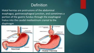
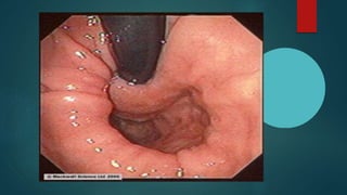
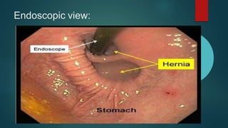
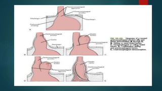
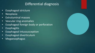
Hiatal Hernia
Submitted by: Faizan Ali
Submitted to: DR Hafiz Manzoor
Outline
 Definition
 Pathophysiology
 Diagnosis
 Physical examination
 Diagnostic imaging
 Differential diagnosis
 Surgical treatment
 Post operative care
 Complications
 Prognosis
What is hiatal hernia?
• The hiatus is an opening in the diaphragm
which is the muscular wall separating the chest
cavity from the abdomen
• Normally, the esophagus (food pipe) goes
through the hiatus and attaches to the stomach
• In a hiatal hernia (also called hiatus hernia) the
stomach bulges up into the chest through that
opening
Definition
Hiatal hernias are protrusions of the abdominal
esophagus, gastroesophageal junction, and sometimes a
portion of the gastric fundus through the esophageal
hiatus into the caudal mediastinum cranial to the
diaphragm
PATHOPYSIOLOGY
 Hiatal hernias usually are caused by congenital
abnormalities of the hiatus that allow cranial movement of
the abdominal esophagus and stomach
 The phrenicoesophageal ligament is stretched and allows
the gastroesophageal junction to be displaced through the
hiatus into the caudal mediastinum
 Malpositioning or lack of support of the gastroesophageal
sphincter reduces gastroesophageal sphincter pressure
and leads to gastroesophageal reflux
 Gastroesophageal reflux and subsequent esophagitis and
megaesophagus are responsible for most of clinical signs
 Hiatal hernia occasionally occurs secondary to trauma and
has occurred concurrently with severe respiratory distress
 Trauma may damage diaphragmatic nerves and muscles,
resulting in hiatal laxity and subsequent herniation
 In patients with upper respiratory obstruction, reduced
intrathoracic pressure during inspiration has been theorized
to contribute to esophageal reflux and visceral herniation
 Hiatal hernia has been reported with tetanus
What causes hiatal hernia?
• Most of the time, the cause is not known
• An animal may be born with a larger hiatal opening
• Increased pressure in the abdomen such as from
pregnancy, obesity, coughing, or straining during
bowel movements may also play a role
DIAGNOSIS
Signalment:
Hiatal hernias may occur in a variety of dog and cat breeds;
however, males and Shar Peis and English Bulldogs appear to be predisposed to
this condition. Most symptomatic animals have signs relating to congenital hiatal
hernia before reaching 1 year of age, although diagnosis may occur later
Patients with acquired hernias may develop signs at any age
HISTORY:
Regurgitation is the primary clinical sign in symptomatic individuals,
but many patients are asymptomatic. Other signs may include vomiting,
hypersalivation, dysphagia, respiratory distress, hematemesis, anorexia, and
weight loss
Severe dyspnea or trauma may have occurred
Physical examination
• Affected patients may be thin on physical examination
• Abnormal lung sounds are auscultated
• Aspiration pneumonia
• Hunched back
• Abdominal pain
• Vomiting with or without blood
• Diarrhea
• Regurgitation
• Difficulty breathing
• Drooling
Diagnostic imaging
• Hiatal hernias usually appear as a soft tissue or soft tissue/ gas–filled mass
near the esophageal hiatus in the caudodorsal thoracic region on survey
radiographs
• However, with sliding hernias, several radiographs may be necessary to
identify the herniation, which may be intermittent
• The presence of gas in the herniated portion aids in identification of the mass
as herniated stomach
• A positive contrast esophagram should show the gastroesophageal junction,
rugal folds, or both cranial to the hiatus
• Compressing the abdomen during fluoroscopy may help identify hernia
• Some hiatal hernias are intermittent (sliding) and require multiple radiographs
or fluoroscopy (or both) to diagnose
Endoscopic view:
Differential diagnosis
• Esophageal stricture
• Neoplasia
• Extraluminal masses
• Vascular ring anomalies
• Esophageal foreign body or perforation
• Esophagitis
• Esophageal intussusception
• Esophageal diverticulum
• Megaesophagus
Medical Treatment:
• Neutralizing acid or inhibiting acid production by antacids:
• Omeprazole
• Esomeprazole
• Cimitidine
• Enhancing esophageal and gastric motility:
• -Domperidone
• -Metoclopromide
SURGICAL TREATMENT
Surgery generally is recommended in symptomatic, young animals with
congenital disease that does not respond to 30 days of appropriate medical
treatment
A number of surgical techniques have been described for correcting hiatal
hernias like Diaphragmatic hiatal reduction and plication, esophagopexy, and
left-sided fundic gastropexy
Gastropexy is probably the most important step in the repair
If esophagitis is severe and oral intake is to be withheld for several days, a
gastrostomy tube allows early alimentation without further esophageal
irritation. Some surgeons perform sphincter-enhancing procedures, such as a
Nissen fundoplication (antireflux procedure)
Preoperative management
• Reflux esophagitis and aspiration pneumonia should be treated before
induction of anesthesia
• Feeding frequent, small meals of high-protein/low-fat foods may be
beneficial
• If megaesophagus is present, feeding affected animals in a standing,
upright position may reduce regurgitation
Anesthesia
• Positive-pressure ventilation may be necessary if
pneumothorax is created during hiatal manipulations.
Nitrous oxide should not be used in these patients
• Negative intrathoracic pressure is reestablished by
thoracentesis or tube thoracostomy after hiatal
manipulations are complete
Surgical anatomy
The esophageal hiatus is one of three openings in the diaphragm
The esophageal hiatus is more centrally located than the caval foramen (located
ventrally) or aortic hiatus (located dorsally). The esophagus passes through the
esophageal hiatus, along with the vagal nerve trunks and esophageal vessels.
The esophageal hiatus is surrounded by the phrenicoesophageal ligament, the
thickened collagen fibers of which are weakened, stretched, or in some way
defective in hiatal hernias. The terminal 1 to 2 cm of the esophagus is expected
to lie within the abdominal cavity caudal to the diaphragm. The esophagogastric
junction and gastroesophageal sphincter, which are in the abdomen,
regulate movement of ingesta between the esophagus and
the stomach.
Surgery
1. Make a cranial ventral midline incision extending caudal to the umbilicus to
expose the diaphragm and stomach
2. Retract the left lobes of the liver medially to expose the esophageal hiatus
3. Pass a stomach tube to help identify and manipulate the esophagus
4. Grasp the stomach and reduce the hernia with gentle traction
5. Examine the hiatus
6. Dissect the phrenicoesophageal membrane, freeing the esophagus from the
diaphragm ventrally
7. Preserve the vagal trunks and esophageal vessels during dissection
8. Place an umbilical tape sling around the abdominal esophagus to displace it
caudally and facilitate manipulations
11.Perform plication around a large stomach tube
12.Reduce the hiatus to 1 or 2 cm, a size that allows passage of one finger
13. Accomplish esophagopexy by placing sutures (3-0 or 2-0 polydioxanone
or polypropylene) from the remaining margin of the hiatus through the
adventitia and muscular layers of the abdominal esophagus
14. Complete the repair with either a left-sided tube gastropexy or incisional
gastropexy
9. Perform a diaphragmatic hiatal plicationreduction, esophagopexy,
and left-sided fundic gastropexy
10.Accomplish diaphragmatic hiatal plication-reduction by excoriating
or debriding the margins of the hiatus and then place three to five
sutures (2-0 polydioxanone or polypropylene) to appose the edges and
narrow the hiatus
Surgical approaches
• Nissen fundoplication: This procedure involves a 360° fundic wrap
around the gastroesophageal junction. The diaphragmatic hiatus also
is repaired
• Belsey fundoplication: This operation involves a 270° wrap in an
attempt to reduce the incidence of gas bloating and postoperative
dysphagia. It also is preferred when minimal esophageal dysmotility is
suspected
• Hill repair: the cardia of the stomach is anchored to the posterior
abdominal areas
Post operative care
• Patients should be monitored after surgery for dyspnea caused by
pneumothorax, and air should be evacuated from the thorax as necessary
• Nasal oxygen may benefit dyspneic animals
• Analgesics should be provided as necessary to control pain
• Affected animals may continue to regurgitate after surgery because of
persistent esophagitis or reflux
• Feed small portions of low-fat, high protein softened or liquefied food three
to five times a day
Prognosis
• The prognosis without surgery is good in asymptomatic patients and those
that respond to medical therapy; however, symptomatic patients that do not
respond to medical therapy and are not surgically repaired may develop
severe esophagitis and stricture
• The prognosis is good; however, aspiration pneumonia must be controlled
for a favorable outcome
• Patients with gastroesophageal sphincter incompetence may benefit from
additional antireflux procedures
Hiatal hernia

Hiatal hernia

  • 1.
    Hiatal Hernia Submitted by:Faizan Ali Submitted to: DR Hafiz Manzoor
  • 2.
    Outline  Definition  Pathophysiology Diagnosis  Physical examination  Diagnostic imaging  Differential diagnosis  Surgical treatment  Post operative care  Complications  Prognosis
  • 3.
    What is hiatalhernia? • The hiatus is an opening in the diaphragm which is the muscular wall separating the chest cavity from the abdomen • Normally, the esophagus (food pipe) goes through the hiatus and attaches to the stomach • In a hiatal hernia (also called hiatus hernia) the stomach bulges up into the chest through that opening
  • 4.
    Definition Hiatal hernias areprotrusions of the abdominal esophagus, gastroesophageal junction, and sometimes a portion of the gastric fundus through the esophageal hiatus into the caudal mediastinum cranial to the diaphragm
  • 5.
    PATHOPYSIOLOGY  Hiatal herniasusually are caused by congenital abnormalities of the hiatus that allow cranial movement of the abdominal esophagus and stomach  The phrenicoesophageal ligament is stretched and allows the gastroesophageal junction to be displaced through the hiatus into the caudal mediastinum  Malpositioning or lack of support of the gastroesophageal sphincter reduces gastroesophageal sphincter pressure and leads to gastroesophageal reflux  Gastroesophageal reflux and subsequent esophagitis and megaesophagus are responsible for most of clinical signs  Hiatal hernia occasionally occurs secondary to trauma and has occurred concurrently with severe respiratory distress
  • 6.
     Trauma maydamage diaphragmatic nerves and muscles, resulting in hiatal laxity and subsequent herniation  In patients with upper respiratory obstruction, reduced intrathoracic pressure during inspiration has been theorized to contribute to esophageal reflux and visceral herniation  Hiatal hernia has been reported with tetanus
  • 7.
    What causes hiatalhernia? • Most of the time, the cause is not known • An animal may be born with a larger hiatal opening • Increased pressure in the abdomen such as from pregnancy, obesity, coughing, or straining during bowel movements may also play a role
  • 8.
    DIAGNOSIS Signalment: Hiatal hernias mayoccur in a variety of dog and cat breeds; however, males and Shar Peis and English Bulldogs appear to be predisposed to this condition. Most symptomatic animals have signs relating to congenital hiatal hernia before reaching 1 year of age, although diagnosis may occur later Patients with acquired hernias may develop signs at any age HISTORY: Regurgitation is the primary clinical sign in symptomatic individuals, but many patients are asymptomatic. Other signs may include vomiting, hypersalivation, dysphagia, respiratory distress, hematemesis, anorexia, and weight loss Severe dyspnea or trauma may have occurred
  • 9.
    Physical examination • Affectedpatients may be thin on physical examination • Abnormal lung sounds are auscultated • Aspiration pneumonia • Hunched back • Abdominal pain • Vomiting with or without blood • Diarrhea • Regurgitation • Difficulty breathing • Drooling
  • 10.
    Diagnostic imaging • Hiatalhernias usually appear as a soft tissue or soft tissue/ gas–filled mass near the esophageal hiatus in the caudodorsal thoracic region on survey radiographs • However, with sliding hernias, several radiographs may be necessary to identify the herniation, which may be intermittent • The presence of gas in the herniated portion aids in identification of the mass as herniated stomach • A positive contrast esophagram should show the gastroesophageal junction, rugal folds, or both cranial to the hiatus • Compressing the abdomen during fluoroscopy may help identify hernia • Some hiatal hernias are intermittent (sliding) and require multiple radiographs or fluoroscopy (or both) to diagnose
  • 15.
  • 17.
    Differential diagnosis • Esophagealstricture • Neoplasia • Extraluminal masses • Vascular ring anomalies • Esophageal foreign body or perforation • Esophagitis • Esophageal intussusception • Esophageal diverticulum • Megaesophagus
  • 18.
    Medical Treatment: • Neutralizingacid or inhibiting acid production by antacids: • Omeprazole • Esomeprazole • Cimitidine • Enhancing esophageal and gastric motility: • -Domperidone • -Metoclopromide
  • 19.
    SURGICAL TREATMENT Surgery generallyis recommended in symptomatic, young animals with congenital disease that does not respond to 30 days of appropriate medical treatment A number of surgical techniques have been described for correcting hiatal hernias like Diaphragmatic hiatal reduction and plication, esophagopexy, and left-sided fundic gastropexy Gastropexy is probably the most important step in the repair If esophagitis is severe and oral intake is to be withheld for several days, a gastrostomy tube allows early alimentation without further esophageal irritation. Some surgeons perform sphincter-enhancing procedures, such as a Nissen fundoplication (antireflux procedure)
  • 20.
    Preoperative management • Refluxesophagitis and aspiration pneumonia should be treated before induction of anesthesia • Feeding frequent, small meals of high-protein/low-fat foods may be beneficial • If megaesophagus is present, feeding affected animals in a standing, upright position may reduce regurgitation
  • 21.
    Anesthesia • Positive-pressure ventilationmay be necessary if pneumothorax is created during hiatal manipulations. Nitrous oxide should not be used in these patients • Negative intrathoracic pressure is reestablished by thoracentesis or tube thoracostomy after hiatal manipulations are complete
  • 22.
    Surgical anatomy The esophagealhiatus is one of three openings in the diaphragm The esophageal hiatus is more centrally located than the caval foramen (located ventrally) or aortic hiatus (located dorsally). The esophagus passes through the esophageal hiatus, along with the vagal nerve trunks and esophageal vessels. The esophageal hiatus is surrounded by the phrenicoesophageal ligament, the thickened collagen fibers of which are weakened, stretched, or in some way defective in hiatal hernias. The terminal 1 to 2 cm of the esophagus is expected to lie within the abdominal cavity caudal to the diaphragm. The esophagogastric junction and gastroesophageal sphincter, which are in the abdomen, regulate movement of ingesta between the esophagus and the stomach.
  • 23.
    Surgery 1. Make acranial ventral midline incision extending caudal to the umbilicus to expose the diaphragm and stomach 2. Retract the left lobes of the liver medially to expose the esophageal hiatus 3. Pass a stomach tube to help identify and manipulate the esophagus 4. Grasp the stomach and reduce the hernia with gentle traction 5. Examine the hiatus 6. Dissect the phrenicoesophageal membrane, freeing the esophagus from the diaphragm ventrally 7. Preserve the vagal trunks and esophageal vessels during dissection 8. Place an umbilical tape sling around the abdominal esophagus to displace it caudally and facilitate manipulations
  • 24.
    11.Perform plication arounda large stomach tube 12.Reduce the hiatus to 1 or 2 cm, a size that allows passage of one finger 13. Accomplish esophagopexy by placing sutures (3-0 or 2-0 polydioxanone or polypropylene) from the remaining margin of the hiatus through the adventitia and muscular layers of the abdominal esophagus 14. Complete the repair with either a left-sided tube gastropexy or incisional gastropexy 9. Perform a diaphragmatic hiatal plicationreduction, esophagopexy, and left-sided fundic gastropexy 10.Accomplish diaphragmatic hiatal plication-reduction by excoriating or debriding the margins of the hiatus and then place three to five sutures (2-0 polydioxanone or polypropylene) to appose the edges and narrow the hiatus
  • 25.
    Surgical approaches • Nissenfundoplication: This procedure involves a 360° fundic wrap around the gastroesophageal junction. The diaphragmatic hiatus also is repaired • Belsey fundoplication: This operation involves a 270° wrap in an attempt to reduce the incidence of gas bloating and postoperative dysphagia. It also is preferred when minimal esophageal dysmotility is suspected • Hill repair: the cardia of the stomach is anchored to the posterior abdominal areas
  • 26.
    Post operative care •Patients should be monitored after surgery for dyspnea caused by pneumothorax, and air should be evacuated from the thorax as necessary • Nasal oxygen may benefit dyspneic animals • Analgesics should be provided as necessary to control pain • Affected animals may continue to regurgitate after surgery because of persistent esophagitis or reflux • Feed small portions of low-fat, high protein softened or liquefied food three to five times a day
  • 27.
    Prognosis • The prognosiswithout surgery is good in asymptomatic patients and those that respond to medical therapy; however, symptomatic patients that do not respond to medical therapy and are not surgically repaired may develop severe esophagitis and stricture • The prognosis is good; however, aspiration pneumonia must be controlled for a favorable outcome • Patients with gastroesophageal sphincter incompetence may benefit from additional antireflux procedures