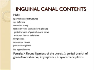
COMPOSITION OF HERNIA
COMPOSITIONOF HERNIA
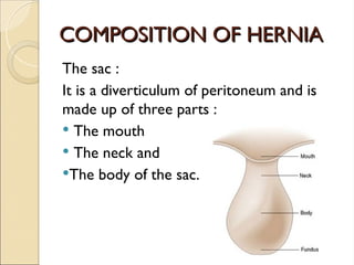
The sac :
It is a diverticulum of peritoneum and is
made up of three parts :
The mouth
The neck and
The body of the sac.
6.
COMPOSITION OF HERNIA
COMPOSITIONOF HERNIA
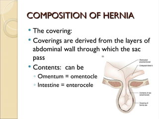
The covering:
Coverings are derived from the layers of
abdominal wall through which the sac
pass
Contents: can be
◦ Omentum = omentocle
◦ Intestine = enterocele
7.
COMPOSITION OF HERNIA
COMPOSITIONOF HERNIA
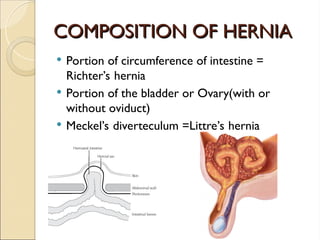
Portion of circumference of intestine =
Richter’s hernia
Portion of the bladder or Ovary(with or
without oviduct)
Meckel’s diverteculum =Littre’s hernia
ETIOLOGY
ETIOLOGY
Hernias occurat sites of weakness in the
wall This weakness may be :
Normal (physiological) weakness, related
to the anatomical causes.
Congenital abnormality.
Acquired
Traumatic
Diseases
Repeated INCREASE in
RepeatedINCREASE in
abdominal pressure is usually
abdominal pressure is usually
due to
due to
Chronic cough
Straining
Bladder neck or urethral obstruction
Pregnancy
Vomiting
Severe muscular effort
Ascitic fluid
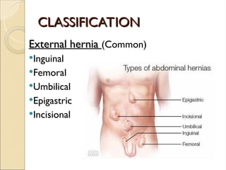
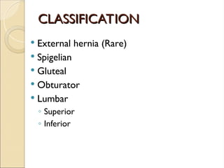
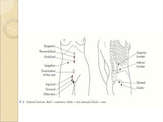
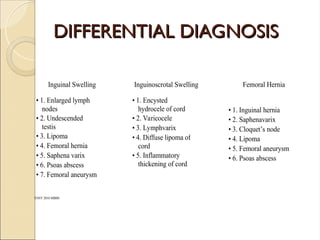
VARIETIES
VARIETIES
Reducible
Reducible contentsof the sac reduced
spontaneously or can be pushed back
manually. A reducible hernia imparts an
expansile impulse on coughing
Irreducible
Irreducible contents cannot be
returned to the peritoneal cavity either
because there are:
adhesions between the sac and contents,
because of the narrow neck of the sac.
IRREDUCIBLE HERNIAS
IRREDUCIBLE HERNIAS
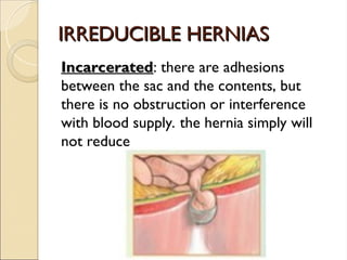
Obstructed
Obstructed: a hollow viscus is trapped
within the sac and obstruction occurs.
The blood supply remains intact. This is a
common cause of small bowel
obstruction.
17.
IRREDUCIBLE HERNIAS
IRREDUCIBLE HERNIAS
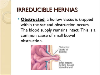
Strangulated
Strangulated: the arterial blood supply
to the contents of the sac is
compromised, in such a hernia unless
surgical relief is undertaken the contents
of the sac will become gangrenous.
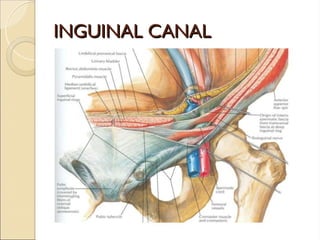
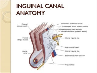
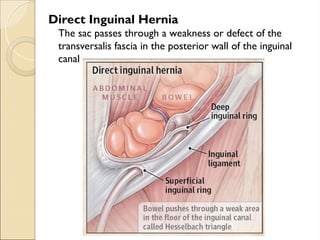
Direct Inguinal Hernia
Thesac passes through a weakness or defect of the
transversalis fascia in the posterior wall of the inguinal
canal
30.
Inguinal hernia
Inguinal hernia
History:
1.Age ( young vs. old)
2.Occupation ( nature ?? )
3.Local symptoms: Swelling, discomfort and
pain
4.Systemic symptoms: if there is
obstruction or strangulation
5.Precipitating factors
31.
Inguinal hernia
Inguinal hernia
Examination:
1.Inspection for site, size, shape and color.
2.Palpation for surface, temp, tenderness,
composition and reducibility.
3.Expansible cough impulse.
4.General exam: for common causes of
increase intra abdominal pressure
32.
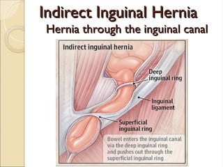
Indirect Versus Directinguinal
Indirect Versus Direct inguinal
hernias
hernias
Indirect is the most common form of
hernia and its usually congenital due to
patent processus viginalis
Direct usually acquired occur in old men
with weak abdominal muscles.
33.
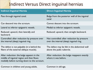
Indirect Versus Directinguinal hernias
Indirect Inguinal Hernia Direct Inguinal Hernia
Pass through inguinal canal. Bulge from the posterior wall of the inguinal
canal
Can descend into the scrotum. Cannot descent into the scrotum.
Lateral to inferior epigastric vessels. Medial to inferior epigastric vessels.
Reduced: upward, then laterally and
backward.
Reduced: upward, then straight backward.
Controlled: after reduction by pressure over
the internal (deep) inguinal ring.
Not controlled: after reduction by pressure
over the internal (deep) inguinal ring.
The defect is not palpable (it is behind the
fibers of the external oblique muscle).
The defect may be felt in the abdominal wall
above the pubic tubercle.
After reduction: the bulge appears in the
middle of inguinal region and then flows
medially before turning down to the scrotum.
After reduction: the bulge reappears exactly
where it was before.
Common in children and young adults. Common in old age.
34.
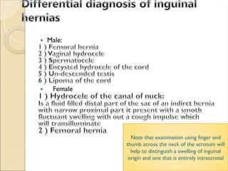
Note that examinationusing finger and
thumb across the neck of the scrotum will
help to distinguish a swelling of inguinal
origin and one that is entirely intrascrotal
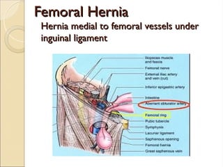
Femoral Canal
Femoral Canal
Themajor feature of the femoral canal is the femoral sheath. This
sheath is a condensation of the deep fascia (fascia lata) of the thigh
and contains, from lateral to medial, the femoral artery, femoral
vein, and femoral canal. The femoral canal is a space medial to the
vein that allows for venous expansion and contains a lymph node
(node of Cloquet). Other features of the femoral triangle include
the femoral nerve, which lies lateral to the sheath,
37.
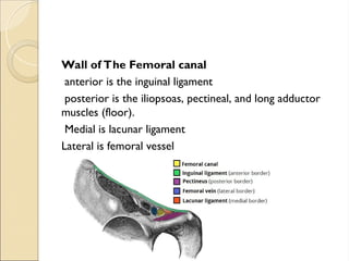
Wall of TheFemoral canal
anterior is the inguinal ligament
posterior is the iliopsoas, pectineal, and long adductor
muscles (floor).
Medial is lacunar ligament
Lateral is femoral vessel
38.
Femoral hernia
Femoral hernia
Smallfemoral hernia may be unnoticed by
the patient or disregarded for years
perhaps until the day it strangulates.
Adherence of the greater omentum
sometimes causes a dragging pain. Rarely
a large sac is present .
39.
Femoral hernia
Femoral hernia
History
Age ; uncommon in children , most common
in old age female .
Sex; women > men (but still commonest
hernia in women the inguinal hernia )
The patient came with local symptoms
1- discomfort and pain
2- swelling in the groin
General ; femoral hernia is more likely to be
strangulated than the inguinal hernia
Multiplicity ; often bilateral
40.
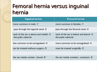
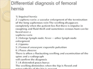
Femoral hernia versusinguinal
Femoral hernia versus inguinal
hernia
hernia
Inguinal hernia Femoral hernia
1
-
more common in male 1
-
more common in females
2
-
pass through the inguinal canal 2
-
pass through the femoral canal
3
-
neck of the sac is above and medial
the pubic tubercle
3
-
neck of the sac is below and lateral
the pubic tubercle
4
-
less common to be strangulated 4
-
more common to be strangulated
5
-
can be treated without surgery 5
-
must be treated surgically
6
-
the sac mainly contain ; bowel 6
-
the sac mainly contains ; omentum
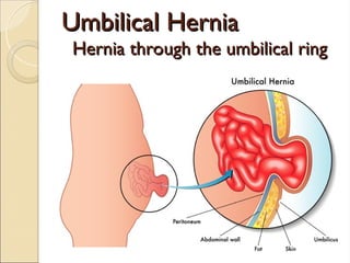
Umbilical hernia
Umbilical hernia
Signs and symptoms
Age ; doesn’t appear until the umbilical
cord has separated and healed .
No specific symptoms
Have wide neck and reduce easily , rarely
give intestinal obstruction.
Nature history ; 90 % disappear
spontaneously during the first year.
44.
Examination
Inspection
Site ; in the center of the umbilicus
Size and shape ; size can vary from vary small to
very large . Shape is usually hemispherical.
Palpation
Composition ; contain bowel , which makes it
resonant to percussion . They reduce
spontaneously when the child lies down .
Reducibility ; easy
Cough impulse; invariably present .
45.
Acquired umbilical hernia
Acquiredumbilical hernia
Hernia through the umbilical scar , so it is a
true umbilical hernia.
Not common and is usually secondary to
increase intra abdominal pressure.
The most common causes
1- pregnancy
2- ascitis
3- ovarian cyst
4- fibrodis
5- bowel distention
46.
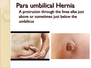
Para umbilical Hernia
Paraumbilical Hernia
A protrusion through the linea alba just
A protrusion through the linea alba just
above or sometimes just below the
above or sometimes just below the
umbilicus
umbilicus
47.
PARAUMBLICAL HERNIA
PARAUMBLICAL HERNIA
It occurs just above or just below the
umbilicus, and is more common in obese
females.
Predisposing factors
◦ multiple pregnancies and
◦ obesity.
48.
PARAUMBLICAL HERNIA
PARAUMBLICAL HERNIA
The neck of the sac is usually narrow and
therefore there is a high risk of
strangulation.
The most common content is
◦ omentum then
◦ transverse colon and
◦ small intestine
49.
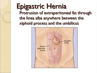
Epigastric Hernia
Epigastric Hernia
Protrusionof extraperitoneal fat through
Protrusion of extraperitoneal fat through
the linea alba anywhere between the
the linea alba anywhere between the
xiphoid process and the umbilicus
xiphoid process and the umbilicus
50.
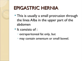
EPIGASTRIC HERNIA
EPIGASTRIC HERNIA
This is usually a small protrusion through
the linea Alba in the upper part of the
abdomen
It consists of :
◦ extraperitoneal fat only, but
◦ may contain omentum or small bowel.
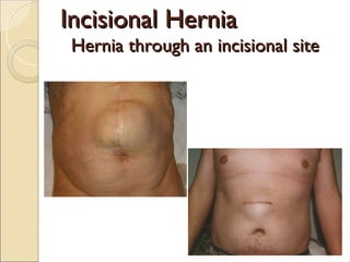
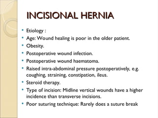
INCISIONAL HERNIA
INCISIONAL HERNIA
Etiology :
Age: Wound healing is poor in the older patient.
Obesity.
Postoperative wound infection.
Postoperative wound haematoma.
Raised intra-abdominal pressure postoperatively, e.g.
coughing, straining, constipation, ileus.
Steroid therapy.
Type of incision: Midline vertical wounds have a higher
incidence than transverse incisions.
Poor suturing technique: Rarely does a suture break
54.
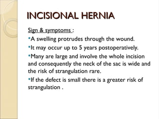
INCISIONAL HERNIA
INCISIONAL HERNIA
Sign& symptoms :
A swelling protrudes through the wound.
It may occur up to 5 years postoperatively.
Many are large and involve the whole incision
and consequently the neck of the sac is wide and
the risk of strangulation rare.
If the defect is small there is a greater risk of
strangulation .
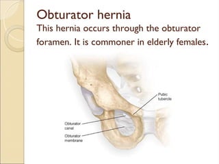
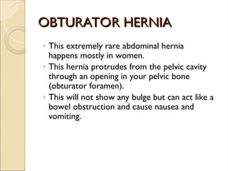
OBTURATOR HERNIA
OBTURATOR HERNIA
◦This extremely rare abdominal hernia
happens mostly in women.
◦ This hernia protrudes from the pelvic cavity
through an opening in your pelvic bone
(obturator foramen).
◦ This will not show any bulge but can act like a
bowel obstruction and cause nausea and
vomiting.
59.
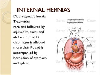
INTERNAL HERNIAS
INTERNAL HERNIAS
Diaphragmatichernia
Traumatic:
rare and followed by
injuries to chest and
abdomen. The Lt
diaphragm is affected
more than Rt and is
accompanied by
herniation of stomach
and spleen.
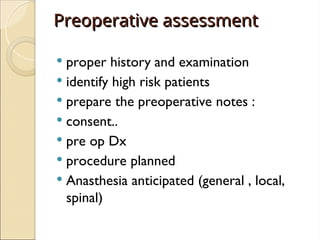
Preoperative assessment
Preoperative assessment
proper history and examination
identify high risk patients
prepare the preoperative notes :
consent..
pre op Dx
procedure planned
Anasthesia anticipated (general , local,
spinal)
70.
Preoperative assessment
Preoperative assessment
Investigation data ( pre operative tests ) :
1. Lab :
* CBC : to check hemoglobin level anemia and WBCs
infections
* U&E : to check for any electrolyte imbalance
* LFTs : indicated in jaundiced patients and suspected hepatitis
or any clotting problems
* PT & PTT
* ABG
* grouping and cross matching
2. Imaging :
* Chest X ray : for all patients
3. ECG : for any patient who is more than 40 years of age
71.
TREATMENT
TREATMENT
Treat the precipitatingcause of hernia first:
Benign prostatic hyperplasia
Tuberculosis
COPD
Constipation
Stop smoking
Conservative Treatment
Only in old patients with direct hernia
Truss
Surgery
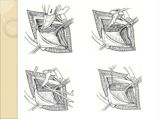
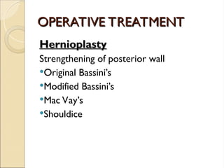
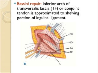
TREATMENT
TREATMENT
Herniotomy
Herniotomy:
Dissecting out andopening of hernia
sac,reducing any contents ,transfixing
neck of sac & removing the remainder
NO NEED TO OPEN UP CANAL IN CHILDREN BECAUSE
SUPERFICIAL AND DEEP RING ARE SUPERIMPOSED ……
THERE FORE NO NEED OF REPAIR
HENCE DONE ALONE IN CHILDREN,ADOLESCENT
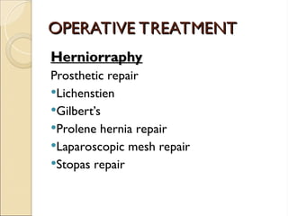
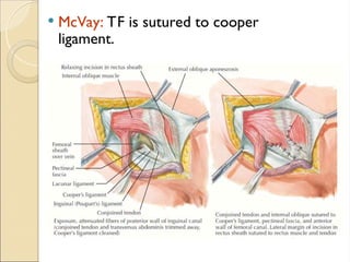
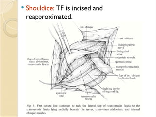
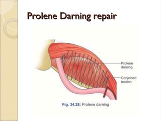
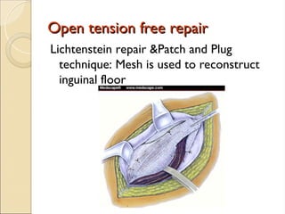
Open tension freerepair
Open tension free repair
Lichtenstein repair &Patch and Plug
technique: Mesh is used to reconstruct
inguinal floor
82.
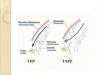
Laproscopic &
Laproscopic &
preperitonealrepairs
preperitoneal repairs
TAPP (transabdominal prepeitoneal procedure): peritoneal space
entered by conventional lap at umbilicus and peritoneum
overlaying inguinal floor is dissected away as flap.
TEP (Total extraperitoneal repair): preperitoneal space is
developed with a balloon inserted between posterior rectus
sheath and peritoneum balloon inflated to dissect the peritoneal
flaps awau from posterior abdomianl wall and the direct and
indirect spaces, other ports inserted into this preperitoneal space
without entering peritoneal cavity.
After lap. Dissection and reduction of hernia sac , a large piece of
mesh is placed over inguinal floor
84.
Femoral hernia repair
Femoralhernia repair
• Femoral hernias should be repaired very soon after the
diagnosis has been made because of the high risk of
strangulation.
• There is no place for a truss for a femoral hernia.
• Different approaches :
Open VS Laparoscopic
85.
Open surgery
Open surgery
Threeapproaches have been described for
open surgery :
1. Infra-inguinal approach (Lookwood)
2. Supra-inguinal approach ( McEvedy)
3. Trans-inguinal approach ( Lotheissen)


![PERI-PROSTHETIC FRACTURE NAIL-PLATE CONSTRUCT [NPC].pptx](https://cdn.slidesharecdn.com/ss_thumbnails/drarunkumardrmohamedashrafperiprostheticfrasturenail-plateconstructnpc-260209164459-7e9d15a1-thumbnail.jpg?width=640&height=640&fit=bounds)

![ONFH[AVN HIP] -TRIPLE REGIME -A NOVAL SURGICAL CONCEPT .pptx](https://cdn.slidesharecdn.com/ss_thumbnails/onfhavnhip2026koaconcalicutdrgokuldevdrmashraf-260210064517-213ec005-thumbnail.jpg?width=640&height=640&fit=bounds)

