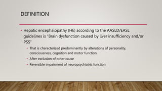
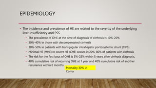
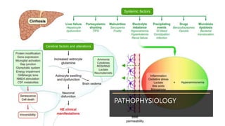
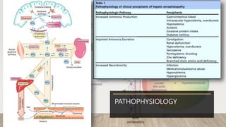
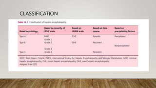
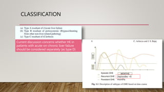
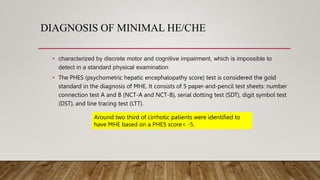
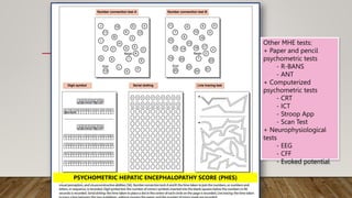
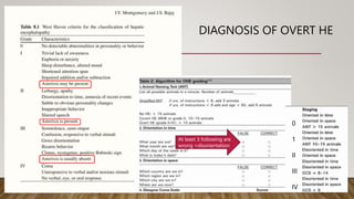
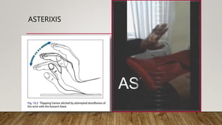
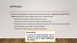
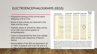
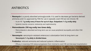
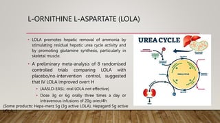
3. DEFINITION
⢠Hepatic encephalopathy (HE) according to the AASLD/EASL
guidelines is âBrain dysfunction caused by liver insufficiency and/or
PSSâ
⢠That is characterized predominantly by alterations of personality,
consciousness, cognition and motor function.
⢠After exclusion of other cause
⢠Reversible impairment of neuropsychiatric function
4. EPIDEMIOLOGY
⢠The incidence and prevalence of HE are related to the severity of the underlying
liver insufficiency and PSS
⢠The prevalence of OHE at the time of diagnosis of cirrhosis is 10%-20%
⢠30%-40% in those with decompensated cirrhosis
⢠10%-50% in patients with trans jugular intrahepatic portosystemic shunt (TIPS)
⢠Minimal HE (MHE) or covert HE (CHE) occurs in 20%-80% of patients with cirrhosis
⢠The risk for the first bout of OHE is 5%-25% within 5 years after cirrhosis diagnosis,
⢠40% cumulative risk of recurring OHE at 1 year and 40% cumulative risk of another
recurrence within 6 months
Mortality 30% in
Coma
9. DIAGNOSIS OF MINIMAL HE/CHE
⢠characterized by discrete motor and cognitive impairment, which is impossible to
detect in a standard physical examination
⢠The PHES (psychometric hepatic encephalopathy score) test is considered the gold
standard in the diagnosis of MHE. It consists of 5 paper-and-pencil test sheets: number
connection test A and B (NCT-A and NCT-B), serial dotting test (SDT), digit symbol test
(DST), and line tracing test (LTT).
Around two third of cirrhotic patients were identified to
have MHE based on a PHES score< -5.
10. PSYCHOMETRIC HEPATIC ENCEPHALOPATHY SCORE (PHES)
Other MHE tests:
+ Paper and pencil
psychometric tests
- R-BANS
- ANT
+ Computerized
psychometric tests
- CRT
- ICT
- Stroop App
- Scan Test
+ Neurophysiological
tests
- EEG
- CFF
- Evoked potential
11. DIAGNOSIS OF OVERT HE
0
1
II
III
IV
At least 3 following are
wrong =disorientation
13. AMMONIA
⢠Blood ammonia levels correlate with the severity of HE, increased in about 90 %of
patients with HEď lower plasma ammonia in improving HE
⢠Without liver disease can display hyperammonemia
⢠Normal blood ammonia level has negative predictive value
⢠Normal ammonia in a patient with cirrhosis and delirium ď work up other cause delirium
⢠Ammonia levels are not used to monitor therapy
⢠Arterial NH3>100 mmol/L(or two fold) is abnormal
14. ELECTROENCEPHALOGRAMS (EEGS)
⢠Progressive slowing of the normal alpha
frequency of 8 to13 Hz
⢠Bursts of slow activity are observed in the
theta (4â8 Hz) range
⢠Triphasic waves or arrhythmic delta activity
occur with more severe grades of
encephalopathy
⢠Coma is characterized by slow, low-voltage
delta activity with sequences of electric
silence
⢠Abnormalities of the EEG are reported in 43
to 100% of patients with overt HE and in 8
to 40% of clinically unimpaired patients with
cirrhosis
15. MAGNETIC RESONANCE IMAGING (MRI)
⢠T1-weighted imaging :
Bilateral, symmetric, high
signal intensity in the
Substantia nigraand globus
pallidus(basal ganglia)
⢠Diffuse white matter signal
intensities involving the
hemispheric white matter
and the corticospinal tract
on T2-weighted imaging
16. DIAGNOSIS OF HE
Grade Characteristics Asterixis EEG(cycle/sec) NCT-A(sec) NH3(g/L)
0 or
minimal
No No 8-12 15-30 Normal 0.7-1.2
I
Trivial lack of awareness
Euphoria or anxiety,
Shortened attention span,
Impairment of addition or
subtraction , Altered sleep
rhythm
Mild
(1-2/30ââ)
7-8 31-50 1-1.33
II
Lethargy or apathy
Disorientation for time
Obvious personality change
Inappropriate behavior
Dyspraxia , Asterixis
Moderate
(3-4/30ââ)
5-7 51-80 1.33-1.67
III
Somnolence to semistupor
Responsive to stimuli Confused
Gross disorientation Bizarre
behavior
Severe
(5-30/30ââ)
3-5 81-120 1.67-2
17. DIFFERENTIAL DIAGNOSIS OF HE
⢠Diabetic (hypoglycemia, ketoacidosis,
hyperosmolar, lactate acidosis)
⢠Alcohol (intoxication, withdrawal, Wernicke)
⢠Drugs (benzodiazepines, neuroleptics,
opioids)
⢠Neuroinfections
⢠Electrolyte disorders (hyponatremia and
hypercalcemia)
⢠Nonconvulsive epilepsy
⢠Psychiatric disorders
⢠Intracranial bleeding and stroke
⢠Severe medical stress (organ failure and
inflammation)
⢠Other presentations
⢠Dementia (primary and secondary)
⢠Brain lesions (traumatic, neoplasms, normal
pressure hydrocephalus)
⢠Obstructive sleep apnea
19. MANAGEMENT OF HE
⢠Patients with overt HE grade 3 and 4 are at
risk of aspiration and should be treated in
the ICU
⢠Treatments are outlined below based on the
mechanism of action.
⢠The goal is to maximize the bodyâs ability to
remove ammonia from the bloodstream
while reducing ammonia production, which
remains the major priority.
⢠The first step in therapy is to address any
potential precipitating causes. Nearly 90% of
patients can be treated with just correction
of the precipitating factor
⢠A more individualized course of therapy will
need to be created for the patients.
20. NON-ABSORBABLE DISACCHARIDES AND
POLYETHYLENE GLYCOL
⢠Lactulose (β-galactosidofructose): the mechanisms is
reduction of intestinal pH by production of acetic and
lactic acid that converts ammonia(NH3)ď
ammonium(NH4+) and then by an osmotic laxative
effect that flushes the NH4+ out.
⢠Dose 20g/30mlâ30g/45ml 3â4/day for 2â3 bowel
movements a day orally/NG or 300ml in 700mL water
of enemas 3â4/day.
⢠Prophylaxis/outpatient: 15â45 mL orally 2 or 3 times
daily for 2â3 bowel movements a day.
⢠Complication: GI sideâeffects 30% (anorexia, flatulence,
bloating, and abdominal discomfort), aspiration,
dehydration, hyperNa and overuse can even precipitate
HE.
Lactulose was approved by the US FDA in
1977
⢠Lactilol (β-galactosidosorbitol) is a
secondâgeneration disaccharide,
which is easily produced in a
chemically pure crystalline form and
can be dispensed as a powder.
⢠The dosage required to ensure daily
passage of two semiâsoft stools
ranges from 10 to 90g (0,3-
0,5g/kg/day)
⢠Lactitol is more tolerated owing to
less diarrhea and flatulence
21. NON-ABSORBABLE DISACCHARIDES AND
POLYETHYLENE GLYCOL
⢠Polyethylene glycol(PEG): PEG can be used for
management of acute OHE. Its mechanism of
action is a flushing-out effect of ammonia from
the gut-like lactulose.
⢠A single RCT comparing PEG (4-l dose Ă1 over 4h
orally/NG) to lactulose only (20â30 g/day via
oral/NG or 200 g/day rectal) has proven it to be
superior in terms of clinical improvement over a
24-h period, documented by the HE scoring
algorithm (91% vs. 52%, P<0.01)
⢠PEG has not been FDA-approved for this
indication but could be a viable alternative to
lactulose.
22. ANTIBIOTICS
⢠Antibiotics can be used to selectively eliminate ureaseâ producing organisms from
the intestinal tract (block RNA synthesis in bacteria). This reduces the production of
ammonia
⢠Rifaximin, it has a very low rate of systemic absorption (0.4%).
⢠A recently published study showed that there was no difference in the efficacy of
traditional rifaximin dosing (400 mg three times daily) compared to newer dosing (550
mg twice daily) for the prevention of recurrence of HE. However the 550 mg regimen
had a lower acquisition cost.
⢠Rifaximin decreased the risk of recurrence of overt HE, decreased the risk of
hospitalization, had a beneficial effect on the secondary prevention of overt HE (RR 1.32;
95% CI 1.06 to 1.65)
23. ANTIBIOTICS
⢠Neomycin is a poorly absorbed aminoglycoside. It is used to decrease gut bacteria-derived
ammonia and it is approved by FDA for use in episodic overt HE but not chronic HE
⢠Acute HE: 1 g orally every 6 hours for up to 6 days. Outpatient: 1â2 g orally daily
⢠Systemic toxicity concerns: ototoxicity and nephrotoxicity
⢠Metronidazole: 250 mg orally two times daily,
⢠Metronidazole is absorbed and long-term use can cause peripheral neuropathy and other CNS
toxicities
⢠Vancomycin: vancomycin-resistant enterococci colonization limit its long-term use
⢠Vancomycin 1â2 g daily in divided doses
⢠Probiotics: reduced ammonia and reduced systemic inflammation
⢠Efficacy in reversing CHE (reduce progression from minimal to overt HE) and decrease hospitalization
rates.
⢠No current evidence for probiotics use in acute OHE (only for secondary prophylaxis- Not approved by
24. L-ORNITHINE L-ASPARTATE (LOLA)
⢠LOLA promotes hepatic removal of ammonia by
stimulating residual hepatic urea cycle activity and
by promoting glutamine synthesis, particularly in
skeletal muscle.
⢠A preliminary meta-analysis of 8 randomised
controlled trials comparing LOLA with
placebo/no-intervention control, suggested
that IV LOLA improved overt H
⢠(AASLD-EASL: oral LOLA not effective)
⢠Dose 3g or 6g orally three times a day or
intravenous infusions of 20g over/4h
(Some products: Hepa-merz 5g (3g active LOLA), Hepagard 5g active
LOLAâŚ)
25. BRANCHED-CHAIN AMINO ACIDS (BCAAS)
⢠BCAAs are reduced in patients with cirrhosis (such as valine, leucine, and
isoleucine) while plasma aromatic amino acids are increased
⢠These BCAAs are essential for detoxification of ammonia in skeletal muscles
during the process of glutamine synthesis
⢠A sum of 16 RCTs consisting of 827 participants was included. Among 16, 08 trials
were with BCAA supplements, and 07 trials were with IV BCAA
⢠Results showed a beneficial effect of BCAA on HE (AASLD-EASL: no effect of IV BCAA)
⢠No significant change in mortality, QOL, and nutritional parameters
⢠Initial dose10g-20g/day, increase 10g in every 3-5days.
⢠Some products: Morihepamine 200ml , Aminoleben 500ml..
26. NUTRITION
⢠Guidelines recommend daily energy intakes of 35 to 45kcal/kg and daily protein
intakes of 1.2 to 1.5g/kg. Increase protein intake from vegetable sources (is better
tolerated than animal protein; the benefits relate to the effects of dietary fibre on
colonic function).
should avoid fasting for longer than 3 to 6h during the
daytime
27. NOVEL DRUGS (THERAPEUTIC TARGET) FOR HE
⢠Albumin (inflammation modulator)
⢠AST-120 (Inhibition of intestinal NH3
absorption)
⢠Acetyl-L-carnitine (Amino-acid prepation)
⢠Flumazenil (neuro-transmitter modulation)
⢠Glycerol phenyl-butyrate (provide alternat
pathway to urea)
⢠Nitazoxanide (alteration gut microbiome)
⢠Naloxone(neuro-transmitter modulation)
⢠Bromocriptine 7.5mg daily (if no fluid
retention)
⢠Sodium benzoate has been used for patients
with urea cycle disorders, in which it fixes
ammonia to form hippurate for excretion
⢠Dose 5g twice daily (if no fluid retention)
⢠Zinc is a critical cofactor in the activity of urea
cycle enzymes in the liver and of glutamine
synthetase in muscle. Zinc reduced in patients
with HE and correlate inversely with blood NH3
⢠Zinc acetate or sulphate 600mg/day/3ms
Avoiding drug in HE: Tramadol, Pethidine/meperidine, NSAIDs,
BenzodiazepinesâŚ
28. SECONDARY PROPHYLAXIS FOR OHE
⢠Lactulose with rifaximin has been shown to be superior than just lactulose in this
regard (22.1% vs. 45.9% over 6 months) and superior to just rifaximin
monotherapy (13.9% vs. 24.8% over 6 months)
30. REFERENCE
1. American Association for the Study of Liver Diseases, European Association for the Study of the Liver. Hepatic encephalopathy in
chronic liver disease: 2014 practice guideline by the European Association for the Study of the Liver and the American Association
for the Study of Liver Diseases. J Hepatol 2014 Sep;61(3):642â659
2. Acharya C, Bajaj JS. Current Management of Hepatic Encephalopathy. Am J Gastroenterol. 2018 Nov;113(11):1600-1612. doi:
10.1038/s41395-018-0179-4. PMID: 30002466.
3. European Association for the Study of the Liver. Electronic address: easloffice@easloffice.eu; European Association for the Study of
the Liver. EASL Clinical Practice Guidelines on the management of hepatic encephalopathy. J Hepatol. 2022 Sep;77(3):807-824. doi:
10.1016/j.jhep.2022.06.001. Epub 2022 Jun 17. PMID: 35724930.
4. Ferenci P, Lockwood A, Mullen K, Tarter R, Weissenborn K, Blei AT. Hepatic encephalopathy â definition, nomenclature, diagnosis,
and quantification: final report of the working party at the 11th World Congresses of Gastroenterology, Vienna, 1998. Hepatology
2002;35(3):716â21.
5. Rajpurohit S, Musunuri B, Shailesh, Basthi Mohan P, Shetty S. Novel Drugs for the Management of Hepatic Encephalopathy: Still a
Long Journey to Travel. J Clin Exp Hepatol. 2022 Jul-Aug;12(4):1200-1214. doi: 10.1016/j.jceh.2022.01.012. Epub 2022 Jan 31. PMID:
35814520; PMCID: PMC9257922.
6. Rudler M, Weiss N, Bouzbib C, Thabut D. Diagnosis and Management of Hepatic Encephalopathy. Clin Liver Dis. 2021
May;25(2):393-417. doi: 10.1016/j.cld.2021.01.008. Epub 2021 Mar 11. PMID: 33838857.
7. Sherlockâs Diseases of the Liver and Biliary System, Thirteenth Edition. Edited by James S. Dooley, Anna S. F. Lok,
8. Schiffâs Diseases of the Liver, Twelfth Edition. Edited by Eugene R. Schiff, Willis C. Maddrey and K. Rajender Reddy
Fig. 2. Pathogenesis and pathophysiology of hepatic encephalopathy. Chronic liver disease leads to hepatocyte dysfunction, portal hypertension, portalsystemic shunting, altered microbiota, bacterial translocation, malnutrition, sarcopenia, electrolyte imbalance as well as constipation and gastrointestinal bleeding. Consequently, pathogenic factors are generated including hyperammonemia, systemic inflammation/oxidative stress as well as increased blood manganese, circulating bile acids and lactate. These systemic factors influence the blood-brain barrier (BBB) by increasing its permeability (increased signaling across the BBB, physical breakdown of the BBB which allows for an increased influx of molecules which normally do and do not cross the BBB). Independent of BBB status, ammonia passes freely into the brain which is exclusively removed by astrocytes via glutamine synthetase. The generation of glutamine renders the astrocyte hypertonic resulting in swelling and impaired function and brain oedema. Astrocyte swelling leads to compromised neuronal communication leading to neuronal dysfunction. Alterations of cerebrospinal fluid (CSF) metabolites are observed, as well as alterations in neurotransmission such as increased GABergic tone potentiated with neurosteroids and glutamate-induced N-methyl-D-aspartate (NMDA) stimulation. Blood derived increase in brain ammonia is central in the pathophysiological mechanisms underlying the development of HE. Neuroinflammation and microglia activation are significant modulators in the onset of neurological decline. Astrocyte senescence as well as neuronal cell death may be key features in the irreversibility of HE. However, the extent and underlying causes of neuronal cell death remain to be defined. BBB, blood-brain barrier; CSF, cerebrospinal fluid; HE, hepatic encephalopathy; NMDA, N-methyl-D-aspartate; TIPS, transjugular intrahepatic portosystemic shunt