
More Related Content
Similar to hemorrhoids_fistula.pptx
Similar to hemorrhoids_fistula.pptx (20)
Recently uploaded
Recently uploaded (20)
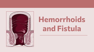
hemorrhoids_fistula.pptx
- 2. ● Cushions of submucosal tissue containing venules, arterioles, and smooth muscle fibers that are located in the anal canal ● Function as part of the continence mechanism and aid in complete closure of the anal canal at rest ● 3 hemorrhoidal cushions: ○ Left lateral (3 o’clock) ○ Right posterior (7 o’clock) ○ Right anterior (11 o’clock) ● Excessive straining, increased abdominal pressure, and hard stools increase venous engorgement of the hemorrhoidal plexus and cause prolapse of the hemorrhoidal tissue ● Bleeding, thrombosis, and symptomatic prolapse Hemorrhoids
- 4. Pathophysiology ● Thompson’s vascular cushion theory ○ Normal hemorrhoidal tissue represents discrete masses of submucosa ○ During straining, the vascular cushions can become engorged and possibly prevent the escape of fecal material or gas ○ With passage of time, the anatomic structures supporting the muscular submucosa weaken, allowing hemorrhoidal tissue to slip or prolapse, leading to hemorrhoidal symptoms ● Matrix metalloproteinases (MMPs) ○ Enzymes present in extracellular space and can degrade collagen, elastin, and fibronectin ○ MMP-9: overexpressed in hemorrhoid tissue in association with breakdown of elastic fibers
- 6. History ● Changes in bowel habits ● Rectal bleeding ○ Nature ○ Color ○ Intensity ● Pain ○ Intensity ○ Frequency ○ Duration ● Protrusion or swelling in the rectal area
- 7. Clinical Manifestations TYPE DESCRIPTION AND MANIFESTATIONS Internal hemorrhoids ● Located proximal to the dentate line ● Covered by insensate anorectal mucosa ● May prolapse or bleed, but rarely become painful unless they develop thrombosis and necrosis ● Graded according to the extent of prolapse External hemorrhoids ● Located distal to the dentate line ● Covered by anoderm ● A skin tag may remain after healing ● External hemorrhoids and skin tags may cause itching and difficulty with hygiene if they are large. Combined internal and external hemorrhoids ● Straddle the dentate line and have characteristics of both internal and external hemorrhoids ● Hemorrhoidectomy: often required for large, symptomatic, combined hemorrhoids
- 8. Internal Hemorrhoids GRADE DESCRIPTION I Protrudes through anal canal, but not beyond the anal verge II Protrusion, but with spontaneous reduction III Protrusion requiring manual reduction IV Protrusion that can’t be reduced (at risk for strangulation) Luchtefeld, M., Hoedema, R.E. (2016). Hemorrhoids. In: Steele, S., Hull, T., Read, T., Saclarides, T., Senagore, A., Whitlow, C. (eds) The ASCRS Textbook of Colon and Rectal Surgery. Springer, Cham. https://doi.org/10.1007/978-3-319-25970-3_12
- 9. Clinical Manifestations TYPE DESCRIPTION AND MANIFESTATIONS Internal hemorrhoids ● Located proximal to the dentate line ● Covered by insensate anorectal mucosa ● May prolapse or bleed, but rarely become painful unless they develop thrombosis and necrosis ● Graded according to the extent of prolapse External hemorrhoids ● Located distal to the dentate line ● Covered by anoderm ● A skin tag may remain after healing ● External hemorrhoids and skin tags may cause itching and difficulty with hygiene if they are large. Combined internal and external hemorrhoids ● Straddle the dentate line and have characteristics of both internal and external hemorrhoids ● Hemorrhoidectomy: often required for large, symptomatic, combined hemorrhoids
- 10. Physical Examination ● Focus on the abdomen, groin and perianal area ● Supine →prone jack knife or left lateral position ● Inspection ● Digital rectal exam ○ Masses ○ Pain ○ Sphincter tone Luchtefeld, M., Hoedema, R.E. (2016). Hemorrhoids. In: Steele, S., Hull, T., Read, T., Saclarides, T., Senagore, A., Whitlow, C. (eds) The ASCRS Textbook of Colon and Rectal Surgery. Springer, Cham. https://doi.org/10.1007/978-3-319-25970-3_12
- 11. Diagnostics ● Anoscopy, rigid proctosigmoidoscopy, and/or flexible sigmoidoscopy ● American Society for Gastrointestinal Endoscopy and the Society for Surgery of the Alimentary Tract guidelines: bright red rectal bleeding → anoscopy and flexible sigmoidoscopy
- 12. Diagnostics
- 13. Anoscopy ● Examination of the anal canal and the distal rectum ● Best way to evaluate the anoderm, dentate line, internal and external hemorrhoids, papillae, fissures, anal masses, and distal rectal mucosa ● Anoscope ○ Obturator ○ Scope ○ Light source ● The examination is initiated only after DRE has been performed. ● Enema is not warranted ● Prone jackknife or left lateral position Davis, K., Valente, M.A. (2016). Endoscopy. In: Steele, S., Hull, T., Read, T., Saclarides, T., Senagore, A., Whitlow, C. (eds) The ASCRS Textbook of Colon and Rectal Surgery. Springer, Cham. https://doi.org/10.1007/978-3-319-25970-3_4
- 14. Rigid Proctoscopy ● Suitable to examine the rectum ● Proctoscope needs to hold air so the rectum can be distended ● Enema preparation within 2 hours of the procedure ● Use has declined in recent years due to flexible endoscopy ● Indications ○ Identification and precise localization of rectal lesions ○ Evaluation of rectal bleeding ● Contraindications: painful anorectal conditions Davis, K., Valente, M.A. (2016). Endoscopy. In: Steele, S., Hull, T., Read, T., Saclarides, T., Senagore, A., Whitlow, C. (eds) The ASCRS Textbook of Colon and Rectal Surgery. Springer, Cham. https://doi.org/10.1007/978-3-319- 25970-3_4
- 15. Flexible Sigmoidoscopy ● Sigmoidoscope is inserted through the anus, the distal colonic mucosa is examined ● Preparation ○ Bowel cleaning ○ Medications
- 16. Management
- 17. Management Medical Therapy ● First- and second-degree hemorrhoidal bleeding: dietary fiber, stool softeners, increased fluid intake, avoidance of straining ● Pruritus: improved hygiene Rubber Band Ligation ● Persistent bleeding from first-, second-, and selected third-degree hemorrhoids ● Complications: severe pain if rubber band is placed at or distal to the dentate line, urinary retention, infection, and bleeding Infrared Photocoagulation ● Small first- and second-degree hemorrhoids ● Larger hemorrhoids with a significant amount of prolapse are not effectively treated with this technique Sclerotherapy ● First-, second-, and selected third-degree hemorrhoids ● 1-3 ml of sclerosing solution is injected into submucosa of each hemorrhoid ● Few complications
- 18. Management Excision of Thrombosed External Hemorrhoids ● Thrombosis can be effectively treated with an elliptical excision under local anesthesia ● Sitz bath and analgesics Operative Hemorrhoidectomy ● Elective resection of symptomatic hemorrhoids ● Based on decreasing blood flow to the hemorrhoidal plexus and excising redundant anoderm and mucosa ● Closed submucosal hemorrhoidectomy: Parks or Ferguson hemorrhoidectomy ● Open hemorrhoiddectomy: Milligan and Morgan hemorrhoidectomy ● Whiteheads’s hemorrhoidectomy ● Procedure for prolapse and hemorrhoids/Stapled Hemorrhoidectomy ● Doppler-guided hemorrhoidal artery ligation
- 19. ● The fistula usually originates in the infected crypt (internal opening) and tracks to the external opening, usually the site of prior drainage. ● Majority of fistulas are cryptoglandular in origin ● Trauma, Crohn’s disease, malignancy, radiation, or unusual infections may also produce fistulas Fistula-in-ano
- 20. Diagnosis
- 21. Diagnosis ● Persistent drainage from the internal and /or external openings ● Indurated tract is often palpable ● External opening is often easily identifiable ● Goodsall’s rule can be used as a guide in determining the location of the internal opening ○ Fistulas with an external opening anteriorly connect to the internal opening by a short, radial tract ○ Fistulas with an external opening posteriorly track in a curvilinear fashion to the posterior midline ○ Exception: if an anterior external opening is >3cm from the anal margin, such fistulas usuually track to the posterior midline
- 22. Parks Classification of Fistula in Ano Classification Description Intersphincteric ● Due to a perianal abscess ● Tracks through the distal internal sphincter and intersphincteric space to an external opening near the anal verge Transsphincteric ● Usually results from an ischiorectal fossa abscess ● Extends through both the internal and external sphincters Suprasphincteric ● Usually from a supralevator abscess ● Originates in the intersphincteric plane ● Tracks up and around the entire external sphincter
- 23. Parks Classification of Fistula in Ano Extrasphincteric ● May arise from foreign body penetration of the rectum, penetrating injury to the perineum or carcinoma ● Originates in the rectal wall ● Tracks around both sphincters to exit laterally, usually in the ischiorectal fossa Classification Description
- 24. Diagnostics ● Anoscopy: May be required to identify the internal opening of the fistula ● MRI: Diagnostic imaging of choice for the diagnosis of fistula-in-ano ● Most patients can undergo surgery even without an imaging modality
- 25. Management
- 26. Management Technique Description Fistulotomy ● Useful in the Majority ● Probe is inserted through the fistula (both openings); then skin and sphincteric muscles are divided, thereby opening (unroofing) the tract ● Fistulotomy is closed by secondary intention Seton Placement ● Thick suture placed through fistula tract to allow slow transection of sphincter muscle ● Made from large, silk suture that is threaded through the fistula tract to: ○ Allow direct visualization of the tract ○ Allow drainage and promotes fibrosis ○ Cuts through the fistula ● Advantage: avoids complication of incontinence ( in contrast to fistulotomy)
- 27. Preferred Techniques ● Simple fistula-in-ano: fistulotomy or unroofing of the fistolous tract ● Complex or high lying fistula-in-ano: seton placement ● LIFT ( Ligation of Intersphincteric Fistula Tract): new procedure that ligates the fistula at the intersphincteric plane (Rojanasakul procedure)
- 28. Mini Quiz
- 29. Question 1. Type of hemorrhoid that is located proximal to the dentate line and covered by insensate anorectal mucosa a. Internal hemorrhoid b. External hemorrhoid c. Combined internal and external hemorrhoid d. All of the above
- 30. Question 2. Excessive straining, increased abdominal pressure, and hard stools increase venous engorgement of the hemorrhoidal plexus and cause prolapse of the hemorrhoidal tissue a. True b. False
- 31. Question 3. Prolapse through the anal canal and require manual reduction a. First-degree b. Second-degree c. Third-degree d. Fourth-degree
- 32. Question 4. Located distal to the dentate line and covered by anoderm a. Internal hemorrhoid b. External hemorrhoid c. Combined internal and external hemorrhoid d. All of the above
- 33. Question 5. According to Thompson’s vascular cushion theory, normal hemorrhoidal tissue represents discrete masses of submucosa. a. True b. False
- 34. Question 6. Seton placement avoids complication of incontinence a. True b. False
- 35. Question 7. Usually results from an ischiorectal fossa abscess and extends through both the internal and external sphincters a. Intersphincteric b. Extrasphincteric c. Transsphincteric d. Suprasphincteric
- 36. Question 8. Diagnostic imaging of choice for the diagnosis of fistula-in-ano a. MRI b. Anoscopy c. Both d. None of the above
- 37. Question 9. According to Goodsall’s rule, Fistulas with an external opening anteriorly connect to the internal opening by a short, radial tract a. True b. False
- 38. Question 10. Usually from a supralevator abscess and tracks up and around the entire external sphincter a. Intersphincteric b. Extrasphincteric c. Transsphincteric d. Suprasphincteric
Editor's Notes
- Cushions of submucosal tissue containing venules, arterioles, and smooth muscle fibers that are located in the anal canal Function as part of the continence mechanism and aid in complete closure of the anal canal at rest 3 hemorrhoidal cushions: Left lateral (3 o’clock) Right posterior (7 o’clock) Right anterior (11 o’clock) Excessive straining, increased abdominal pressure, and hard stools increase venous engorgement of the hemorrhoidal plexus and cause prolapse of the hemorrhoidal tissue Bleeding, thrombosis, and symptomatic prolapse may result.
- Thompson’s vascular cushion theory states that normal hemorrhoidal tissue represents discrete masses of submucosa. During straining, the vascular cushions can become engorged and possibly prevent the escape of fecal material or gas. With the passage of time, how- ever, the anatomic structures supporting the muscular submu- cosa weaken, allowing the hemorrhoidal tissue to slip or prolapse, leading to typical hemorrhoidal symptoms. Haas et al. noted that supporting tissues can be shown microscopi- cally to deteriorate by the third decade of life Studies have investigated why this degradation occurs and what are the changes in the local microvasculature. Matrix metalloproteinases (MMPs) are enzymes present in the extracellular space and can degrade collagen, elastin, and fibronectin. MMP-9 has been found to be overexpressed in hemorrhoid tissue in association with breakdown of elastic fibers. Once the hemorrhoids start to prolapse, the inter- nal sphincter can slow the rate of venous return and increase the hemorrhoid engorgement.
- A careful history should be done to guide the clinician to an accurate diagnosis. In addition, it is helpful to know which symptoms bother the patient the most. Part of the history should include the patient’s bowel habits. If a patient has constipation, treatment of the consti- pation will be an important part of the treatment plan. Ulcerative colitis and Crohn’s disease need to be considered in patients that have had significant diarrhea. If there has been a significant change in bowel habits, one also has to consider the many possibilities that can lead to this change. For patients with rectal bleeding, the nature, color, and intensity of the bleeding should be noted. If also accompa- nied by a change in bowel habits, one needs to be suspicious of a malignancy or inflammatory bowel disease. If pain is a significant component of the presentation, the intensity, frequency, and duration of the pain should be noted. If the pain is severe and described as a tearing sensa- tion primarily at the time of the bowel movement, an anal fissure should be considered. Pain that is constant and has been present for days at a time should elicit consideration of a thrombosed hemorrhoid or perianal abscess as the underlying diagnosis. Protrusion or swelling in the rectal area can be many differ- ent things. If the protrusion has been present constantly for weeks, months, or even years, it can be something as simple as a skin tag. However, one needs to also be mindful of diag- noses such as condyloma and neoplasm in this situation.
- Internal hemorrhoids May prolapse or bleed, but rarely become painful unless they develop thrombosis and necrosis (usually related to severe prolapse, incarceration, and/or strangulation) External hemorrhoids Located below or distal to the dentate line Covered by anoderm Because anoderm is richly innervated, thrombosis of an external hemorrhoid may cause significant pain. It is for this reason that external hemorrhoids should not be ligated or excised w/o adequate local anesthetic Enlarges secondary to dilation or thrombosis Skin tag is a redundant fibrotic skin at the anal verge, often persisting as the residua of a thrombosed external hemorrhoid External hemorrhoids and skin tags may cause itching and difficulty with hygiene if they are large. bleeding that occurs with hemorrhoids is typically described as bright red in nature with the frequency ranging from rarely to several times per day. The blood can be seen on the toilet paper and in the toilet water, and sometimes patients even describe the sensation of the blood squirting out of the anus. Typically the frequency and severity will increase over time
- First-degree hemorrhoids bulge into the anal canal and may prolapse beyond the dentate line on straining. Second-degree hemorrhoids prolapse through the anus but reduce spontaneously. Third-degree hemorrhoids prolapse through the anal canal and require manual reduction. Fourth-degree hemorrhoids prolapse but cannot be reduced and are at risk for strangulation
- Internal hemorrhoids May prolapse or bleed, but rarely become painful unless they develop thrombosis and necrosis (usually related to severe prolapse, incarceration, and/or strangulation) External hemorrhoids Located below or distal to the dentate line Covered by anoderm Because anoderm is richly innervated, thrombosis of an external hemorrhoid may cause significant pain. It is for this reason that external hemorrhoids should not be ligated or excised w/o adequate local anesthetic Enlarges secondary to dilation or thrombosis Skin tag is a redundant fibrotic skin at the anal verge, often persisting as the residua of a thrombosed external hemorrhoid External hemorrhoids and skin tags may cause itching and difficulty with hygiene if they are large.
- A general physical examination should be conducted with concentration on the abdomen, groin, and perianal area. Typically the patient will be examined in the supine position first before switching to a prone jackknife or left lateral (Sims) position (Figure 12-4). It is important to be as reas- suring as possible during this examination as it is inherently embarrassing and uncomfortable. It is always helpful to explain the steps of the examination so as to minimize sur- prise and discomfort. The examination begins by gently spreading the buttocks and inspecting the skin, perineum, and the external anal opening. Anal fissures are usually diagnosed just with these simple measures, but if one is not thinking of this possibility, it is easy to miss a fissure. In addition, many other conditions can be identified: dermatitis, fistulas, abscess, anal cancer, skin tags, and condyloma. A digital rectal exam is then per- formed to assess for masses, pain, and sphincter tone. If there is any component of fecal soiling or incontinence, the sphinc- ter tone should also be investigated by asking the patient to voluntarily squeeze during the digital exam.
- Patients with anorectal complaints must undergo Anoscopy, rigid proctosigmoidoscopy, and/or flexible sigmoidoscopy (further work-up depends on physical examination, patient age, and history) American Society for Gastrointestinal Endoscopy and the Society for Surgery of the Alimentary Tract guidelines: suggest anoscopy and flexible sigmoidoscopy for bright red rectal bleeding
- Anoscopy is the examination of the anal canal and the distal rectum. Anoscopy offers the best way to adequately evaluate the anoderm, dentate line, internal and external hemorrhoids, papillae, fissures, anal masses, and distal rectal mucosa. The anoscope is a relatively simple instrument consisting of an obturator, the scope itself, and a light source. There exist several variations in type, size, and length of anoscopes available. Additionally, commercially available anoscopes include slotted or beveled styles, reusable or disposable, and lighted or unlighted. The particular type of instrument and light source used are based on individual preference, expense, and prior training (Figure 4-3). Regardless of the choice of instrument used, the examina- tion is initiated only after a DRE has been performed (if a DRE is unable to be performed secondary to pain, spasm, or stenosis, an anoscopic exam should not be attempted). For most instances, cleansing of the anorectum with an enema is not warranted. The anoscope (with obturator in place) is liberally lubricated and gently and gradually advanced until the instru- ment is fully inserted. It is important to align the anoscope along the anterior–posterior axis of the anus. If unsuccessful due to patient intolerance, remove the scope, reapply lubrica- tion and try again. After successful insertion, the obturator is removed and examination of the anorectum undertaken. The obturator should then be reinserted while the scope still in the anus, and the anoscope is gently rotated to examine a new area. The prone jackknife position offers good visualization and ease of insertion as well does the lateral position, however, an assistant must retract the buttock if the lateral position is uti- lized. During the examination, the patient is asked to strain while the anoscope is withdrawn to visualize any prolapsing anorectal mucosa or hemorrhoidal tissue. During the anoscopic examination, hemorrhoids may be banded or sclerosing agents injected and biopsies of any suspicious lesions may be obtained. Complications are rare, but may include occasional bleeding from hemorrhoids or inadvertently tearing the anoderm.
- Rigid proctoscopy is suitable to examine the rectum, and in some patients, the distal sigmoid colon may also be evaluated. Similar to the anoscope, the proctoscope consists of an obtura- tor, the scope itself, and a light source. Illumination is supplied by a built-in light source and a lens is attached to the external orifice of the scope after the obturator is removed. The main difference between an anoscope is that a proctoscope needs to hold air so the rectum can be distended. This is achieved by having a bellows attached to the scope, which allows for insuf- flation of air to gain better visualization and negotiation of the scope proximally through the rectum. A suction device or cot- ton tipped swabs can be used to remove any endoluminal debris or fluid or to enhance visualization (Figure 4-4). Ideally, the patient should receive an enema preparation within 2 h of the procedure in order to clear any stool, which may make pas- sage of the scope and visualization difficult. Proctoscopes are available in three sizes, all 25 cm in length. Different luminal diameters include 11, 15, and 19 mm (Figure 4-5). The largest scope is suited best for pol- ypectomy or biopsies in which electrocoagulation may be needed. In most patients, the 15 mm×25 cm scope is ideal for a general inspection. There is also a disposable plastic, self-lighted proctoscope which is available for use. The procedure can be performed in either the prone jackknife or left lateral position as previously described. When properly performed, the patient feels little to no dis- comfort. Pain may occur with stretching of the rectosig- moid mesentery due to over insufflation of air or the scope hitting the rectal wall. An overzealous examiner trying to advance the scope too quickly or too proximal is the main cause of patient discomfort. Unfortunately, the art of using the rigid proctoscope has declined in recent years due to the ubiquity of flexible endoscopy. The proctoscope how- ever, still has important indications, especially in the iden- tification and precise localization of rectal lesions or in the evaluation of rectal bleeding. Contraindications are similar to anoscopy and include painful anorectal condition such as acute fissure, incarcerated hemorrhoids, recent anorec- tal surgery (<1 month), or anal stenosis Many patients should also undergo at least a rigid proctos- copy. This allows the surgeon to rule out malignancies or inflammatory conditions that could be mimicking hemor- rhoids. This is especially true in older patients with bleeding, weight loss, anemia, or change in bowel habits.
- Flexible sigmoidoscopy is a procedure wherein a sigmoidoscope is inserted through the anus, the distal colonic mucosa (up to 60 cm from the anal verge) is examined, and any diagnostic or therapeutic maneuvers performed, as needed Bowel cleaning — The lower part of the colon must be cleaned to permit the endoscopist to see the inside lining of the colon. You will be given specific instructions, with preparation often including a clear liquid diet, laxatives, and use of an enema shortly before the examination. Medications — Some medications, such as iron preparations, may need to be stopped one to two weeks before the examination. Iron coats the colon, making it difficult to see the lining. If you take these medications, you should ask your healthcare provider if they need to be stopped before the procedure. People who take a blood thinning medication, such as warfarin (brand name: Jantoven), should consult with their clinician regarding the need to stop taking this medication temporarily.
- Nonsurgical Main goal of this treatment is to minimize straining of stool Warm sitz bath (40 ℃): most effective topical treatment for relief of symptoms (soaking time of 15 minutes) Supportive: increasing fluid and fiber in the diet, recommending exercise, and adding supplemental fiber agents Medical: phlebotonics, topical steroids (hydrocortisone) Procedures Rubber band ligation (RBL): elastic bands are applied onto an internal hemorrhoid at least 1 cm above the dentate line to cut-off the blood supply (if placed too close to the dentate line, intense pain result postprocedure) Sclerotherapy: injection of a sclerosing agent into the hemorrhoid, causing the veins to collapse Surgical Excision hemorrhoidectomy: surgical excision of hemorrhoids, usually recommended for thrombosed external hemorrhoids Indicated for the following: Failure of conservative management Grade III-IV internal hemorrhoids with severe symptoms Concomitant anorectal conditions (eg. anal fissure or fistula) May be done either closed (Parks-Ferguson) or open (Milligan-Morgan) technique Whitehead hemorrhoidectomy: circumferential excision with mucosal advancement (may result to anal ectropion or whitehead deformity)
- Medical therapy Bleeding from first- and second-degree hemorrhoids often improves with the addition of dietary fiber, stool softeners, increased fluid intake, and avoidance of straining. Associated pruritus often may improve with improved hygiene. Many over-the-counter topical medications are desiccants and are relatively ineffective for treating hemorrhoidal symptoms Rubber band ligation Persistent bleeding from first-, second-, and selected third-degree hemorrhoids may be treated by rubber band ligation. Mucosa located 1 to 2 cm proximal to the dentate line is grasped and pulled into a rubber band applier. After firing the ligator, the rubber band strangulates the underlying tissue, causing scarring and preventing further bleeding or prolapse (Fig. 29-31). In general, only one or two quadrants are banded per visit. Severe pain will occur if the rubber band is placed at or distal to the dentate line where sensory nerves are located. Other complications of rubber band ligation include urinary retention, infection, and bleeding. Urinary retention occurs in approximately 1% of patients and is more likely if the ligation has inadvertently included a portion of the internal sphincter. Necrotizing infection is an uncommon, but life-threatening complication. Severe pain, fever, and urinary retention are early signs of infection and should prompt immediate evaluation of the patient usually with an exam under anesthesia. Treatment includes debridement of necrotic tissue, drainage of associated abscesses, and broad-spectrum antibiotics. Bleeding may occur approximately 7 to 10 days after rubber band ligation, at the time when the ligated pedicle necroses and sloughs. Bleeding is usually self-limited, but persistent hemorrhage may require exam under anesthesia and suture ligation of the pedicle. Infrared photocoagulation Infrared photocoagulation is an effective office treatment for small first- and second-degree hemorrhoids. The instrument is applied to the apex of each hemorrhoid to coagulate the underlying plexus. All three quadrants may be treated during the same visit. Larger hemorrhoids and hemorrhoids with a significant amount of prolapse are not effectively treated with this technique Sclerotherapy The injection of bleeding internal hemorrhoids with sclerosing agents is another effective office technique for treatment of first-, second-, and some third-degree hemorrhoids. One to 3 mL of a sclerosing solution (phenol in olive oil, sodium morrhuate, or quinine urea) is injected into the submucosa of each hemorrhoid. Few complications are associated with sclerotherapy, but infection and fibrosis have been reported
- Excision of Thrombosed External Hemorrhoids Acutely thrombosed external hemorrhoids generally cause intense pain and a palpable perianal mass during the first 24 to 72 hours after thrombosis. The thrombosis can be effectively treated with an elliptical excision performed in the office under local anesthesia. Because the clot is usually loculated, simple incision and drainage is rarely effective. After 72 hours, the clot begins to resorb, and the pain resolves spontaneously. Excision is unnecessary, but sitz baths and analgesics are often helpful. Operative Hemorrhoidectomy A number of surgical procedures have been described for elective resection of symptomatic hemorrhoids. All are based on decreasing blood flow to the hemorrhoidal plexuses and excising redundant anoderm and mucosa Closed Submucosal Hemorrhoidectomy The Parks or Ferguson hemorrhoidectomy involves resection of hemorrhoidal tissue and closure of the wounds with absorbable suture. The procedure may be performed in the prone or lithotomy position under local, regional, or general anesthesia. The anal canal is examined and an anal speculum inserted. The hemorrhoid cushions and associated redundant mucosa are identified and excised using an elliptical incision starting just distal to the anal verge and extending proximally to the anorectal ring. It is crucial to identify the fibers of the internal sphincter and carefully brush these away from the dissection in order to avoid injury to the sphincter. The apex of the hemorrhoidal plexus is then ligated and the hemorrhoid excised. The wound is then closed with a running absorbable suture. All three hemorrhoidal cushions may be removed using this technique; however, care should be taken to avoid resecting a large area of perianal skin in order to avoid postoperative anal stenosis (Fig. 29-32). Open Hemorrhoidectomy This technique, often called the Milligan and Morgan hemorrhoidectomy, follows the same principles of excision described earlier, but the wounds are left open and allowed to heal by secondary intention. Whitehead’s Hemorrhoidectomy Whitehead’s hemorrhoidectomy involves circumferential excision of the hemorrhoidal cushions just proximal to the dentate line. After excision, the rectal mucosa is then advanced and sutured to the dentate line. While some surgeons still use Whitehead’s hemorrhoidectomy, most have abandoned this approach because of the risk of ectropion (Whitehead’s deformity). Procedure for Prolapse and Hemorrhoids/Stapled Hemorrhoidectomy Best suited for patients with second- and third-degree hemorrhoids, this outpatient procedure uses a stapling device similar in appearance and mechanism of action to an end-to-end anastomotic (EEA) stapling device used for rectal surgery Just as with an EEA stapler, proximal and distal tissue donuts, in this case consisting of mucosa and submucosa, are generated by the PPH stapler though the primary means by which this procedure provides relief for internal hemorrhoids is by pexying the redundant hemorrhoidal tissue, ligating the venules feeding the hemorrhoidal plexus and fixing redundant mucosa proximal to the dentate line. Complications associated with this procedure include chronic anal pain, bacteremia, rectovaginal fistula, formation of an obstructing rectal stricture and even rectal perforation Doppler-Guided Hemorrhoidal Artery Ligation Another recent approach to treating symptomatic hemorrhoids is Doppler-guided hemorrhoidal artery ligation (also called transanal hemorrhoidal dearterialization). In this procedure, a Doppler probe is used to identify the artery or arteries feeding the hemorrhoidal plexus. These vessels are then ligated. Early reports have shown promise, but long-term durability remains to be determined
- Management depends on patient symptoms If the pain is intense: excision should be offered If the pain is subsiding: conservative management may suffice (eg. warm sitz baths, analgesics, and bulk-producing fiber supplements) Anoscopy and proctoscopy to rule out associated anorectal disease are postponed to a later date when the patient is not in acute pain
- Drainage of an anorectal abscess results in cure for about 50% of patients. The remaining 50% develop a persistent fistula in ano. The fistula usually originates in the infected crypt (internal opening) and tracks to the external opening, usually the site of prior drainage. The course of the fistula can often be predicted by the anatomy of the previous abscess. While the majority of fistulas are cryptoglandular in origin, trauma, Crohn’s disease, malignancy, radiation, or unusual infections (tuberculosis, actinomycosis, and chlamydia) may also produce fistulas. A complex, recurrent, or nonhealing fistula should raise the suspicion of one of these diagnoses
- a
- true
- c
- b
- True
- true
- c
- a
- a
- d