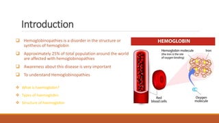
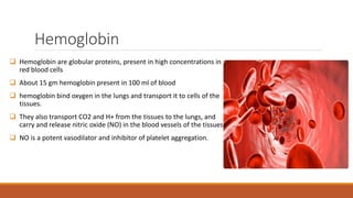
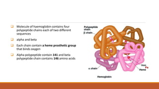
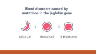
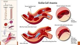
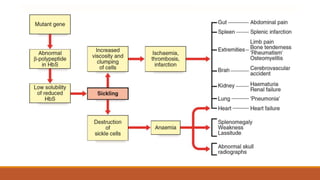
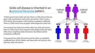
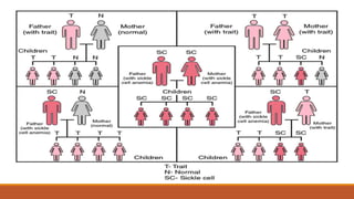
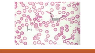
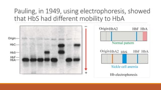
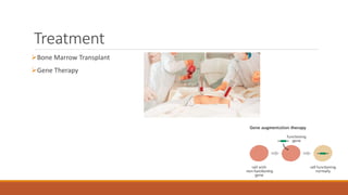
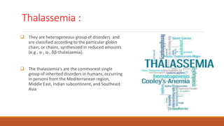
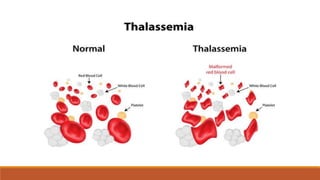
Hemoglobinopathies
Created by –Jadui Hammad group 3
Vaibhav Kushwaha group 1
Tabish Parkar group 1
Overview
Introduction
Hemoglobin
Structure
2,3-BPG
Disorder of Hemoglobin
Structural Disorder-Sickle cell Anemia
Synthesis Disorder-Thalassemia
Introduction
 Hemoglobinopathies is a disorder in the structure or
synthesis of hemoglobin
 Approximately 25% of total population around the world
are affected with hemoglobinopathies
 Awareness about this disease is very important
 To understand Hemoglobinopathies, first we have to
understand
 What is haemoglobin?
 Types of haemoglobin
 Structure of haemoglobin
Hemoglobin
 Hemoglobin are globular proteins, present in high concentrations in
red blood cells
 About 15 gm hemoglobin present in 100 ml of blood
 hemoglobin bind oxygen in the lungs and transport it to cells of the
tissues.
 They also transport CO2 and H+ from the tissues to the lungs, and
carry and release nitric oxide (NO) in the blood vessels of the tissues.
 NO is a potent vasodilator and inhibitor of platelet aggregation.
 Molecule of haemoglobin contains four
polypeptide chains each of two different
sequences
 alpha and beta
 Each chain contain a heme prosthetic group
that binds oxygen
 Alpha polypeptide contain 141 and beta
polypeptide chain contains 146 amino acids
Alpha2 beta2
Alpha2 delta2
Alpha2 gamma2
Structure
 Hemoglobin contains four polypetide chains,each
contain a heme prosthetic group that binds oxygen
 Heme is protoporphyrin lX with an iron atom in its
centre
 Iron is in ferrous(+2) state that can form 5 to 6
covalent bonds depending on oxygen binding
 4 bonds to the pyrrole nitrogen atoms of porphyrin
 5th bond formed from proximal histadine and if
molecule is oxyhemoglobin then 6th bond is bonded
to oxygen otherwise in deoxyhemoglobin it is
unoccupied
Globular protein of
haemoglobin is in the
quaternary structure as
shown in the picture
It consist of multiple alpha
helicles that connected by
turn which help in formation
of spheroidal shape
Generally alpha chain
contains 7 and beta chain
contains 8 alpha helicles
Binding of oxygen to hemoglobin
 Binding of oxygen to haemoglobin involves co-opertivity between subunits
 Binding of first O2 facilitates the binding of another oxygen to next subuits
 Conversly,dissociation of 1 oxygen dissociate all oxygen from molecule as in
deoxyhemoglobin
 R conformational :- the oxy conformation of haemoglobin is relaxed
 T conformational :- the deoxy conformation of haemoglobin is tense
2,3-Biphosphoglycerate(BPG) or 2,3-DPG
 2,3-BPG modulates release of oxygen from haemoglobin
 BPG formed in small amount in all cell during glucose metabolism and its
concentration are equimolar to hemoglobin
 BPG Binds the deoxy haemoglobin and stabilizes the T conformation and increases
its concentration relative to R conformational state
 Then, BPG dissociates as binding of oxygen is done i.e deoxy to oxy hemoglobin
Disorders of hemoglobin
 The disorders of Hb divided into 2 main groups :-
1) Structural globin chain variants such as Sickle cell anaemia
2) Disorders of synthesis of globin chains such as Thalassemia
 Structural variants is due to mutation,there many types of mutation
Point mutation
1) Deletion
2) Insertion
3) Frameshift mutation
4) Chain termination etc.
Sickle Cell Anemia
It is type of Structural Disorder of Human Hemoglobin
Sickle cell anemia is one type of anemia. Anemia is a condition
in which your blood has a lower than normal number of red
blood cells.
This condition also can occur if your red blood cells don’t have
enough hemoglobin.
Sickle cell anemia is a serious disease in which the body makes
sickle-shaped red blood cells. “Sickle-shaped” means that the
red blood cells are shaped like a "C."
Red blood cells are made in the spongy marrow inside the large
bones of the body. Bone marrow is always making new red
blood cells to replace old ones.
Normal red blood cells last about 120 days in the bloodstream
and then die. They carry oxygen and remove carbon dioxide
from your body
In sickle cell anemia, a lower-than-normal number of red blood
cells occurs because sickle cells don’t last very long. Sickle cells
usually die after only about 10 to 20 days. The bone marrow
can’t make new red blood cells fast enough to replace the dying
ones.
Hb S causes red blood cells to become stiff and abnormally shaped. Instead of having a normal round,
disk shape, these red blood cells become sickle-shaped, or crescent-shaped.
These cells don't live as long as normal red blood cells. Because of their shape, they get stuck inside
small blood vessels.
The clumps of sickle cells block blood flow in the blood vessels that lead to the limbs and organs.
Blocked blood vessels can cause pain, serious infections, and organ damage.
 These problems cause symptoms of sickle cell disease.
Signs and Symptoms
Most Common symptom is Fatigue
Shortness of breath
Dizziness
Headache
Coldness in hands and feet
Pale skin
Chest pain
Sickle cell disease is inherited in an
Autosomal Recessive pattern.
If both parents have sickle cell trait, there is a 25% chance that any
given child could be born with sickle cell anemia. There is also a
25% chance that any given child could be completely unaffected.
There is a 50% chance that any given child will get the sickle trait
If one parent has sickle trait and the other has sickle cell anemia,
there is a 50% chance that any given child will get sickle trait and a
50% chance of getting sickle cell anemia. No children will be
completely unaffected
If one parent has sickle cell anemia and the other is completely
unaffected then all the children will have sickle cell trait but none
will have sickle cell anemia.
Individual’s with African,Spanish,Mediterranean,Middle Eastern and Indian ancestry are most
likely to inherit the gene for Sickle cell anemia
The male to female ratio of sickle cell anemia is equal because the gene for sickle cell anemia is
not sex linked.
Heterozygote Advantage
In tropical Africa, where malaria is common:
Homozygous dominant(Normal) HbHb
Reduced Survival or Reproduction from Malaria
Homozygous Recessive HsHs
Reduced Survival and Reproduction from Sickle cell Anemia
Heterozygote Carriers HbHs
Survival and reproduction advantage
Decrease Severity of PLASMODIUM FALCIPARUM Malaria
Sickle cell disease
Sickle cell disease is caused by a mutation in the hemoglobin-Beta gene found on chromosome 11
Sickle cell disease occurs when a person inherits two abnormal copies of the hemoglobin gene, one from
each parent.
Have Life long condition
Sickle cell Trait or Carrier
The heterozygous, or carrier, state for the Sickle Cell allele is known as Sickle cell trait
People with sickle cell trait don’t have the condition,but they have one of the gene that causes condition
 Small increased risk of sudden death associated with strenuous exercise, possible risks from hypoxia on
airplane flights, and anesthesia in pregnant women
The amino acid glutamic acid, at the sixth
position of the β-globin chain, is substituted
by valine .The mutation is therefore a single
base-pair in the triplet code at this point, from
GAG to GTG.
Non Conservative Missense Mutation
Diagnosis
Newborn Blood spot Screen
Blood Smear have Sickled cells
Protein Electrophoresis
Pauling, in 1949, using electrophoresis, showed
that HbS had different mobility to HbA
Treatment
Bone Marrow Transplant
Gene Therapy
Treatment
Use of Hydroxyurea
Increase in Gamma Globin
Increase in fetal Hemoglobin(HbF)
HbF gets in the way of HbS and Prevent Sickling
Thalassemia :
 They are heterogeneous group of disorders and
are classified according to the particular globin
chain, or chains, synthesized in reduced amounts
(e.g., α-, α-, δβ-thalassemia).
 The thalassemia's are the commonest single
group of inherited disorders in humans, occurring
in persons from the Mediterranean region,
Middle East, Indian subcontinent, and Southeast
Asia
α-Thalassemia :
 This results from underproduction of the α-globin chains .
 Occurs most commonly in Southeast Asia but is also prevalent in the Mediterranean, Middle
East, India, and sub-Saharan Africa, with carrier frequencies ranging from 15% to 30%.
 There are two main types of α-thalassemia, with different severity: the severe form, in
which no α chains are produced, is associated with fetal death due to massive edema
secondary to heart failure from severe anemia—hydrops fetalis. Analysis of Hb from such
fetuses reveals a tetramer of γ chains, originally called Hb Bart's.
 In the milder forms of α-thalassemia compatible with survival, although some α chains are
produced, there is still a relative excess of β chains, resulting in production of the β-globin
tetramer Hb H—known as Hb H disease
 Both Hb Bart's and Hb H globin tetramers have an oxygen affinity similar to that of
myoglobin and do not release oxygen as normal to peripheral tissues. Also, Hb H is unstable
and precipitates, resulting in hemolysis of red blood cells.
Mutational Basis of α-Thalassemia :
 The various forms of α-thalassemia are to be mostly the result of deletions of one or more
structural genes for α-Thalassemia present on chromosome 16.
 This result in less production of α globin chain causing α-Thalassemia .
 These deletions are thought to have arisen as a result of unequal crossover events in
meiosis, more likely to occur where genes with homologous sequences are in close
proximity.
 This cause alteration in cells shape.
Symptoms :
 Thalassemia signs and symptoms may include:
 Fatigue
 Weakness
 Pale or yellowish skin
 Facial bone deformities
 Slow growth
 Abdominal swelling
 Dark urine
Treatment :
Individuals with mild forms of alpha thalassemia may not require specific treatment except as
needed for management of low hemoglobin levels. In some patients, supplementation of iron or
folic acid may be useful. Patients with more severe anemia may require lifelong transfusion
therapy. Surgical therapy is considered only in selected cases.
β-Thalassemia :
 Caused due to underproduction of the β-globin chain of Hb.
 Production of β-globin chains may be either reduced (β+) or absent (β0). Individuals
homozygous for β0-thalassemia mutations have severe, transfusion-dependent anemia.
 Children with thalassemia major, or Cooley’s anemia as it was originally known, usually
present in infancy with a severe transfusion-dependent anemia.
 Affected individuals used to die in their teens or early adulthood from complications
resulting from iron overload from repeated transfusions.
Mutational Basis of β-Thalassemia :
 In excess of 100 different mutations have been shown to cause β-thalassemia.
 The various mutations are often unique to certain population groups and can be considered
to fall into six main functional types:
1. Transcriptional mutation
2. m-RNA splicing mutation
3. Polyadenylation signal mutations.
4. RNA modification mutations.
5. Chain termination mutations
Symptoms
Some of the more common symptoms of Beta thalassemia include:
fatigue, weakness, or shortness of breath.
a pale appearance or a yellow color to the skin (jaundice)
irritability.
deformities of the facial bones
slow growth.
a swollen abdomen.
dark urine.
Treatment for beta thalassemia may include:
 Regular blood transfusions.
 Medications (to decrease amount of iron in the body, called chelation therapy)
 Surgical removal of the spleen (if necessary)
 Daily doses of folic acid supplements.
 Monitoring of the gallbladder, liver, and bone density.
 No iron supplements.
Reference
Emery_s Elements of Medical Genetics_14th_Edition
Thomas M. Devlin - Textbook of Biochemistry with Clinical Correlations (2010, John Wiley &
Sons)
 https://emedicine.medscape.com/article/205926-overview
Hemoglobinopathies

Hemoglobinopathies

  • 1.
    Hemoglobinopathies Created by –JaduiHammad group 3 Vaibhav Kushwaha group 1 Tabish Parkar group 1
  • 2.
  • 3.
    Introduction  Hemoglobinopathies isa disorder in the structure or synthesis of hemoglobin  Approximately 25% of total population around the world are affected with hemoglobinopathies  Awareness about this disease is very important  To understand Hemoglobinopathies, first we have to understand  What is haemoglobin?  Types of haemoglobin  Structure of haemoglobin
  • 4.
    Hemoglobin  Hemoglobin areglobular proteins, present in high concentrations in red blood cells  About 15 gm hemoglobin present in 100 ml of blood  hemoglobin bind oxygen in the lungs and transport it to cells of the tissues.  They also transport CO2 and H+ from the tissues to the lungs, and carry and release nitric oxide (NO) in the blood vessels of the tissues.  NO is a potent vasodilator and inhibitor of platelet aggregation.
  • 5.
     Molecule ofhaemoglobin contains four polypeptide chains each of two different sequences  alpha and beta  Each chain contain a heme prosthetic group that binds oxygen  Alpha polypeptide contain 141 and beta polypeptide chain contains 146 amino acids
  • 6.
  • 7.
    Structure  Hemoglobin containsfour polypetide chains,each contain a heme prosthetic group that binds oxygen  Heme is protoporphyrin lX with an iron atom in its centre  Iron is in ferrous(+2) state that can form 5 to 6 covalent bonds depending on oxygen binding  4 bonds to the pyrrole nitrogen atoms of porphyrin  5th bond formed from proximal histadine and if molecule is oxyhemoglobin then 6th bond is bonded to oxygen otherwise in deoxyhemoglobin it is unoccupied
  • 9.
    Globular protein of haemoglobinis in the quaternary structure as shown in the picture It consist of multiple alpha helicles that connected by turn which help in formation of spheroidal shape Generally alpha chain contains 7 and beta chain contains 8 alpha helicles
  • 10.
    Binding of oxygento hemoglobin  Binding of oxygen to haemoglobin involves co-opertivity between subunits  Binding of first O2 facilitates the binding of another oxygen to next subuits  Conversly,dissociation of 1 oxygen dissociate all oxygen from molecule as in deoxyhemoglobin  R conformational :- the oxy conformation of haemoglobin is relaxed  T conformational :- the deoxy conformation of haemoglobin is tense
  • 11.
    2,3-Biphosphoglycerate(BPG) or 2,3-DPG 2,3-BPG modulates release of oxygen from haemoglobin  BPG formed in small amount in all cell during glucose metabolism and its concentration are equimolar to hemoglobin  BPG Binds the deoxy haemoglobin and stabilizes the T conformation and increases its concentration relative to R conformational state  Then, BPG dissociates as binding of oxygen is done i.e deoxy to oxy hemoglobin
  • 12.
    Disorders of hemoglobin The disorders of Hb divided into 2 main groups :- 1) Structural globin chain variants such as Sickle cell anaemia 2) Disorders of synthesis of globin chains such as Thalassemia  Structural variants is due to mutation,there many types of mutation Point mutation 1) Deletion 2) Insertion 3) Frameshift mutation 4) Chain termination etc.
  • 14.
    Sickle Cell Anemia Itis type of Structural Disorder of Human Hemoglobin Sickle cell anemia is one type of anemia. Anemia is a condition in which your blood has a lower than normal number of red blood cells. This condition also can occur if your red blood cells don’t have enough hemoglobin. Sickle cell anemia is a serious disease in which the body makes sickle-shaped red blood cells. “Sickle-shaped” means that the red blood cells are shaped like a "C."
  • 15.
    Red blood cellsare made in the spongy marrow inside the large bones of the body. Bone marrow is always making new red blood cells to replace old ones. Normal red blood cells last about 120 days in the bloodstream and then die. They carry oxygen and remove carbon dioxide from your body In sickle cell anemia, a lower-than-normal number of red blood cells occurs because sickle cells don’t last very long. Sickle cells usually die after only about 10 to 20 days. The bone marrow can’t make new red blood cells fast enough to replace the dying ones.
  • 16.
    Hb S causesred blood cells to become stiff and abnormally shaped. Instead of having a normal round, disk shape, these red blood cells become sickle-shaped, or crescent-shaped. These cells don't live as long as normal red blood cells. Because of their shape, they get stuck inside small blood vessels. The clumps of sickle cells block blood flow in the blood vessels that lead to the limbs and organs. Blocked blood vessels can cause pain, serious infections, and organ damage.  These problems cause symptoms of sickle cell disease.
  • 18.
    Signs and Symptoms MostCommon symptom is Fatigue Shortness of breath Dizziness Headache Coldness in hands and feet Pale skin Chest pain
  • 20.
    Sickle cell diseaseis inherited in an Autosomal Recessive pattern. If both parents have sickle cell trait, there is a 25% chance that any given child could be born with sickle cell anemia. There is also a 25% chance that any given child could be completely unaffected. There is a 50% chance that any given child will get the sickle trait If one parent has sickle trait and the other has sickle cell anemia, there is a 50% chance that any given child will get sickle trait and a 50% chance of getting sickle cell anemia. No children will be completely unaffected If one parent has sickle cell anemia and the other is completely unaffected then all the children will have sickle cell trait but none will have sickle cell anemia.
  • 22.
    Individual’s with African,Spanish,Mediterranean,MiddleEastern and Indian ancestry are most likely to inherit the gene for Sickle cell anemia The male to female ratio of sickle cell anemia is equal because the gene for sickle cell anemia is not sex linked.
  • 23.
    Heterozygote Advantage In tropicalAfrica, where malaria is common: Homozygous dominant(Normal) HbHb Reduced Survival or Reproduction from Malaria Homozygous Recessive HsHs Reduced Survival and Reproduction from Sickle cell Anemia Heterozygote Carriers HbHs Survival and reproduction advantage Decrease Severity of PLASMODIUM FALCIPARUM Malaria
  • 24.
    Sickle cell disease Sicklecell disease is caused by a mutation in the hemoglobin-Beta gene found on chromosome 11 Sickle cell disease occurs when a person inherits two abnormal copies of the hemoglobin gene, one from each parent. Have Life long condition
  • 25.
    Sickle cell Traitor Carrier The heterozygous, or carrier, state for the Sickle Cell allele is known as Sickle cell trait People with sickle cell trait don’t have the condition,but they have one of the gene that causes condition  Small increased risk of sudden death associated with strenuous exercise, possible risks from hypoxia on airplane flights, and anesthesia in pregnant women
  • 26.
    The amino acidglutamic acid, at the sixth position of the β-globin chain, is substituted by valine .The mutation is therefore a single base-pair in the triplet code at this point, from GAG to GTG. Non Conservative Missense Mutation
  • 27.
    Diagnosis Newborn Blood spotScreen Blood Smear have Sickled cells Protein Electrophoresis
  • 29.
    Pauling, in 1949,using electrophoresis, showed that HbS had different mobility to HbA
  • 30.
  • 31.
    Treatment Use of Hydroxyurea Increasein Gamma Globin Increase in fetal Hemoglobin(HbF) HbF gets in the way of HbS and Prevent Sickling
  • 32.
    Thalassemia :  Theyare heterogeneous group of disorders and are classified according to the particular globin chain, or chains, synthesized in reduced amounts (e.g., α-, α-, δβ-thalassemia).  The thalassemia's are the commonest single group of inherited disorders in humans, occurring in persons from the Mediterranean region, Middle East, Indian subcontinent, and Southeast Asia
  • 33.
    α-Thalassemia :  Thisresults from underproduction of the α-globin chains .  Occurs most commonly in Southeast Asia but is also prevalent in the Mediterranean, Middle East, India, and sub-Saharan Africa, with carrier frequencies ranging from 15% to 30%.  There are two main types of α-thalassemia, with different severity: the severe form, in which no α chains are produced, is associated with fetal death due to massive edema secondary to heart failure from severe anemia—hydrops fetalis. Analysis of Hb from such fetuses reveals a tetramer of γ chains, originally called Hb Bart's.
  • 34.
     In themilder forms of α-thalassemia compatible with survival, although some α chains are produced, there is still a relative excess of β chains, resulting in production of the β-globin tetramer Hb H—known as Hb H disease  Both Hb Bart's and Hb H globin tetramers have an oxygen affinity similar to that of myoglobin and do not release oxygen as normal to peripheral tissues. Also, Hb H is unstable and precipitates, resulting in hemolysis of red blood cells.
  • 36.
    Mutational Basis ofα-Thalassemia :  The various forms of α-thalassemia are to be mostly the result of deletions of one or more structural genes for α-Thalassemia present on chromosome 16.  This result in less production of α globin chain causing α-Thalassemia .  These deletions are thought to have arisen as a result of unequal crossover events in meiosis, more likely to occur where genes with homologous sequences are in close proximity.  This cause alteration in cells shape.
  • 38.
    Symptoms :  Thalassemiasigns and symptoms may include:  Fatigue  Weakness  Pale or yellowish skin  Facial bone deformities  Slow growth  Abdominal swelling  Dark urine
  • 40.
    Treatment : Individuals withmild forms of alpha thalassemia may not require specific treatment except as needed for management of low hemoglobin levels. In some patients, supplementation of iron or folic acid may be useful. Patients with more severe anemia may require lifelong transfusion therapy. Surgical therapy is considered only in selected cases.
  • 41.
    β-Thalassemia :  Causeddue to underproduction of the β-globin chain of Hb.  Production of β-globin chains may be either reduced (β+) or absent (β0). Individuals homozygous for β0-thalassemia mutations have severe, transfusion-dependent anemia.  Children with thalassemia major, or Cooley’s anemia as it was originally known, usually present in infancy with a severe transfusion-dependent anemia.  Affected individuals used to die in their teens or early adulthood from complications resulting from iron overload from repeated transfusions.
  • 42.
    Mutational Basis ofβ-Thalassemia :  In excess of 100 different mutations have been shown to cause β-thalassemia.  The various mutations are often unique to certain population groups and can be considered to fall into six main functional types: 1. Transcriptional mutation 2. m-RNA splicing mutation 3. Polyadenylation signal mutations. 4. RNA modification mutations. 5. Chain termination mutations
  • 43.
    Symptoms Some of themore common symptoms of Beta thalassemia include: fatigue, weakness, or shortness of breath. a pale appearance or a yellow color to the skin (jaundice) irritability. deformities of the facial bones slow growth. a swollen abdomen. dark urine.
  • 45.
    Treatment for betathalassemia may include:  Regular blood transfusions.  Medications (to decrease amount of iron in the body, called chelation therapy)  Surgical removal of the spleen (if necessary)  Daily doses of folic acid supplements.  Monitoring of the gallbladder, liver, and bone density.  No iron supplements.
  • 46.
    Reference Emery_s Elements ofMedical Genetics_14th_Edition Thomas M. Devlin - Textbook of Biochemistry with Clinical Correlations (2010, John Wiley & Sons)  https://emedicine.medscape.com/article/205926-overview