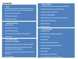
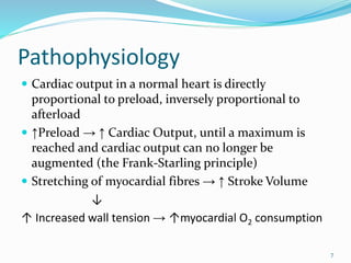
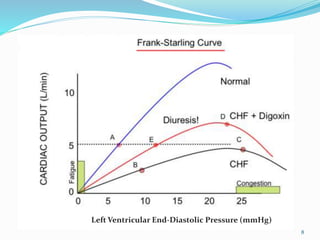
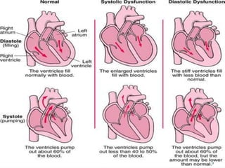
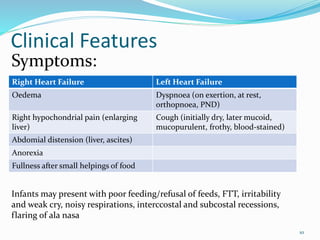
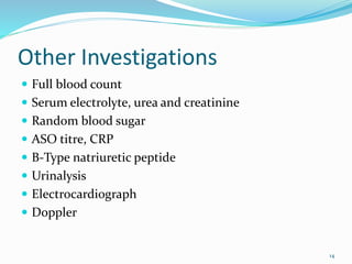
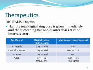
Dr. Eke Eghosasere Paul gave a presentation on pediatric heart failure to the Nelson Club on September 15, 2014. The presentation covered the epidemiology, etiology, pathophysiology, clinical signs and symptoms, diagnosis, treatment and prognosis of heart failure in children. Heart failure occurs when the heart cannot meet the body's metabolic needs due to reduced cardiac output. Compensatory mechanisms initially help maintain function but eventually become ineffective, leading to worsening clinical symptoms. Proper diagnosis and management of the underlying cause are important for treatment.
presentation regarding investigations and treatment of heart failure in pediatrics, including the management of an emergency , and includes brief description about even drugs used
Definition, classification, epidemiology, etiology, diagnosis, prognosis of DCM, HOCM, LVNC
Also review of acute myocarditis in children
R/v of heart failure management
A cyanotic heart defect is a group-type of congenital heart defects (CHDs). The patient appears blue (cyanotic), due to deoxygenated blood bypassing the lungs and entering the systemic circulation. This can be caused by right-to-left or bidirectional shunting, or malposition of the great arteries.
Cyanotic heart defects, which account for approximately 25% of all CHDs, include:
Tetralogy of Fallot (ToF)
Total anomalous pulmonary venous connection
Hypoplastic left heart syndrome (HLHS)
Transposition of the great arteries (d-TGA)
Truncus arteriosus (Persistent)
Tricuspid atresia
Interrupted aortic arch
Pulmonary atresia (PA)
Pulmonary stenosis (critical)
Eisenmenger syndrome(Reversal of Shunt due to Pulmonary Hypertension) .
Patent ductus arteriosus may cause cyanosis in late stage.
Congenital heart disease is one or more problems with the heart's structure that exist since birth. Congenital means that you're born with the defect. Congenital heart disease, also called congenital heart defect, can change the way blood flows through your heart. IF YOU LIKE GIVE YOUR LIKES AND FOLLOW THIS LINK
3. Introduction
DEFINITION OF TERMS
Cardiac Output: the amount of blood the heart pumps
through the circulatory system in a minute
Stroke Volume: the amount of blood put out by the left
ventricle in one contraction
Cardiac Ouput = Stroke Volume X Heart Rate
Preload: the magnitude of the maximal (end-diastolic)
ventricular volume or the end-diastolic pressure
stretching the ventricles
Afterload: the resistance against which the left
ventricle must eject its volume of blood during
contraction 3
4. Introduction Contd.
Heart failure occurs when the heart cannot deliver
adequate cardiac output to meet the metabolic needs
of the body
In the early stages of heart failure, various
compensatory mechanisms are evoked to maintain
normal metabolic function
When these mechanisms become ineffective,
increasingly severe clinical manifestations result
4
5. Epidemiology
Accurately estimating the incidence in children is
problematic
In USA, incidence of heart failure due to congenital
defects is between 1-2 per 1000 live births
Cardiomyopathy contributes significantly to pediatric
cases that present with heart failure (0.87 per 100,000
in the UK)
Data from Nigeria suggests that 7.02% of emergency
paediatric admissions to a tertiary hospital are for
cardiac failure
Over 90% of those cases are from lower socio-
economic groups
5
7. Pathophysiology
Cardiac output in a normal heart is directly
proportional to preload, inversely proportional to
afterload
↑Preload → ↑ Cardiac Output, until a maximum is
reached and cardiac output can no longer be
augmented (the Frank-Starling principle)
Stretching of myocardial fibres → ↑ Stroke Volume
↓
↑ Increased wall tension → ↑myocardial O2 consumption
7
10. Clinical Features
Right Heart Failure Left Heart Failure
Oedema Dyspnoea (on exertion, at rest,
orthopnoea, PND)
Right hypochondrial pain (enlarging
liver)
Cough (initially dry, later mucoid,
mucopurulent, frothy, blood-stained)
Abdomial distension (liver, ascites)
Anorexia
Fullness after small helpings of food
10
Symptoms:
Infants may present with poor feeding/refusal of feeds, FTT, irritability
and weak cry, noisy respirations, interccostal and subcostal recessions,
flaring of ala nasa
11. Right Heart Failure Left Heart Failure
Ankle oedema Dyspnoea
Tender hepatomegaly Basal crepititions + rhonchi
Ascites S3 or S4 gallop rhythm
Raised JVP, pulsatile Pulsus alternans
11
Signs:
• Tachycardia
• Tachypnoea with respiratory distress
• Weak peripheral pulses and/or delayed capillary refill
• Muffled heart sounds
• Murmurs of the original disease
• Arrhythmias may be present
16. Treatment
EMERGENCY MANAGEMENT
ABC, oxygen inhalation
Connect to a cardiac monitor
Secure an IV line
If in shock, intubate and ventilate
Keep fluid input/output chart
Fluid restriction 70% ml/kg/day
If baby is tachypnoeic, consider NG tube feeding
Monitor serum electrolytes frequently (especially
potassium)
16
17. General Management
Bed rest, restriction of activities
Diet: salt and water restriction (older
children), increased caloric intake, NG tube
feeding
17
18. Therapeutics
DIGITALIS: Digoxin
Half the total digitalizing dose is given immediately
and the succeeding two one-quarter doses at 12 hr
intervals later
18
Age (Years) Digitalization
(mg/kg/24hr)
Maintenance (mg/kg/24hr)
< 1 month 0.04 — 0.06 0.01
1 month – 2years 0.04 — 0.08 0.01 — 0.02
> 2 years 0.04 — 0.06 0.01
Adult 0.5 — 1.0
(mg/24hrs)
0.25 — 0.5
(mg/24hrs)
20. Prognosis
Depends on the underlying cause, stage of
presentation at the hospital, early/accurate diagnosis,
speed of instituting correct therapy, socioeconomic
factor, availability of specialized treatment centres for
surgeries
Follow-up
20
21. Conclusion
Heart failure is a common clinical condition in
children which can present at any age
Cases of heart failure should be thoroughly assessed
and investigated for underlying cause for appropriate
diagnosis/treatment
Management is usually multifaceted involving several
departments
Follow up is essential to monitor progress, and ensure
proper development of the child
21
22. References
Nelson Textbook of Paediatrics, 19th Edition; Heart
Failure
Paediatrics and Child Health in a Tropical Region 2nd
Edition, by Azubuike and Nkanginieme; Heart Failure
in Childhood
Medscape Article: Paediatric Congestive Heart Failure
Approach to Paediatric Emergency, by Jaydeep
Choudhury and Jayanta Bandyopadhyay
A Compendium of Clinical Medicine by A.O. Falase
and O.O. Akinkugbe
22