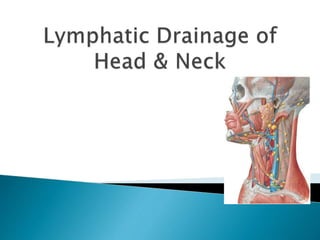
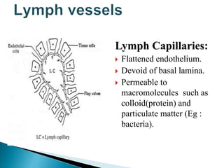
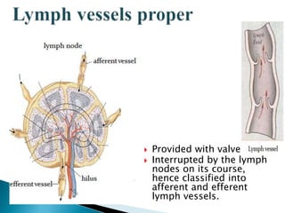
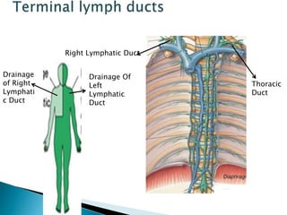
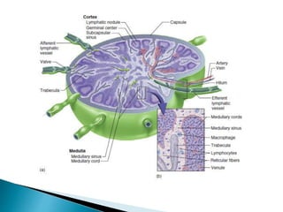
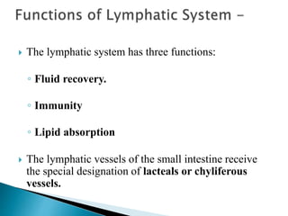
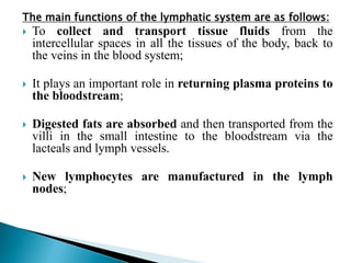
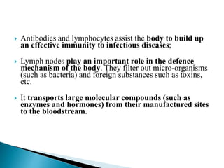
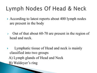
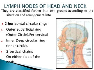
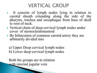
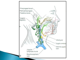
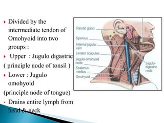
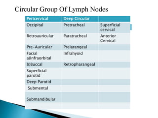
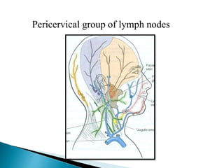
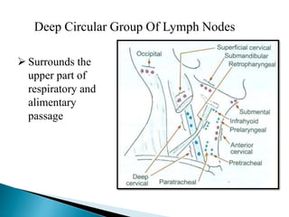
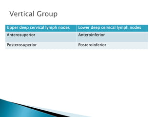
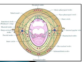
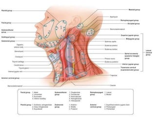
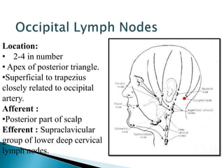
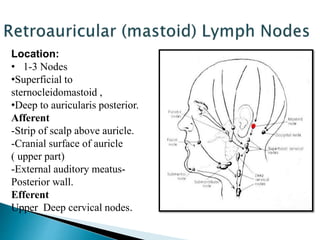
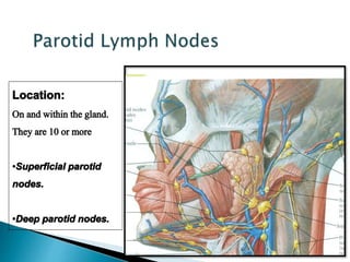
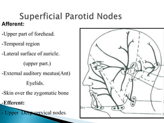
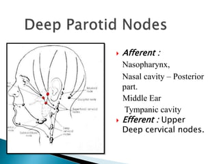
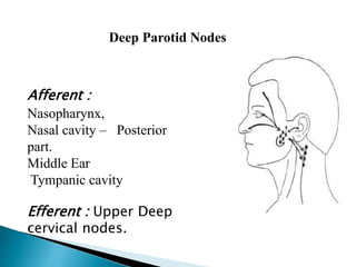
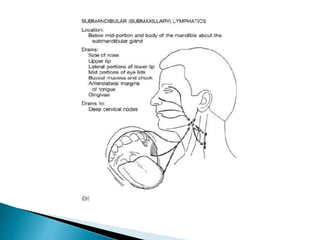
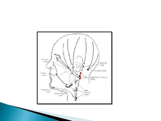
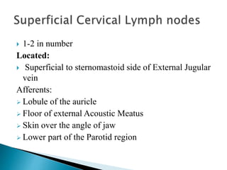
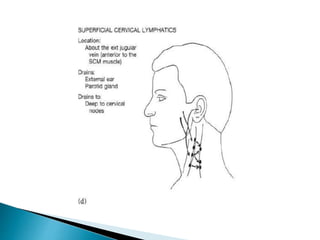
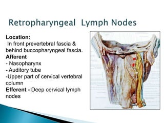
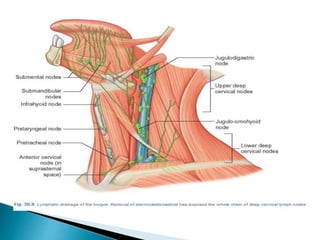
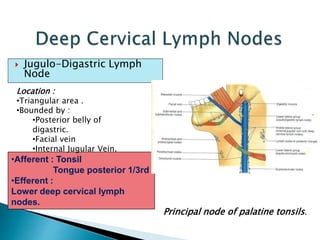
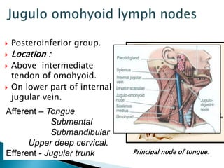
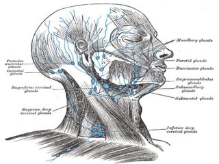
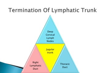
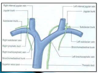
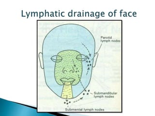
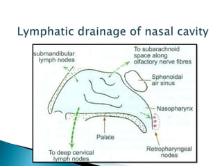
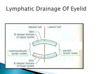
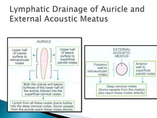
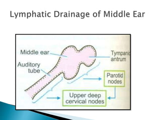
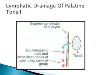
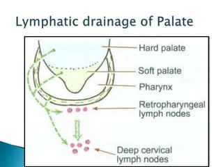
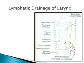
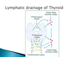
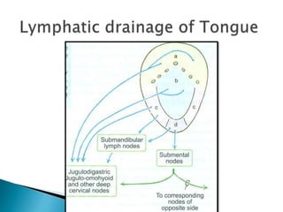
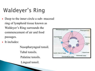
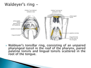
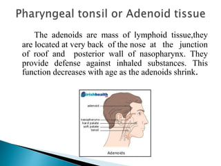
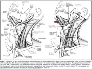
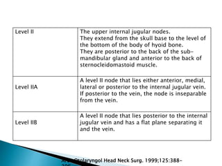
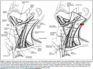
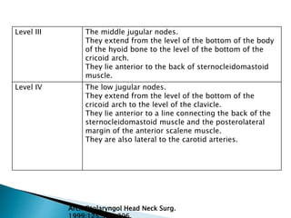
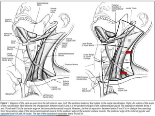
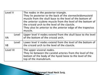
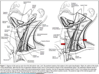
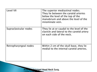
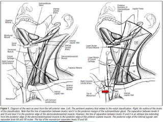
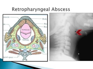
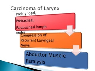
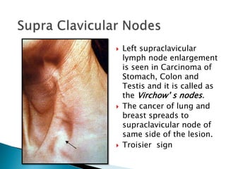
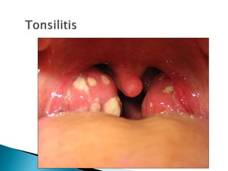
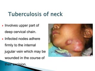
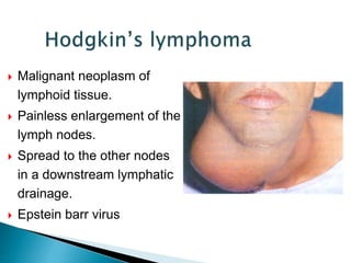
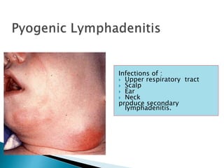
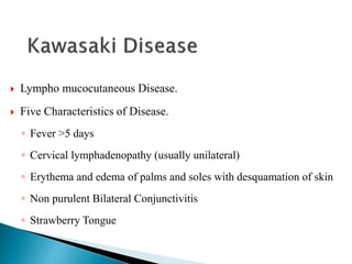
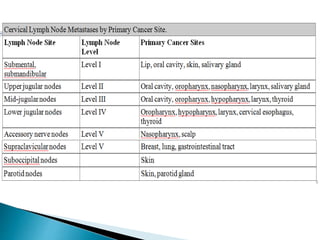
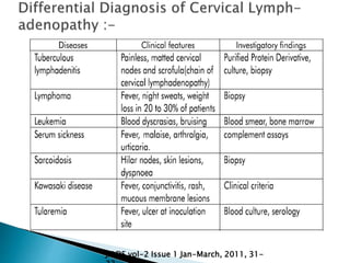
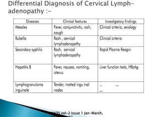
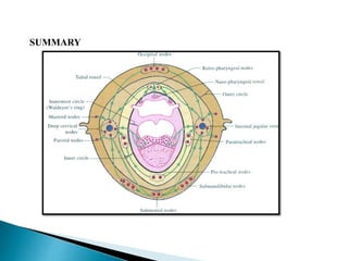
Divya C
 Introduction
 Historical Perspective & Current View
 Embryological Development
 Functions of Lymphatic System
 Lymph Nodes of Head & Neck
 Lymphatic Drainage
 Applied Aspects
 Clinical Assessment
 Laboratory Investigations
 Differential Diagnosis
 Conclusion
 Of all the body systems, the lymphatic system is
perhaps the least familiar to most people. Yet without
it, neither the circulatory system nor the immune
system could function.
 The lymphatic system is an endothelium-lined
network of blind-ended capillaries found in nearly all
tissues .
The components of the lymphatic system are :-
 Lymph, the recovered fluid;
 Lymphatic vessels, which transport the lymph;
 Lymphatic tissue, composed of aggregates of
lymphocytes and macrophages that populate many
organs of the body; and
 lymphatic organs, in which these cells are especially
concentrated and which are set off from surrounding
organs by connective tissue capsules.
Lymph Capillaries:
 Flattened endothelium.
 Devoid of basal lamina.
 Permeable to
macromolecules such as
colloid(protein) and
particulate matter (Eg :
bacteria).
 Provided with valves.
 Interrupted by the lymph
nodes on its course,
hence classified into
afferent and efferent
lymph vessels.
Drainage
of Right
Lymphati
c Duct
Drainage Of
Left
Lymphatic
Duct
Right Lymphatic Duct
Thoracic
Duct
 Lymph nodes serve two functions:
◦ to cleanse the lymph and
◦ alert the immune system to pathogens.
 There are hundreds of lymph nodes in the body.
 They are especially concentrated in the cervical,
axillary, and inguinal regions close to the body
surface, and in thoracic, abdominal, and pelvic
groups deep in the body cavities.
 Most of them are embedded in fat.
 Structure –
 A lymph node is an elongated or bean-shaped
structure, usually less than 3 cm long, often with an
indentation called the hilum on one side.
 It is enclosed in a fibrous capsule with extensions
(trabeculae) that incompletely divide the interior of
the node into compartments.
 The interior consists of
◦ a stroma of reticular connective tissue (reticular
fibers and reticular cells) and
◦ a parenchyma of lymphocytes and antigen-
presenting cells.
 Hippocrates first described vessels containing “white
blood” around 400 B.C.
 Gasparo Aselli re-identified lymphatic vessels in the
1600’s, noting the presence of lipid-filled “milky
veins” in the gut of a “well-fed” dog (Aselli, 1627).
 Historically, the most widely accepted view of
lymphatic development was proposed by Sabin in the
early twentieth century (Sabin 1902, 1904).
 Sabin’s Model :
 The isolated primitive lymph sacs originate from
endothelial cells that bud from the veins during early
development.
 The two jugular lymph sacs were proposed to develop in
the junction of the sub-clavian and anterior cardinal veins
by endothelial budding from the anterior cardinal veins.
 The remaining lymph sacs originate from the mesonephric
vein
 Alternative Model / Centripetal Model :-
 proposed by Huntington and McClure 1910.
 They suggested that the primary lymph sacs arise in the
mesenchyme, independent of the veins, and secondarily
establish venous connections.
 This model was supported by Schneider et al. 1999.
 Perhaps the most definitive evidence for a venous
origin for early lymphatic endothelial cells has come
from the zebra fish (Yaniv et al., 2006).
 Recent studies have shown that the zebra fish
possesses a lymphatic vascular system with many of
the morphological, molecular, and functional
characteristics of the lymphatic's of other vertebrates.
 6 primary Lymph Sacs
2 Jugular
2 Iliac
1 retroperitoneal
1 cisterna chili
 Thoracic duct
 Right Lymphatic duct
 Lymph Nodes
 The lymphatic system has three functions:
◦ Fluid recovery.
◦ Immunity
◦ Lipid absorption
 The lymphatic vessels of the small intestine receive
the special designation of lacteals or chyliferous
vessels.
The main functions of the lymphatic system are as follows:
 To collect and transport tissue fluids from the
intercellular spaces in all the tissues of the body, back to
the veins in the blood system;
 It plays an important role in returning plasma proteins to
the bloodstream;
 Digested fats are absorbed and then transported from the
villi in the small intestine to the bloodstream via the
lacteals and lymph vessels.
 New lymphocytes are manufactured in the lymph
nodes;
 Antibodies and lymphocytes assist the body to build up
an effective immunity to infectious diseases;
 Lymph nodes play an important role in the defence
mechanism of the body. They filter out micro-organisms
(such as bacteria) and foreign substances such as toxins,
etc.
 It transports large molecular compounds (such as
enzymes and hormones) from their manufactured sites
to the bloodstream.
 According to latest reports about 400 lymph nodes
are present in the body
 Out of that about 60-70 are present in the region of
head and neck.
 Lymphatic tissue of Head and neck is mainly
classified into two groups
A) Lymph glands of Head and Neck
B) Waldeyer’s ring
 2 horizontal circular rings
1. Outer superficial ring
(Outer Circle).Pericervical
2. Inner Deep circular ring
(inner circle).
 2 vertical chains
On either side of the
neck.
They are classified further into two groups according to the
situation and arrangement into
 It consists of lymph nodes lying in relation to
carotid sheath extending along the side of the
pharynx, trachea and oesophagus from base of skull
to root of neck.
 Vertical chain of deep cervical lymph nodes under
cover of sternocleidomastoid
 By bifurcation of common carotid artery they are
arbitrarily divided into
a) Upper Deep cervical lymph nodes
b) Lower deep cervical lymph nodes
Both the groups are in relation
with internal jugular vein
 Divided by the
intermediate tendon of
Omohyoid into two
groups :
 Upper : Jugulo digastric
( principle node of tonsil )
 Lower : Jugulo
omohyoid
(principle node of tongue)
• Drains entire lymph from
head & neck
 All lymph vessels of the head and neck drain into
the deep cervical nodes, either directly from the
tissues or indirectly via nodes in outlying groups.
 Lymph is returned to the systemic venous
circulation via either the right lymphatic duct or
the thoracic duct.
Pericervical Deep Circular
Occipital Pretracheal Superficial
cervical
Retroauricular Paratracheal Anterior
Cervical
Pre-Auricular Prelarangeal
Facial
a)Infraorbital
Infrahyoid
b)Buccal Retropharangeal
Superficial
parotid
Deep Parotid
Submental
Submandibular
Circular Group Of Lymph Nodes
Pericervical group of lymph nodes
Deep Circular Group Of Lymph Nodes
 Surrounds the
upper part of
respiratory and
alimentary
passage
Upper deep cervical lymph nodes Lower deep cervical lymph nodes
Anterosuperior Anteroinferior
Posterosuperior Posteroinferior
Location:
• 2-4 in number
• Apex of posterior triangle.
•Superficial to trapezius
closely related to occipital
artery.
Afferent :
•Posterior part of scalp
Efferent : Supraclavicular
group of lower deep cervical
lymph nodes.
Location:
• 1-3 Nodes
•Superficial to
sternocleidomastoid ,
•Deep to auricularis posterior.
Afferent
-Strip of scalp above auricle.
-Cranial surface of auricle
( upper part)
-External auditory meatus-
Posterior wall.
Efferent
Upper Deep cervical nodes.
Afferent:
-Upper part of forehead.
-Temporal region
-Lateral surface of auricle.
(upper part.)
-External auditory meatus(Ant)
Eyelids.
-Skin over the zygomatic bone
-Efferent:
- Upper Deep cervical nodes.
 Afferent :
Nasopharynx,
Nasal cavity – Posterior
part.
Middle Ear
Tympanic cavity
 Efferent : Upper
Deep cervical nodes.
Afferent :
Nasopharynx,
Nasal cavity – Posterior
part.
Middle Ear
Tympanic cavity
Efferent : Upper Deep
cervical nodes.
Deep Parotid Nodes
 Lies in superficial group of parotid nodes
 Lies immediately in front of tragus
 Anterior to ear over parotid fascia
Afferents
1.Drains areas supplied by superficial temporal artery
2.Anterior parietal scalp
3.Anterior surface of ear
Efferents
 Upper deep cervical group of lymph nodes
Superficial
 Upto 12 nodes are present they include
 Buccal
 Mandibular
 Maxillary
 Its distributed along the course of Facial artery and vein
Afferent :
 Skin and mucous membrane of eyelid ,Nose and Cheek
Efferents:
 Upper deep cervical lymph nodes
 Location: On buccinator
muscle
 It lies in relation to the
facial vein
 Afferent: Cheek ,lower
eyelid
 Efferent: Submandibular
lymph nodes
Location:
 Lower border of mandible
 Near Anteronferior Angle of Masseter
 Close relation to Marginal Mandibular Nerve
 Drains the cheek and lower eyelid
 Distributed along course of maxillary artery lateral to
lateral pterygoid muscle
Afferents
 Temporal and infratemporal fossa
Nasal pharynx
Efferents
 Upper deep cervical lymph nodes
 3-4 in number
 Location: On the mylohyoid
muscle between anterior bellies
of both digastric.
 Afferent :
- Tip of tongue.
- Floor of mouth.
- Lower Incisor teeth and
associated gum.
- Central part of lower lip.
- Skin over chin
Efferent :
 Submandibular nodes
 Deep cervical lymph nodes
 Jugulo-omohyoid nodes
Location:
Digastric triangle
on submandibular salivary
gland.
Afferent –
• Center of forehead.
 Medial angle of eye.
 - Nose and adjacent cheek
 - Upper lip
- Lower lip (except center part ).
- Anterior 2/3 of tongue (except tip)
 - Upper and lower teeth (except
lower incisor)
 -Floor of mouth.
 -Frontal, maxillary, ethmoidal air
sinus
 Submental lymph nodes
Efferent -
• Lower Deep cervical lymph nodes
 1-2 in number
Located:
 Superficial to sternomastoid side of External Jugular
vein
Afferents:
 Lobule of the auricle
 Floor of external Acoustic Meatus
 Skin over the angle of jaw
 Lower part of the Parotid region
Efferents:
 Anterior border of sternocleidomastoid to reach
Upper deep cervical group of lymph nodes
 Some passes through external Jugular vein into lower
deep cervical in the subclavian triangle
 Location
 Along anterior jugular
vein.
 Afferent –
 Front of the neck below
the hyoid bone.
 Efferent – Lower deep
cervical lymph nodes.
Anterior cervical
node
 Prelaryngeal :
On Cricovocal membrane.
 Pretracheal :
Infront of trachea.
Above the isthmus of thyroid.
Prelaryngea
l nodes
Pretracheal
nodes
Location:
 Sides of trachea & Esophagus
 Along recurrent laryngeal nerve
Afferent
- Larynx below the vocal fold.
- Trachea
- Esophagus
- Thyroid gland.
Efferent –
 Lower deep cervical lymph nodes
Location:
In front prevertebral fascia &
behind buccopharyngeal fascia.
Afferent
- Nasopharynx
- Auditory tube
-Upper part of cervical vertebral
column
Efferent - Deep cervical lymph
nodes
 Jugulo-Digastric Lymph
Node
•Afferent : Tonsil
Tongue posterior 1/3rd
•Efferent :
Lower deep cervical lymph
nodes.
Location :
•Triangular area .
•Bounded by :
•Posterior belly of
digastric.
•Facial vein
•Internal Jugular Vein.
Principal node of palatine tonsils.
 Posteroinferior group.
 Location :
 Above intermediate
tendon of omohyoid.
 On lower part of internal
jugular vein.
Afferent – Tongue
Submental
Submandibular
Upper deep cervical.
Efferent - Jugular trunk Principal node of tongue.
Deep
Cervical
Lymph
Nodes
Right
Lymphatic
Duct
Jugular
trunk
Thoracic
Duct
 Deep to the inner circle a sub- mucosal
ring of lymphoid tissue known as
Waldeyer’s Ring surrounds the
commencement of air and food
passages.
 It includes:
Nasopharyngeal tonsil.
Tubal tonsils.
Palatine tonsils.
Lingual tonsil.
 Waldeyer's tonsillar ring, consisting of an unpaired
pharyngeal tonsil in the roof of the pharynx, paired
palatine tonsils and lingual tonsils scattered in the
root of the tongue.
The adenoids are mass of lymphoid tissue,they
are located at very back of the nose at the junction
of roof and posterior wall of nasopharynx. They
provide defense against inhaled substances. This
function decreases with age as the adenoids shrink.
 Tubal tonsil: It is a collection of lymphoid nodules
near the pharyngeal opening of the auditory tube
Tubal tonsil
It is an collection of lymphoid tissue deep to the
mucous membrane of the posterior 1/3rd of tongue.
Forms a part of waldayer’s lymphatic ring
Lingual tonsil
 A small oral mass of lymphoid tissue, especially
either of two such masses embedded in the lateral
walls of the opening between the mouth and the
pharynx. They are thought to produce antibodies to
help prevent respiratory and digestive tract infection
but often become infected themselves mostly in
children.
Palatine
tonsil
Level I The sub-mental and sub-mandibular nodes.
They lie above the hyoid bone, below the
mylohoid muscle and anterior to the back of
the sub-mandibular gland.
Level IA The sub-mental nodes.
They lie between the medial margins of the
anterior bellies of the diagastric muscles.
Level IB The sub-mandibular nodes.
On each side, they lie lateral to the level IA
nodes and anterior to the back of each sub-
mandibular gland.
Imaging-based nodal classification :-
1998 modification of the 1991 AAO-HNS (American
Academy of
Otolaryngology – Head and Neck Surgery) classification
Arch Otolaryngol Head Neck Surg. 1999;125:388-
Level II The upper internal jugular nodes.
They extend from the skull base to the level of
the bottom of the body of hyoid bone.
They are posterior to the back of the sub-
mandibular gland and anterior to the back of
sternocleidomastoid muscle.
Level IIA
A level II node that lies either anterior, medial,
lateral or posterior to the internal jugular vein.
If posterior to the vein, the node is inseparable
from the vein.
Level IIB
A level II node that lies posterior to the internal
jugular vein and has a flat plane separating it
and the vein.
Arch Otolaryngol Head Neck Surg. 1999;125:388-
Level III The middle jugular nodes.
They extend from the level of the bottom of the body
of the hyoid bone to the level of the bottom of the
cricoid arch.
They lie anterior to the back of sternocleidomastoid
muscle.
Level IV The low jugular nodes.
They extend from the level of the bottom of the
cricoid arch to the level of the clavicle.
They lie anterior to a line connecting the back of the
sternocleidomastoid muscle and the posterolateral
margin of the anterior scalene muscle.
They are also lateral to the carotid arteries.
Arch Otolaryngol Head Neck Surg.
Level V The nodes in the posterior triangle.
They lie posterior to the back of the sternocleidomastoid
muscle from the skull base to the level of the bottom of
the anterior scalene muscle from the level of the bottom of
the cricoid arch to the level of the clavicle.
They also lie anterior to the anterior edge of the trapezius
muscle.
Level
VA
Upper level V nodes extend from the skull base to the level
of the bottom of the cricoid arch.
Level
VB
Lower level V nodes extend from the level of the bottom of
the cricoid arch to the level of the clavicle.
Level VI The upper visceral nodes.
They lie between the carotid arteries from the level of the
bottom of the body of the hyoid bone to the level of the
top of the manubrium.
Arch Otolaryngol Head Neck Surg.
Level VII The superior mediastinal nodes.
They lie between the carotid arteries
below the level of the top of the
manubrium and above the level of the
innominate vein.
Supraclavicular nodes They lie at or caudal to the level of the
clavicle and lateral to the carotid artery
on each side of the neck.
Retropharyngeal nodes Within 2 cm of the skull base, they lie
medial to the internal carotid arteries.
Arch Otolaryngol Head Neck Surg.
 When a lymph node is under challenge from a
foreign antigen, it may become swollen and painful
to the touch— a condition called lymphadenitis.
 Commonly palpated and accessible lymph nodes are
- the cervical, axillary, and inguinal.
 Lymph nodes are common sites of metastatic cancer
because cancer cells from almost any organ can
break loose, enter the lymphatic capillaries, and
lodge in the nodes.
 Lymphadenopathy is a collective term for all
lymph node diseases
 Left supraclavicular
lymph node enlargement
is seen in Carcinoma of
Stomach, Colon and
Testis and it is called as
the Virchow’ s nodes.
 The cancer of lung and
breast spreads to
supraclavicular node of
same side of the lesion.
 Troisier sign
 Involves upper part of
deep cervical chain.
 Infected nodes adhere
firmly to the internal
jugular vein which may be
wounded in the course of
their excision.
 Malignant neoplasm of
lymphoid tissue.
 Painless enlargement of the
lymph nodes.
 Spread to the other nodes
in a downstream lymphatic
drainage.
 Epstein barr virus
Infections of :
 Upper respiratory tract
 Scalp
 Ear
 Neck
produce secondary
lymphadenitis.
 Lympho mucocutaneous Disease.
 Five Characteristics of Disease.
◦ Fever >5 days
◦ Cervical lymphadenopathy (usually unilateral)
◦ Erythema and edema of palms and soles with desquamation of skin
◦ Non purulent Bilateral Conjunctivitis
◦ Strawberry Tongue
 Lymphadenopathy - enlargement of the lymph
nodes.
 It may be an incidental finding in patients being
examined for various reasons, or it may be a
presenting sign or symptom of the patient's illness.
 Soft, flat, submandibular nodes (<1 cm) are often
palpable in healthy children and young adults;
 .
 The physician will be aided in the pursuit of an
explanation for the lymph-adenopathy by
◦ a careful medical history,
◦ physical examination,
◦ selected laboratory tests, and
◦ an excisional lymph node biopsy.
 Medical History :-
◦ It should reveal the setting in which
lymphadenopathy is occurring.
◦ Symptoms such as sore throat, cough, fever, night
sweats, fatigue, weight loss, or pain in the nodes
should be sought.
◦ The patient's age, sex, occupation, exposure to
pets, sexual behavior, and use of drugs such as
diphenylhydantoin are other important historic
points.
 Physical examination :-
 It can provide useful clues such as
◦ the extent of lymphadenopathy (localized or
generalized),
◦ size of nodes,
◦ texture,
◦ presence or absence of nodal tenderness,
◦ signs of inflammation over the node,
◦ skin lesions, and
◦ splenomegaly.
 The texture of lymph nodes may be described as
soft, firm, rubbery, hard, discrete, matted.
 It may be tender or non-tender.
 It may be movable or fixed.
 Tenderness is found when the capsule is stretched
during rapid enlargement, usually secondary to an
inflammatory process.
 Some malignant diseases such as acute leukemia
may produce rapid enlargement and pain in the
nodes.
 PALPATION OF LYMPH NODES –
 Lymph node and chain palpation starts with the parotid
and preauricular area which may also be palpated
bimanually.
 Palpating with light finger pressure against underlying
firm tissues (bone or muscle), or bimanually where
appropriate.
 The head and neck lymph examination continues down
the mandible to the submandibular region where
bilateral palpation proceeds forward to the submental
nodes just under the chin.
 With the patient seated upright, head tipped slightly
forward, the cervical lymphatic chains are palpated
against the sternocleidomastoid muscle.
 Superficial cervicals lymph nodes are found along
the anterior border, and deep superior and inferior
chains found along the posterior border.
 The laboratory investigation of patients with
lymphadenopathy must be tailored to elucidate the
etiology suspected from the patient's history and
physical findings.
 Complete Blood Count, CBC
provide useful data for the diagnosis of
◦ acute or chronic leukemias,
◦ EBV or CMV mononucleosis,
◦ lymphoma with a leukemic component,
◦ pyogenic infections, or
◦ immune cytopenias in illnesses such as SLE.
 Serologic studies – may demonstrate
◦ antibodies specific to components of EBV, CMV,
HIV, and other viruses;
◦ Toxoplasma gondii;
◦ Brucella;
◦ antinuclear and anti-DNA antibody in case of SLE.
 Chest x-ray –
◦ usually negative
◦ the presence of a pulmonary infiltrate or
mediastinal lymphadenopathy would suggest
tuberculosis,primary lung cancer, or metastatic
cancer
 Lymph node biopsy –
◦ The indications for biopsy are imprecise, yet it is a
valuable diagnostic tool.
◦ Prompt biopsy should occur if the patient's history
and physical findings suggest a malignancy;
E.g. a solitary, hard, nontender cervical node in an
older patient who is a chronic user of tobacco;
◦ supraclavicular adenopathy; and
◦ solitary or generalized adenopathy that is firm,
movable, and suggestive of lymphoma.
 Fine-needle aspiration –
◦ It should not be performed as the first diagnostic
procedure.
◦ Fine-needle aspiration should be reserved for thyroid
nodules and for confirmation of relapse in patients
whose primary diagnosis is known.
 Normal cervical nodes appear sonographically as
somewhat flattened hypoechoic structures with
varying amounts of hilar fat.
◦ US appearance of normal lymph node. Image
shows flattened hypoechoic cigar-shaped structure
(arrow).
JIADS vol-2 Issue 1 Jan-March, 2011,
JIADS vol-2 Issue 1 Jan-March, 2011, 31-
JIADS vol-2 Issue 1 Jan-March,
SUMMARY
 Gray’s Anatomy , 40th Edition - Susan Strandring
 Clinical Anatomy by Regions – Richard . S. Snell
 Clinically Oriented Anatomy – Keith . L . Moore
 Text Book of Anatomy – Cunningham’s
 Clinical Anatomy for Students – Neetha Kulkarni
 Essential Clinical Anatomy – Keith . L . Moore
 Textbook of head and neck anatomy- hiath
gartner
 Text book Of Anatomy – I.B.Singh
 Essentials of Human Anatomy – A.K. Dutta
 Cunningham’s Manual Of Practical Anatomy – G.J.
Romanes.
THANK YOU

Head and neck lymphatic drainage.

  • 1.
  • 2.
     Introduction  HistoricalPerspective & Current View  Embryological Development  Functions of Lymphatic System  Lymph Nodes of Head & Neck  Lymphatic Drainage  Applied Aspects  Clinical Assessment  Laboratory Investigations  Differential Diagnosis  Conclusion
  • 3.
     Of allthe body systems, the lymphatic system is perhaps the least familiar to most people. Yet without it, neither the circulatory system nor the immune system could function.  The lymphatic system is an endothelium-lined network of blind-ended capillaries found in nearly all tissues .
  • 4.
    The components ofthe lymphatic system are :-  Lymph, the recovered fluid;  Lymphatic vessels, which transport the lymph;  Lymphatic tissue, composed of aggregates of lymphocytes and macrophages that populate many organs of the body; and  lymphatic organs, in which these cells are especially concentrated and which are set off from surrounding organs by connective tissue capsules.
  • 5.
    Lymph Capillaries:  Flattenedendothelium.  Devoid of basal lamina.  Permeable to macromolecules such as colloid(protein) and particulate matter (Eg : bacteria).
  • 6.
     Provided withvalves.  Interrupted by the lymph nodes on its course, hence classified into afferent and efferent lymph vessels.
  • 7.
    Drainage of Right Lymphati c Duct DrainageOf Left Lymphatic Duct Right Lymphatic Duct Thoracic Duct
  • 9.
     Lymph nodesserve two functions: ◦ to cleanse the lymph and ◦ alert the immune system to pathogens.  There are hundreds of lymph nodes in the body.  They are especially concentrated in the cervical, axillary, and inguinal regions close to the body surface, and in thoracic, abdominal, and pelvic groups deep in the body cavities.  Most of them are embedded in fat.
  • 10.
     Structure – A lymph node is an elongated or bean-shaped structure, usually less than 3 cm long, often with an indentation called the hilum on one side.  It is enclosed in a fibrous capsule with extensions (trabeculae) that incompletely divide the interior of the node into compartments.  The interior consists of ◦ a stroma of reticular connective tissue (reticular fibers and reticular cells) and ◦ a parenchyma of lymphocytes and antigen- presenting cells.
  • 11.
     Hippocrates firstdescribed vessels containing “white blood” around 400 B.C.  Gasparo Aselli re-identified lymphatic vessels in the 1600’s, noting the presence of lipid-filled “milky veins” in the gut of a “well-fed” dog (Aselli, 1627).  Historically, the most widely accepted view of lymphatic development was proposed by Sabin in the early twentieth century (Sabin 1902, 1904).
  • 12.
     Sabin’s Model:  The isolated primitive lymph sacs originate from endothelial cells that bud from the veins during early development.  The two jugular lymph sacs were proposed to develop in the junction of the sub-clavian and anterior cardinal veins by endothelial budding from the anterior cardinal veins.  The remaining lymph sacs originate from the mesonephric vein
  • 13.
     Alternative Model/ Centripetal Model :-  proposed by Huntington and McClure 1910.  They suggested that the primary lymph sacs arise in the mesenchyme, independent of the veins, and secondarily establish venous connections.  This model was supported by Schneider et al. 1999.
  • 14.
     Perhaps themost definitive evidence for a venous origin for early lymphatic endothelial cells has come from the zebra fish (Yaniv et al., 2006).  Recent studies have shown that the zebra fish possesses a lymphatic vascular system with many of the morphological, molecular, and functional characteristics of the lymphatic's of other vertebrates.
  • 15.
     6 primaryLymph Sacs 2 Jugular 2 Iliac 1 retroperitoneal 1 cisterna chili
  • 16.
     Thoracic duct Right Lymphatic duct  Lymph Nodes
  • 17.
     The lymphaticsystem has three functions: ◦ Fluid recovery. ◦ Immunity ◦ Lipid absorption  The lymphatic vessels of the small intestine receive the special designation of lacteals or chyliferous vessels.
  • 18.
    The main functionsof the lymphatic system are as follows:  To collect and transport tissue fluids from the intercellular spaces in all the tissues of the body, back to the veins in the blood system;  It plays an important role in returning plasma proteins to the bloodstream;  Digested fats are absorbed and then transported from the villi in the small intestine to the bloodstream via the lacteals and lymph vessels.  New lymphocytes are manufactured in the lymph nodes;
  • 19.
     Antibodies andlymphocytes assist the body to build up an effective immunity to infectious diseases;  Lymph nodes play an important role in the defence mechanism of the body. They filter out micro-organisms (such as bacteria) and foreign substances such as toxins, etc.  It transports large molecular compounds (such as enzymes and hormones) from their manufactured sites to the bloodstream.
  • 20.
     According tolatest reports about 400 lymph nodes are present in the body  Out of that about 60-70 are present in the region of head and neck.  Lymphatic tissue of Head and neck is mainly classified into two groups A) Lymph glands of Head and Neck B) Waldeyer’s ring
  • 21.
     2 horizontalcircular rings 1. Outer superficial ring (Outer Circle).Pericervical 2. Inner Deep circular ring (inner circle).  2 vertical chains On either side of the neck. They are classified further into two groups according to the situation and arrangement into
  • 22.
     It consistsof lymph nodes lying in relation to carotid sheath extending along the side of the pharynx, trachea and oesophagus from base of skull to root of neck.  Vertical chain of deep cervical lymph nodes under cover of sternocleidomastoid  By bifurcation of common carotid artery they are arbitrarily divided into a) Upper Deep cervical lymph nodes b) Lower deep cervical lymph nodes Both the groups are in relation with internal jugular vein
  • 24.
     Divided bythe intermediate tendon of Omohyoid into two groups :  Upper : Jugulo digastric ( principle node of tonsil )  Lower : Jugulo omohyoid (principle node of tongue) • Drains entire lymph from head & neck
  • 25.
     All lymphvessels of the head and neck drain into the deep cervical nodes, either directly from the tissues or indirectly via nodes in outlying groups.  Lymph is returned to the systemic venous circulation via either the right lymphatic duct or the thoracic duct.
  • 26.
    Pericervical Deep Circular OccipitalPretracheal Superficial cervical Retroauricular Paratracheal Anterior Cervical Pre-Auricular Prelarangeal Facial a)Infraorbital Infrahyoid b)Buccal Retropharangeal Superficial parotid Deep Parotid Submental Submandibular Circular Group Of Lymph Nodes
  • 27.
  • 28.
    Deep Circular GroupOf Lymph Nodes  Surrounds the upper part of respiratory and alimentary passage
  • 29.
    Upper deep cervicallymph nodes Lower deep cervical lymph nodes Anterosuperior Anteroinferior Posterosuperior Posteroinferior
  • 32.
    Location: • 2-4 innumber • Apex of posterior triangle. •Superficial to trapezius closely related to occipital artery. Afferent : •Posterior part of scalp Efferent : Supraclavicular group of lower deep cervical lymph nodes.
  • 33.
    Location: • 1-3 Nodes •Superficialto sternocleidomastoid , •Deep to auricularis posterior. Afferent -Strip of scalp above auricle. -Cranial surface of auricle ( upper part) -External auditory meatus- Posterior wall. Efferent Upper Deep cervical nodes.
  • 35.
    Afferent: -Upper part offorehead. -Temporal region -Lateral surface of auricle. (upper part.) -External auditory meatus(Ant) Eyelids. -Skin over the zygomatic bone -Efferent: - Upper Deep cervical nodes.
  • 36.
     Afferent : Nasopharynx, Nasalcavity – Posterior part. Middle Ear Tympanic cavity  Efferent : Upper Deep cervical nodes.
  • 37.
    Afferent : Nasopharynx, Nasal cavity– Posterior part. Middle Ear Tympanic cavity Efferent : Upper Deep cervical nodes. Deep Parotid Nodes
  • 38.
     Lies insuperficial group of parotid nodes  Lies immediately in front of tragus  Anterior to ear over parotid fascia Afferents 1.Drains areas supplied by superficial temporal artery 2.Anterior parietal scalp 3.Anterior surface of ear
  • 39.
    Efferents  Upper deepcervical group of lymph nodes
  • 40.
    Superficial  Upto 12nodes are present they include  Buccal  Mandibular  Maxillary  Its distributed along the course of Facial artery and vein Afferent :  Skin and mucous membrane of eyelid ,Nose and Cheek Efferents:  Upper deep cervical lymph nodes
  • 41.
     Location: Onbuccinator muscle  It lies in relation to the facial vein  Afferent: Cheek ,lower eyelid  Efferent: Submandibular lymph nodes
  • 42.
    Location:  Lower borderof mandible  Near Anteronferior Angle of Masseter  Close relation to Marginal Mandibular Nerve  Drains the cheek and lower eyelid
  • 43.
     Distributed alongcourse of maxillary artery lateral to lateral pterygoid muscle Afferents  Temporal and infratemporal fossa Nasal pharynx Efferents  Upper deep cervical lymph nodes
  • 44.
     3-4 innumber  Location: On the mylohyoid muscle between anterior bellies of both digastric.  Afferent : - Tip of tongue. - Floor of mouth. - Lower Incisor teeth and associated gum. - Central part of lower lip. - Skin over chin
  • 45.
    Efferent :  Submandibularnodes  Deep cervical lymph nodes  Jugulo-omohyoid nodes
  • 47.
  • 48.
    Afferent – • Centerof forehead.  Medial angle of eye.  - Nose and adjacent cheek  - Upper lip - Lower lip (except center part ). - Anterior 2/3 of tongue (except tip)  - Upper and lower teeth (except lower incisor)  -Floor of mouth.  -Frontal, maxillary, ethmoidal air sinus  Submental lymph nodes Efferent - • Lower Deep cervical lymph nodes
  • 51.
     1-2 innumber Located:  Superficial to sternomastoid side of External Jugular vein Afferents:  Lobule of the auricle  Floor of external Acoustic Meatus  Skin over the angle of jaw  Lower part of the Parotid region
  • 53.
    Efferents:  Anterior borderof sternocleidomastoid to reach Upper deep cervical group of lymph nodes  Some passes through external Jugular vein into lower deep cervical in the subclavian triangle
  • 54.
     Location  Alonganterior jugular vein.  Afferent –  Front of the neck below the hyoid bone.  Efferent – Lower deep cervical lymph nodes. Anterior cervical node
  • 56.
     Prelaryngeal : OnCricovocal membrane.  Pretracheal : Infront of trachea. Above the isthmus of thyroid. Prelaryngea l nodes Pretracheal nodes
  • 57.
    Location:  Sides oftrachea & Esophagus  Along recurrent laryngeal nerve Afferent - Larynx below the vocal fold. - Trachea - Esophagus - Thyroid gland. Efferent –  Lower deep cervical lymph nodes
  • 58.
    Location: In front prevertebralfascia & behind buccopharyngeal fascia. Afferent - Nasopharynx - Auditory tube -Upper part of cervical vertebral column Efferent - Deep cervical lymph nodes
  • 60.
     Jugulo-Digastric Lymph Node •Afferent: Tonsil Tongue posterior 1/3rd •Efferent : Lower deep cervical lymph nodes. Location : •Triangular area . •Bounded by : •Posterior belly of digastric. •Facial vein •Internal Jugular Vein. Principal node of palatine tonsils.
  • 61.
     Posteroinferior group. Location :  Above intermediate tendon of omohyoid.  On lower part of internal jugular vein. Afferent – Tongue Submental Submandibular Upper deep cervical. Efferent - Jugular trunk Principal node of tongue.
  • 63.
  • 75.
     Deep tothe inner circle a sub- mucosal ring of lymphoid tissue known as Waldeyer’s Ring surrounds the commencement of air and food passages.  It includes: Nasopharyngeal tonsil. Tubal tonsils. Palatine tonsils. Lingual tonsil.
  • 76.
     Waldeyer's tonsillarring, consisting of an unpaired pharyngeal tonsil in the roof of the pharynx, paired palatine tonsils and lingual tonsils scattered in the root of the tongue.
  • 77.
    The adenoids aremass of lymphoid tissue,they are located at very back of the nose at the junction of roof and posterior wall of nasopharynx. They provide defense against inhaled substances. This function decreases with age as the adenoids shrink.
  • 78.
     Tubal tonsil:It is a collection of lymphoid nodules near the pharyngeal opening of the auditory tube Tubal tonsil
  • 79.
    It is ancollection of lymphoid tissue deep to the mucous membrane of the posterior 1/3rd of tongue. Forms a part of waldayer’s lymphatic ring Lingual tonsil
  • 80.
     A smalloral mass of lymphoid tissue, especially either of two such masses embedded in the lateral walls of the opening between the mouth and the pharynx. They are thought to produce antibodies to help prevent respiratory and digestive tract infection but often become infected themselves mostly in children. Palatine tonsil
  • 81.
    Level I Thesub-mental and sub-mandibular nodes. They lie above the hyoid bone, below the mylohoid muscle and anterior to the back of the sub-mandibular gland. Level IA The sub-mental nodes. They lie between the medial margins of the anterior bellies of the diagastric muscles. Level IB The sub-mandibular nodes. On each side, they lie lateral to the level IA nodes and anterior to the back of each sub- mandibular gland. Imaging-based nodal classification :- 1998 modification of the 1991 AAO-HNS (American Academy of Otolaryngology – Head and Neck Surgery) classification Arch Otolaryngol Head Neck Surg. 1999;125:388-
  • 83.
    Level II Theupper internal jugular nodes. They extend from the skull base to the level of the bottom of the body of hyoid bone. They are posterior to the back of the sub- mandibular gland and anterior to the back of sternocleidomastoid muscle. Level IIA A level II node that lies either anterior, medial, lateral or posterior to the internal jugular vein. If posterior to the vein, the node is inseparable from the vein. Level IIB A level II node that lies posterior to the internal jugular vein and has a flat plane separating it and the vein. Arch Otolaryngol Head Neck Surg. 1999;125:388-
  • 85.
    Level III Themiddle jugular nodes. They extend from the level of the bottom of the body of the hyoid bone to the level of the bottom of the cricoid arch. They lie anterior to the back of sternocleidomastoid muscle. Level IV The low jugular nodes. They extend from the level of the bottom of the cricoid arch to the level of the clavicle. They lie anterior to a line connecting the back of the sternocleidomastoid muscle and the posterolateral margin of the anterior scalene muscle. They are also lateral to the carotid arteries. Arch Otolaryngol Head Neck Surg.
  • 87.
    Level V Thenodes in the posterior triangle. They lie posterior to the back of the sternocleidomastoid muscle from the skull base to the level of the bottom of the anterior scalene muscle from the level of the bottom of the cricoid arch to the level of the clavicle. They also lie anterior to the anterior edge of the trapezius muscle. Level VA Upper level V nodes extend from the skull base to the level of the bottom of the cricoid arch. Level VB Lower level V nodes extend from the level of the bottom of the cricoid arch to the level of the clavicle. Level VI The upper visceral nodes. They lie between the carotid arteries from the level of the bottom of the body of the hyoid bone to the level of the top of the manubrium. Arch Otolaryngol Head Neck Surg.
  • 89.
    Level VII Thesuperior mediastinal nodes. They lie between the carotid arteries below the level of the top of the manubrium and above the level of the innominate vein. Supraclavicular nodes They lie at or caudal to the level of the clavicle and lateral to the carotid artery on each side of the neck. Retropharyngeal nodes Within 2 cm of the skull base, they lie medial to the internal carotid arteries. Arch Otolaryngol Head Neck Surg.
  • 91.
     When alymph node is under challenge from a foreign antigen, it may become swollen and painful to the touch— a condition called lymphadenitis.  Commonly palpated and accessible lymph nodes are - the cervical, axillary, and inguinal.  Lymph nodes are common sites of metastatic cancer because cancer cells from almost any organ can break loose, enter the lymphatic capillaries, and lodge in the nodes.  Lymphadenopathy is a collective term for all lymph node diseases
  • 96.
     Left supraclavicular lymphnode enlargement is seen in Carcinoma of Stomach, Colon and Testis and it is called as the Virchow’ s nodes.  The cancer of lung and breast spreads to supraclavicular node of same side of the lesion.  Troisier sign
  • 98.
     Involves upperpart of deep cervical chain.  Infected nodes adhere firmly to the internal jugular vein which may be wounded in the course of their excision.
  • 99.
     Malignant neoplasmof lymphoid tissue.  Painless enlargement of the lymph nodes.  Spread to the other nodes in a downstream lymphatic drainage.  Epstein barr virus
  • 100.
    Infections of : Upper respiratory tract  Scalp  Ear  Neck produce secondary lymphadenitis.
  • 101.
     Lympho mucocutaneousDisease.  Five Characteristics of Disease. ◦ Fever >5 days ◦ Cervical lymphadenopathy (usually unilateral) ◦ Erythema and edema of palms and soles with desquamation of skin ◦ Non purulent Bilateral Conjunctivitis ◦ Strawberry Tongue
  • 104.
     Lymphadenopathy -enlargement of the lymph nodes.  It may be an incidental finding in patients being examined for various reasons, or it may be a presenting sign or symptom of the patient's illness.  Soft, flat, submandibular nodes (<1 cm) are often palpable in healthy children and young adults;  .
  • 105.
     The physicianwill be aided in the pursuit of an explanation for the lymph-adenopathy by ◦ a careful medical history, ◦ physical examination, ◦ selected laboratory tests, and ◦ an excisional lymph node biopsy.
  • 106.
     Medical History:- ◦ It should reveal the setting in which lymphadenopathy is occurring. ◦ Symptoms such as sore throat, cough, fever, night sweats, fatigue, weight loss, or pain in the nodes should be sought. ◦ The patient's age, sex, occupation, exposure to pets, sexual behavior, and use of drugs such as diphenylhydantoin are other important historic points.
  • 107.
     Physical examination:-  It can provide useful clues such as ◦ the extent of lymphadenopathy (localized or generalized), ◦ size of nodes, ◦ texture, ◦ presence or absence of nodal tenderness, ◦ signs of inflammation over the node, ◦ skin lesions, and ◦ splenomegaly.
  • 108.
     The textureof lymph nodes may be described as soft, firm, rubbery, hard, discrete, matted.  It may be tender or non-tender.  It may be movable or fixed.  Tenderness is found when the capsule is stretched during rapid enlargement, usually secondary to an inflammatory process.  Some malignant diseases such as acute leukemia may produce rapid enlargement and pain in the nodes.
  • 109.
     PALPATION OFLYMPH NODES –  Lymph node and chain palpation starts with the parotid and preauricular area which may also be palpated bimanually.  Palpating with light finger pressure against underlying firm tissues (bone or muscle), or bimanually where appropriate.  The head and neck lymph examination continues down the mandible to the submandibular region where bilateral palpation proceeds forward to the submental nodes just under the chin.
  • 110.
     With thepatient seated upright, head tipped slightly forward, the cervical lymphatic chains are palpated against the sternocleidomastoid muscle.  Superficial cervicals lymph nodes are found along the anterior border, and deep superior and inferior chains found along the posterior border.
  • 114.
     The laboratoryinvestigation of patients with lymphadenopathy must be tailored to elucidate the etiology suspected from the patient's history and physical findings.  Complete Blood Count, CBC provide useful data for the diagnosis of ◦ acute or chronic leukemias, ◦ EBV or CMV mononucleosis, ◦ lymphoma with a leukemic component, ◦ pyogenic infections, or ◦ immune cytopenias in illnesses such as SLE.
  • 115.
     Serologic studies– may demonstrate ◦ antibodies specific to components of EBV, CMV, HIV, and other viruses; ◦ Toxoplasma gondii; ◦ Brucella; ◦ antinuclear and anti-DNA antibody in case of SLE.  Chest x-ray – ◦ usually negative ◦ the presence of a pulmonary infiltrate or mediastinal lymphadenopathy would suggest tuberculosis,primary lung cancer, or metastatic cancer
  • 116.
     Lymph nodebiopsy – ◦ The indications for biopsy are imprecise, yet it is a valuable diagnostic tool. ◦ Prompt biopsy should occur if the patient's history and physical findings suggest a malignancy; E.g. a solitary, hard, nontender cervical node in an older patient who is a chronic user of tobacco; ◦ supraclavicular adenopathy; and ◦ solitary or generalized adenopathy that is firm, movable, and suggestive of lymphoma.
  • 117.
     Fine-needle aspiration– ◦ It should not be performed as the first diagnostic procedure. ◦ Fine-needle aspiration should be reserved for thyroid nodules and for confirmation of relapse in patients whose primary diagnosis is known.
  • 118.
     Normal cervicalnodes appear sonographically as somewhat flattened hypoechoic structures with varying amounts of hilar fat. ◦ US appearance of normal lymph node. Image shows flattened hypoechoic cigar-shaped structure (arrow).
  • 119.
    JIADS vol-2 Issue1 Jan-March, 2011,
  • 120.
    JIADS vol-2 Issue1 Jan-March, 2011, 31-
  • 121.
    JIADS vol-2 Issue1 Jan-March,
  • 122.
  • 123.
     Gray’s Anatomy, 40th Edition - Susan Strandring  Clinical Anatomy by Regions – Richard . S. Snell  Clinically Oriented Anatomy – Keith . L . Moore  Text Book of Anatomy – Cunningham’s  Clinical Anatomy for Students – Neetha Kulkarni  Essential Clinical Anatomy – Keith . L . Moore  Textbook of head and neck anatomy- hiath gartner  Text book Of Anatomy – I.B.Singh  Essentials of Human Anatomy – A.K. Dutta  Cunningham’s Manual Of Practical Anatomy – G.J. Romanes.
  • 124.