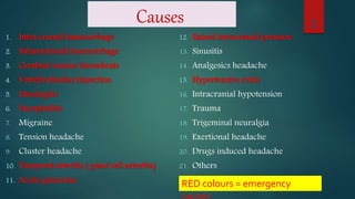
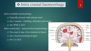
Headache
Causes
1. Intra cranial haemorrhage
2. Subarachnoid haemorrhage
3. Cerebral venous thrombosis
4. Vertebrobasilar dissection
5. Meningitis
6. Encephalitis
7. Migraine
8. Tension headache
9. Cluster headache
10. Temporal arteritis ( giant cell arteritis)
11. Acute glaucoma
12. Raised intracranial pressure
13. Sinusitis
14. Analgesics headache
15. Hypertensive crisis
16. Intracranial hypotension
17. Trauma
18. Trigeminal neuralgia
19. Exertional headache
20. Drugs induced headache
21. Others
2
RED colours = emergency
 Intra cerebellar haemorrhage :
 Typically present with abrupt onset
 Ass. è nausea , vomiting, dizziness and ataxia
 Ass. With  GCS
 Intra ventricular / intracerebral haemorrhage :
 The onset is usu. Over minutes to hours
 Ass. è focal neurological signs
 Ass. è  GCS
3 Intra cranial haemorrhage
Subarachnoid haemorrhage
 Potentially, devastating bleed( typically aneurysm ) into subarachnoid
space.
 Rapid onset (<2 min) , severe, continuous ( > 2h)
 Occipital ( hit around the back of the head)
 Ass. è Vomiting, dizziness, seizures
 Neck stiffness, drowsy, photophobia, focal neurological signs, GCS
 Urgent CT ; but CT mey be normal in small bleeds (20%)
 If CT is normal, LP( >12 h after onset ) for xanthochromia
4
Cerebral venous thrombosis
 Thunderclap, throbbing, band-like
 Ass. è nausea , vomiting, seizure, cranial nerve palsies,
hemiparesis, ataxia, GCS
5
Vertebrobasilar dissection
 Acute onset
 Occipital/posterior neck pain
 Brainstem signs and symptoms
6
Meningitis
 Fever (>38°C ), meningism
 Headache develop over hours rather than sudden
 Viral meningitis is self-limiting
 Bacterial meningitis; life-threatening, GCS, shock, purpuric
rash, focal neurological signs
 LP & blood culture & throat swab is diagnostic
7
Encephalitis
 Brain inflammation, usually viral
 Rare and easily missed in the early stages
 Drowsy, confusion, vomiting, seizures, preceding flu-like illness,
non-specific symptoms
 Pyrexia, GCS, confusion, focal neurological signs, neck
stiffness, photophobia
 Do an urgent CT head and LP to look for signs of infection.
8
Migraine
 Recurrent, severe
 Lasting several hours to a few days
 Ass. è photophobia, nausea, vomiting , focal neurological features (
aura e.g. flashing light, zigzags, visual loss )
 Typically; intense, throbbing, unilateral, causing pt. to cease normal
activities in favour of bed- rest in quiet, darkened room
 Triggers; cheese, chocolate, alcohol , oral contraceptives pill
First attack Aged > 40 is uncommon
 Consider MRI head if new and > 55 year.
9
Tension headache
 Bilateral ( generalised or frontal)
 Duration 30 min. to 7 days.
 Dull, tight, pressing, in nature
 Worse when stressed
 Nausea and photophobia are usu. Absent
 ± scalp muscle tenderness
 Pt continue with normal activities
10
11
Cluster headache
 Rapid-onset of excruciating pain around one eye that may become watery
and bloodshot with lid swelling, lacrimation
 facial flushing, rhinorrhoea, miosis ± ptosis (20% of attacks).
 Restlessness, agitation
 Pain is strictly unilateral and almost always affects the same side.
 It lasts 15–180min, occurs once or twice a day, and is often nocturnal.
 Clusters last 4–12wks and are followed by pain-free periods of months or
even 1–2yrs before the next cluster. Sometimes it is chronic, not episodic.
 Occur in clusters lasting days to weeks , separated by months without
symptoms
 Male : female = 5:1, commoner in smokers.
12
Temporal arteritis ( giant cell arteritis)
 Large vessel vasculitis
 Ass. é polymyalgia Rheumatica( proximal pain, stiffness)
 More common in women
 Unusual in pt. < 50 years
 Localized headache ( temporal / occipital)
 Scalp tenderness, jaw claudication , visual loss
 Constitutional upset ( malaise, sweating, pyrexia, wt. loss)
 Abnormal temporal artery ( inflamed, tender, non pulsatile, nodular )
 Raised ESR, CRP, plts, ALP with anaemia
 The potential for rapid onset irreversible visual loss necessitates urgent treatment
with steroids.
 Temporal artery biopsy may confirm the diagnosis in < 1 wk. of starting therapy.
13
Acute glaucoma
 Ophthalmological emergency
 Dt sudden increase in intraocular pressure
 Pt is long-sighted, middle-aged or elderly, often occurs at night
 conjunctival injection
 clouding of the cornea
 irregular/non-reactive pupil
 ↓visual acuity or blurred vision
 sees coloured ‘halos’ around lights.
14
Raised intracranial pressure
 Caused by CVA, tumours, trauma, infection( abscess), cerebral oedema, ( post-
hypoxia ), electrolyte imbalance, coagulopathy, idiopathic.
 Suspected è  GCS, focal neurological signs, new onset seizures
 Cushing’s reflex ( HR, BP )
 Tend to be worse in the morning and on lying flat, coughing, straining
 Ass. è Vomiting without nausea +/- papilloedema
 CT brain is diagnostic
 consider HIV
15
Sinusitis
 Inflammation of the mucosa of the paranasal sinuses due to bacteria, virus, fungi;
mey become chronic.
 Dull, throbbing
 Focal pain over the sinuses
 Tend to be worse on bending forward
 Ethmoid or sphenoid sinus pain is felt deep in the midline at the root of the nose.
 Temp mey be normal
 Invariable ass. è Nasal symptoms, Look for at least two of:
• nasal blockage/congestion
• rhinorrhoea/discharge
• loss of smell
• facial pressure or tenderness.
 Sinusitis lasting > 8 wks requires CT to confirm dx
16
Analgesics headache
 Ass. è chronic analgesic use esp. Opioids
 Bilateral
 Occur prior to the next dose of analgesia
17
Hypertensive crisis
 Hypertension usually represents to a response to pain of the
headache and respond to the treatment of the headache
 But, BP more than 200/120 is the cause of the headache
18
Intracranial hypotension
 CSF leakage, e.g. Iatrogenic after LP or epidural anaesthesia.
 Suspect if headaches worse on standing
 usu. Present within 4-5 days of the procedure
19
Trauma
 Extradural haemorrhage, Subdural haemorrhage, Concussion.
 Commonly causes localized pain but can be more generalized.
 It lasts ~2wks; often resistant to analgesia.
 Do CT to exclude subdural or extradural haemorrhage if
drowsiness ± lucid interval, or focal signs
20
Trigeminal neuralgia
 Brief, repetitive episodes of intense shooting, stabbing, electric shock - like
pain in 2nd and 3rd divisions of trigeminal nerve
 Paroxysms of intense, stabbing pain, lasting seconds.
 The face screws up with pain (hence tic douloureux).
 Triggers: Washing affected area, shaving, eating, talking, dental prostheses.
 Typical patient: >50yrs old; in Asians F:M ≈ 2:1.
 Secondary causes: Compression of the trigeminal root by anomalous or
aneurysmal intracranial vessels or a tumour, chronic meningeal
inflammation, MS, zoster, skull base malformation (e.g. Chiari).
 MRI: Is necessary to exclude secondary causes (~14% of cases).
21
22Trigeminal neuralgia
Exertional headache
 Sudden, bilateral, explosive pain
 Typical on exercise or orgasm
 May mimic migraine or SAH, but meningism is absent
 Consider CT / LP to rule out SAH
23
Drugs induced headache
 Nitrate
 Ca channel blocker
 Metronidazole with alcohol
 Recreational drugs e.g. Solvents
24
others
 Carbon monoxide
poisoning
 Hypercapnia
 Post-coital headache
 Dehydration
 Hypoglycaemia
 Hyponatraemia
 Eyes disorders
 Ear disorders
 Sepsis
 Tropical illness
e.g. ( malaria &
typhus )
25
Nice to know
 All sudden onset, severe headaches should, ideally, be discussed
with a neurologist.
 Headaches that recur tend to be benign: migraine, tension headache,
trigeminal neuralgia.
 Chronic, progressive headaches can indicate increased ICP .
 Consider benign intracranial hypertension in patients with features
of ↑intracranial pressure but no mass on neuroimaging.
 Ask about stress or recent life events; may not explain the pathology,
but will help you appreciate the context in which symptoms are.
26
Red flags
 New-onset headache/change in headache in patients over 50 years
 Persistent visual disturbance Focal CNS signs, ataxia or new cognitive or
behavioural disturbance
 Headache that changes with posture or wakes the patient up
 Headache brought on by physical exertion
 Papilloedema
 New-onset headache in a patient with known HIV or active malignancy
27
👉 Arrange CT brain ± MRI and, if normal, seek input from a neurologist.
Sources
 MacLeod's clinical diagnosis, 2013
 Oxford Handbook of Clinical Medicine,10th edition
 Oxford Handbook for the Foundation Programme, 5th edition
28
Headache,ppt

Headache,ppt

  • 1.
  • 2.
    Causes 1. Intra cranialhaemorrhage 2. Subarachnoid haemorrhage 3. Cerebral venous thrombosis 4. Vertebrobasilar dissection 5. Meningitis 6. Encephalitis 7. Migraine 8. Tension headache 9. Cluster headache 10. Temporal arteritis ( giant cell arteritis) 11. Acute glaucoma 12. Raised intracranial pressure 13. Sinusitis 14. Analgesics headache 15. Hypertensive crisis 16. Intracranial hypotension 17. Trauma 18. Trigeminal neuralgia 19. Exertional headache 20. Drugs induced headache 21. Others 2 RED colours = emergency
  • 3.
     Intra cerebellarhaemorrhage :  Typically present with abrupt onset  Ass. è nausea , vomiting, dizziness and ataxia  Ass. With  GCS  Intra ventricular / intracerebral haemorrhage :  The onset is usu. Over minutes to hours  Ass. è focal neurological signs  Ass. è  GCS 3 Intra cranial haemorrhage
  • 4.
    Subarachnoid haemorrhage  Potentially,devastating bleed( typically aneurysm ) into subarachnoid space.  Rapid onset (<2 min) , severe, continuous ( > 2h)  Occipital ( hit around the back of the head)  Ass. è Vomiting, dizziness, seizures  Neck stiffness, drowsy, photophobia, focal neurological signs, GCS  Urgent CT ; but CT mey be normal in small bleeds (20%)  If CT is normal, LP( >12 h after onset ) for xanthochromia 4
  • 5.
    Cerebral venous thrombosis Thunderclap, throbbing, band-like  Ass. è nausea , vomiting, seizure, cranial nerve palsies, hemiparesis, ataxia, GCS 5
  • 6.
    Vertebrobasilar dissection  Acuteonset  Occipital/posterior neck pain  Brainstem signs and symptoms 6
  • 7.
    Meningitis  Fever (>38°C), meningism  Headache develop over hours rather than sudden  Viral meningitis is self-limiting  Bacterial meningitis; life-threatening, GCS, shock, purpuric rash, focal neurological signs  LP & blood culture & throat swab is diagnostic 7
  • 8.
    Encephalitis  Brain inflammation,usually viral  Rare and easily missed in the early stages  Drowsy, confusion, vomiting, seizures, preceding flu-like illness, non-specific symptoms  Pyrexia, GCS, confusion, focal neurological signs, neck stiffness, photophobia  Do an urgent CT head and LP to look for signs of infection. 8
  • 9.
    Migraine  Recurrent, severe Lasting several hours to a few days  Ass. è photophobia, nausea, vomiting , focal neurological features ( aura e.g. flashing light, zigzags, visual loss )  Typically; intense, throbbing, unilateral, causing pt. to cease normal activities in favour of bed- rest in quiet, darkened room  Triggers; cheese, chocolate, alcohol , oral contraceptives pill First attack Aged > 40 is uncommon  Consider MRI head if new and > 55 year. 9
  • 10.
    Tension headache  Bilateral( generalised or frontal)  Duration 30 min. to 7 days.  Dull, tight, pressing, in nature  Worse when stressed  Nausea and photophobia are usu. Absent  ± scalp muscle tenderness  Pt continue with normal activities 10
  • 11.
  • 12.
    Cluster headache  Rapid-onsetof excruciating pain around one eye that may become watery and bloodshot with lid swelling, lacrimation  facial flushing, rhinorrhoea, miosis ± ptosis (20% of attacks).  Restlessness, agitation  Pain is strictly unilateral and almost always affects the same side.  It lasts 15–180min, occurs once or twice a day, and is often nocturnal.  Clusters last 4–12wks and are followed by pain-free periods of months or even 1–2yrs before the next cluster. Sometimes it is chronic, not episodic.  Occur in clusters lasting days to weeks , separated by months without symptoms  Male : female = 5:1, commoner in smokers. 12
  • 13.
    Temporal arteritis (giant cell arteritis)  Large vessel vasculitis  Ass. é polymyalgia Rheumatica( proximal pain, stiffness)  More common in women  Unusual in pt. < 50 years  Localized headache ( temporal / occipital)  Scalp tenderness, jaw claudication , visual loss  Constitutional upset ( malaise, sweating, pyrexia, wt. loss)  Abnormal temporal artery ( inflamed, tender, non pulsatile, nodular )  Raised ESR, CRP, plts, ALP with anaemia  The potential for rapid onset irreversible visual loss necessitates urgent treatment with steroids.  Temporal artery biopsy may confirm the diagnosis in < 1 wk. of starting therapy. 13
  • 14.
    Acute glaucoma  Ophthalmologicalemergency  Dt sudden increase in intraocular pressure  Pt is long-sighted, middle-aged or elderly, often occurs at night  conjunctival injection  clouding of the cornea  irregular/non-reactive pupil  ↓visual acuity or blurred vision  sees coloured ‘halos’ around lights. 14
  • 15.
    Raised intracranial pressure Caused by CVA, tumours, trauma, infection( abscess), cerebral oedema, ( post- hypoxia ), electrolyte imbalance, coagulopathy, idiopathic.  Suspected è  GCS, focal neurological signs, new onset seizures  Cushing’s reflex ( HR, BP )  Tend to be worse in the morning and on lying flat, coughing, straining  Ass. è Vomiting without nausea +/- papilloedema  CT brain is diagnostic  consider HIV 15
  • 16.
    Sinusitis  Inflammation ofthe mucosa of the paranasal sinuses due to bacteria, virus, fungi; mey become chronic.  Dull, throbbing  Focal pain over the sinuses  Tend to be worse on bending forward  Ethmoid or sphenoid sinus pain is felt deep in the midline at the root of the nose.  Temp mey be normal  Invariable ass. è Nasal symptoms, Look for at least two of: • nasal blockage/congestion • rhinorrhoea/discharge • loss of smell • facial pressure or tenderness.  Sinusitis lasting > 8 wks requires CT to confirm dx 16
  • 17.
    Analgesics headache  Ass.è chronic analgesic use esp. Opioids  Bilateral  Occur prior to the next dose of analgesia 17
  • 18.
    Hypertensive crisis  Hypertensionusually represents to a response to pain of the headache and respond to the treatment of the headache  But, BP more than 200/120 is the cause of the headache 18
  • 19.
    Intracranial hypotension  CSFleakage, e.g. Iatrogenic after LP or epidural anaesthesia.  Suspect if headaches worse on standing  usu. Present within 4-5 days of the procedure 19
  • 20.
    Trauma  Extradural haemorrhage,Subdural haemorrhage, Concussion.  Commonly causes localized pain but can be more generalized.  It lasts ~2wks; often resistant to analgesia.  Do CT to exclude subdural or extradural haemorrhage if drowsiness ± lucid interval, or focal signs 20
  • 21.
    Trigeminal neuralgia  Brief,repetitive episodes of intense shooting, stabbing, electric shock - like pain in 2nd and 3rd divisions of trigeminal nerve  Paroxysms of intense, stabbing pain, lasting seconds.  The face screws up with pain (hence tic douloureux).  Triggers: Washing affected area, shaving, eating, talking, dental prostheses.  Typical patient: >50yrs old; in Asians F:M ≈ 2:1.  Secondary causes: Compression of the trigeminal root by anomalous or aneurysmal intracranial vessels or a tumour, chronic meningeal inflammation, MS, zoster, skull base malformation (e.g. Chiari).  MRI: Is necessary to exclude secondary causes (~14% of cases). 21
  • 22.
  • 23.
    Exertional headache  Sudden,bilateral, explosive pain  Typical on exercise or orgasm  May mimic migraine or SAH, but meningism is absent  Consider CT / LP to rule out SAH 23
  • 24.
    Drugs induced headache Nitrate  Ca channel blocker  Metronidazole with alcohol  Recreational drugs e.g. Solvents 24
  • 25.
    others  Carbon monoxide poisoning Hypercapnia  Post-coital headache  Dehydration  Hypoglycaemia  Hyponatraemia  Eyes disorders  Ear disorders  Sepsis  Tropical illness e.g. ( malaria & typhus ) 25
  • 26.
    Nice to know All sudden onset, severe headaches should, ideally, be discussed with a neurologist.  Headaches that recur tend to be benign: migraine, tension headache, trigeminal neuralgia.  Chronic, progressive headaches can indicate increased ICP .  Consider benign intracranial hypertension in patients with features of ↑intracranial pressure but no mass on neuroimaging.  Ask about stress or recent life events; may not explain the pathology, but will help you appreciate the context in which symptoms are. 26
  • 27.
    Red flags  New-onsetheadache/change in headache in patients over 50 years  Persistent visual disturbance Focal CNS signs, ataxia or new cognitive or behavioural disturbance  Headache that changes with posture or wakes the patient up  Headache brought on by physical exertion  Papilloedema  New-onset headache in a patient with known HIV or active malignancy 27 👉 Arrange CT brain ± MRI and, if normal, seek input from a neurologist.
  • 28.
    Sources  MacLeod's clinicaldiagnosis, 2013  Oxford Handbook of Clinical Medicine,10th edition  Oxford Handbook for the Foundation Programme, 5th edition 28