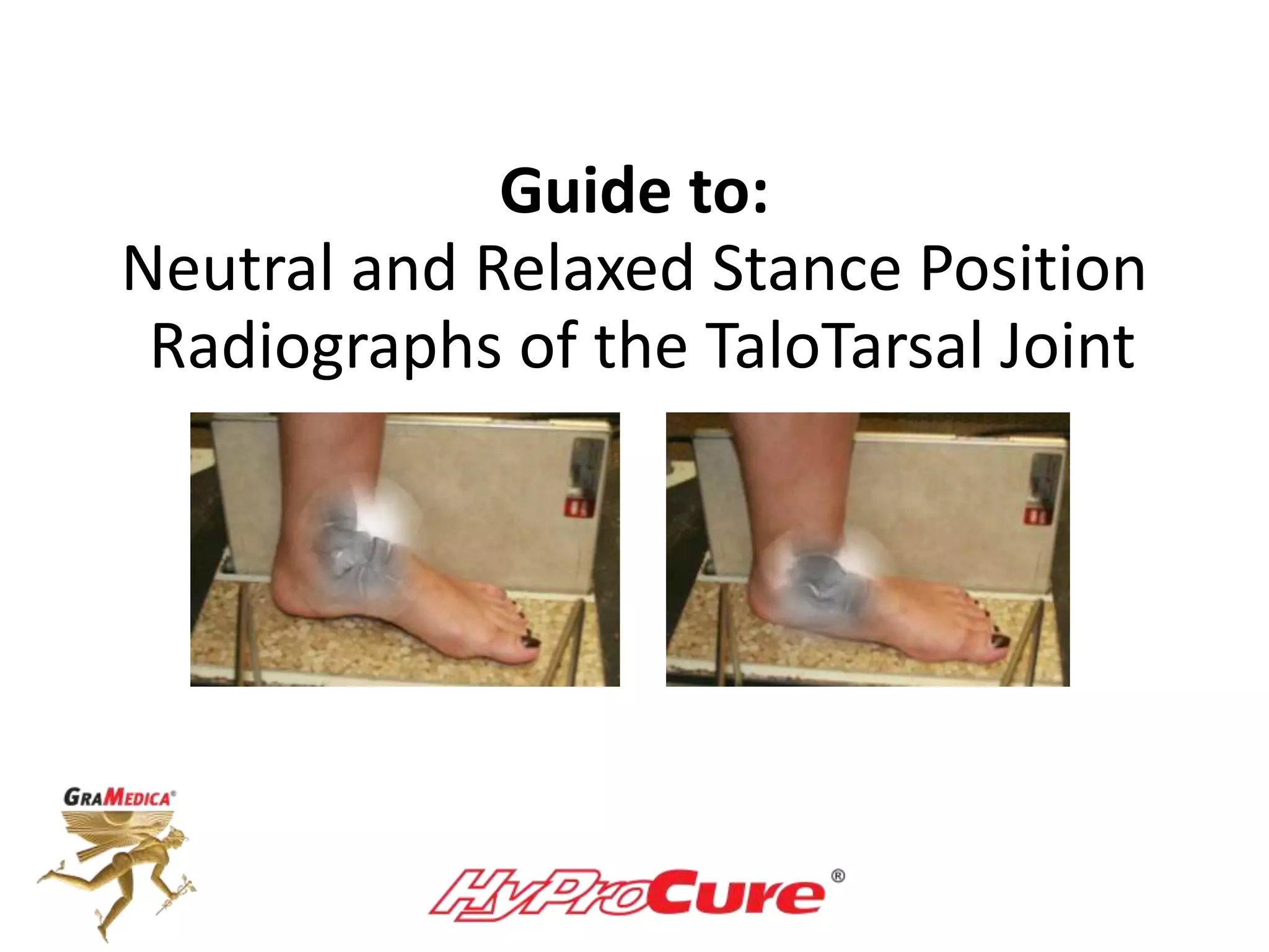
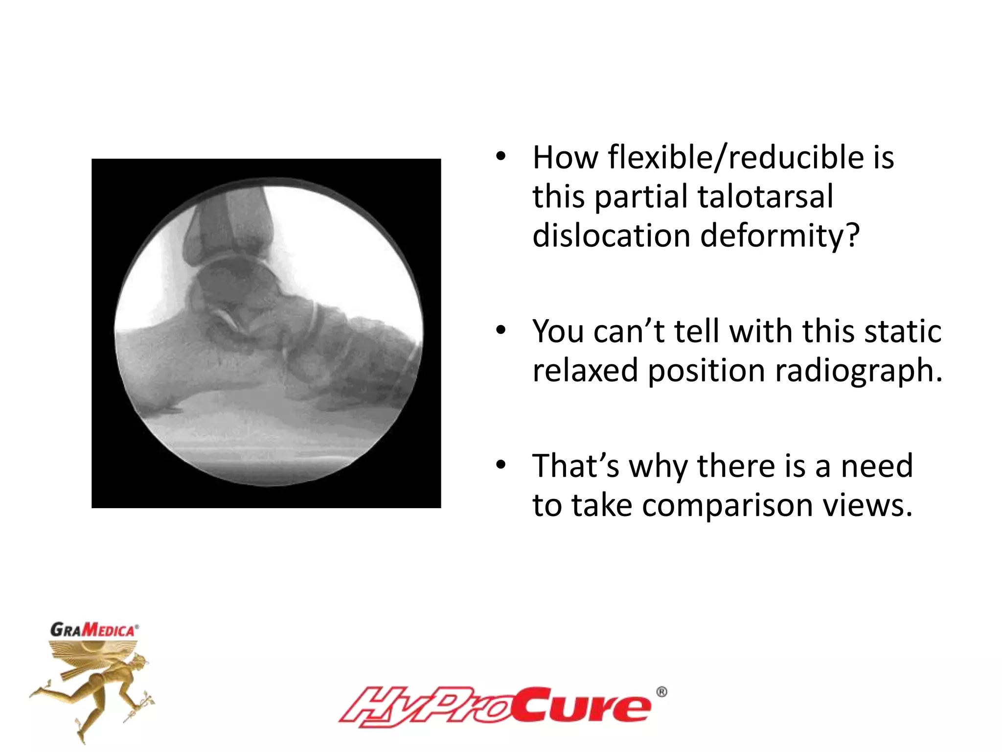
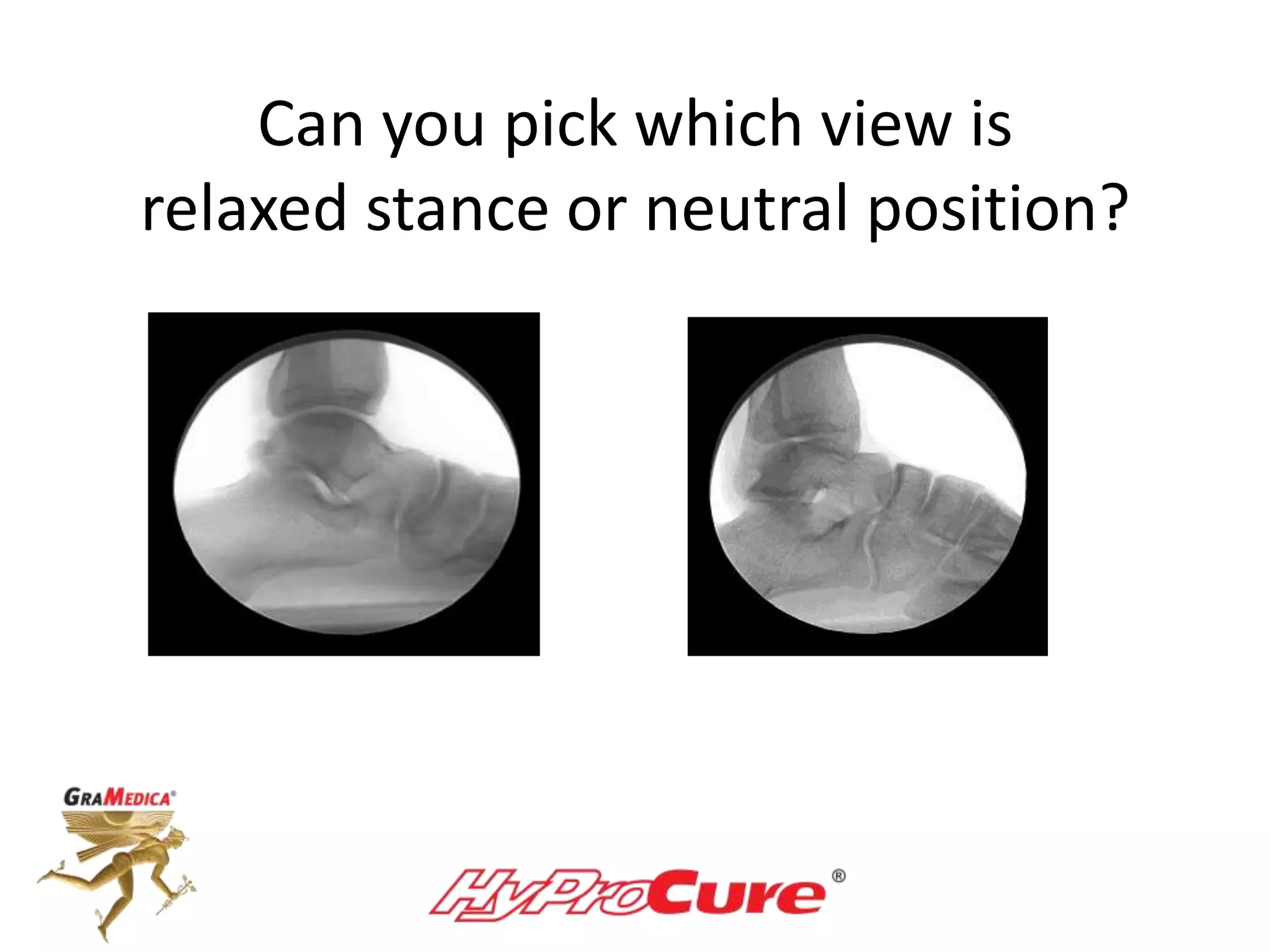
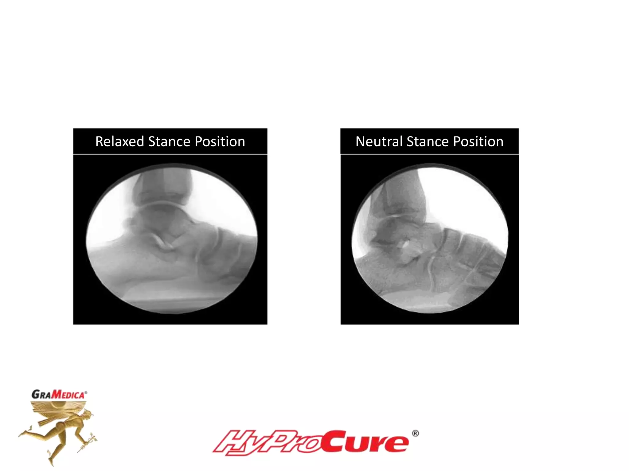
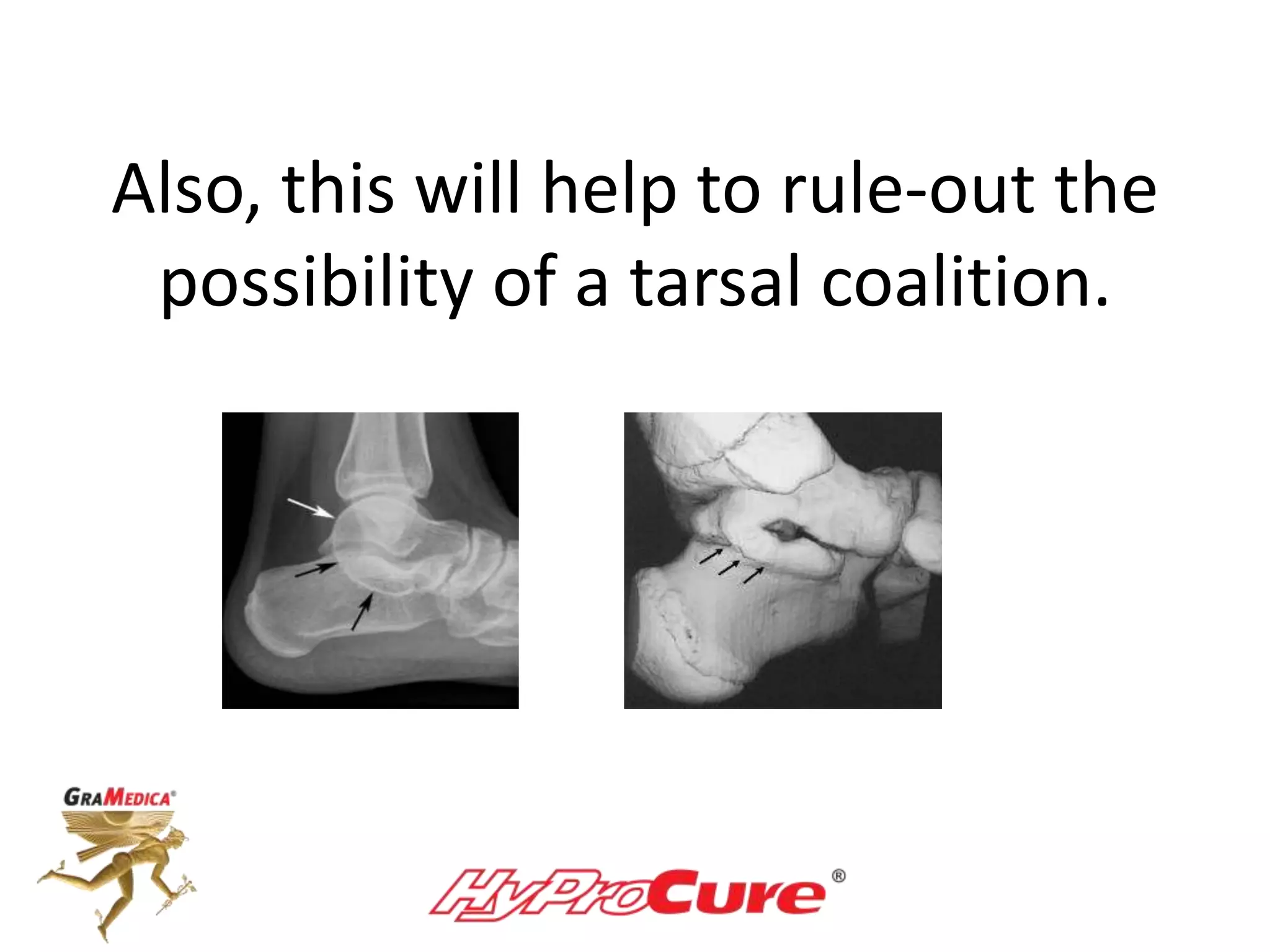
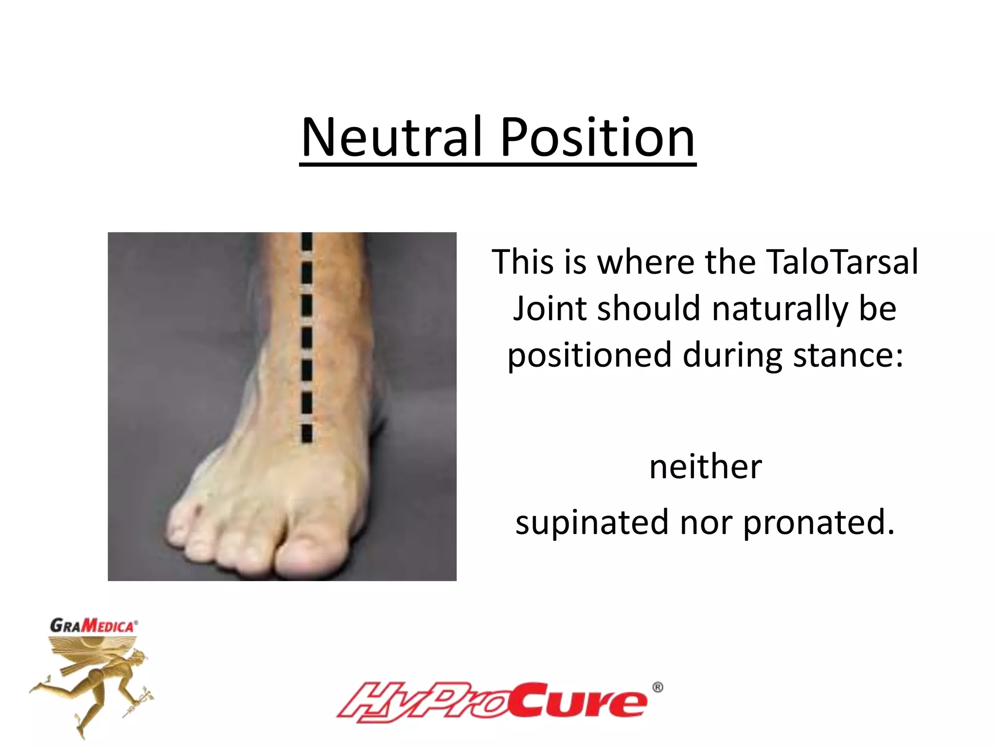
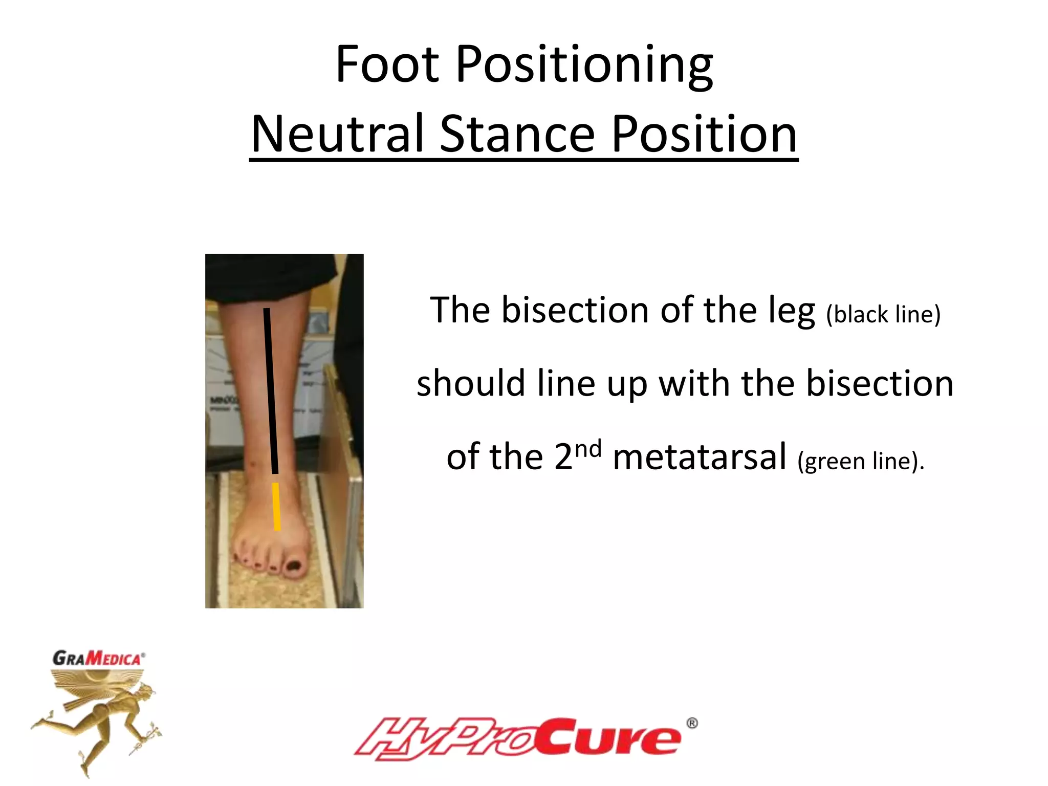
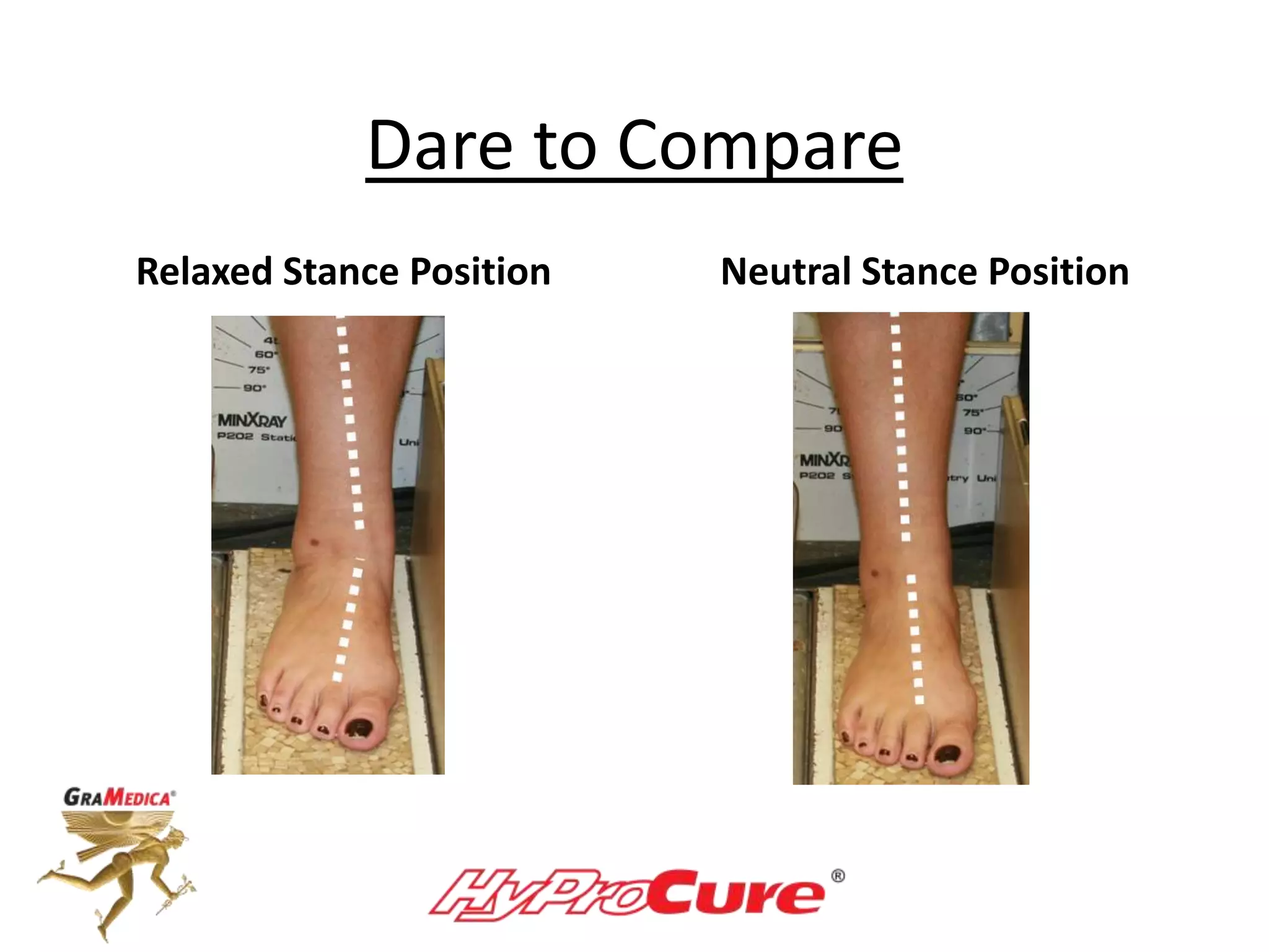
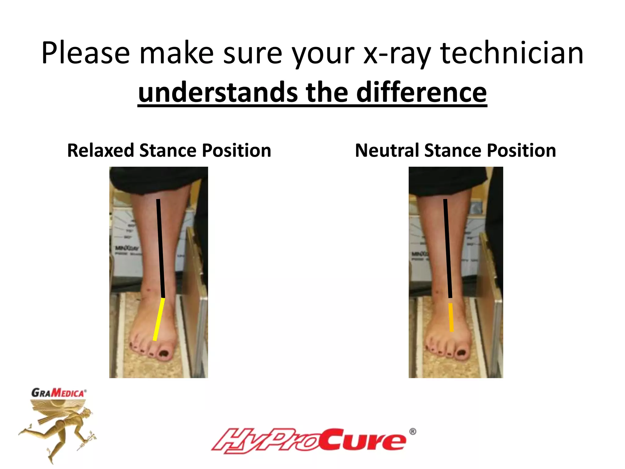
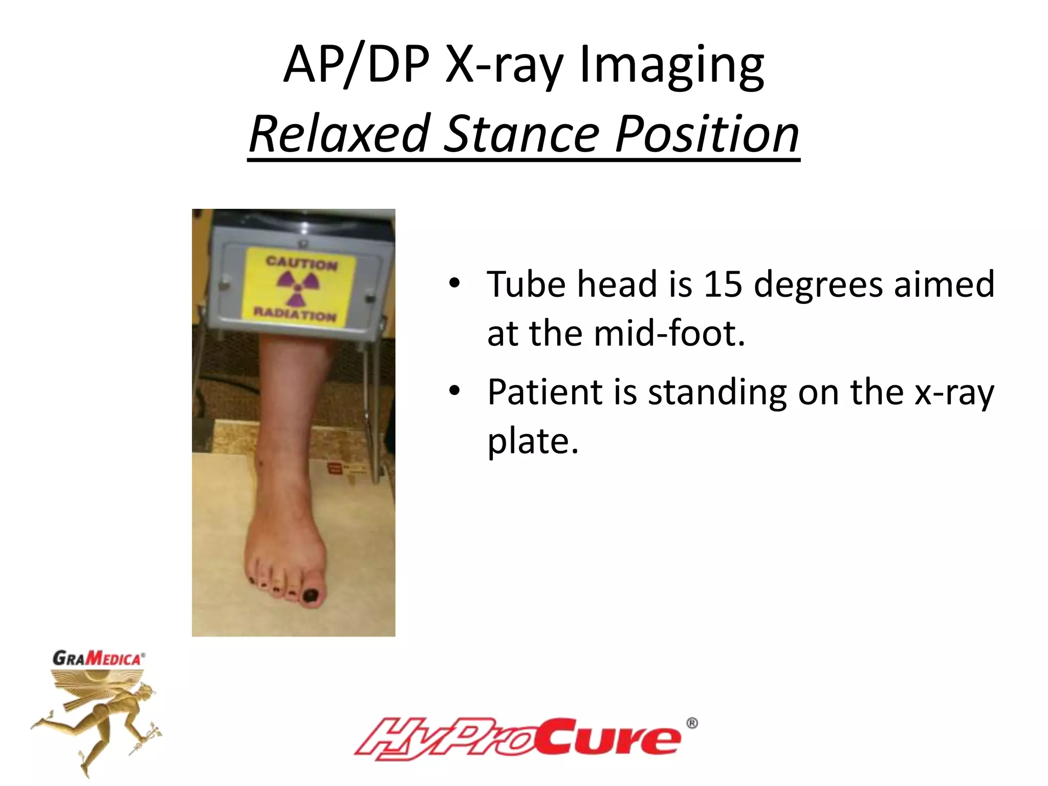
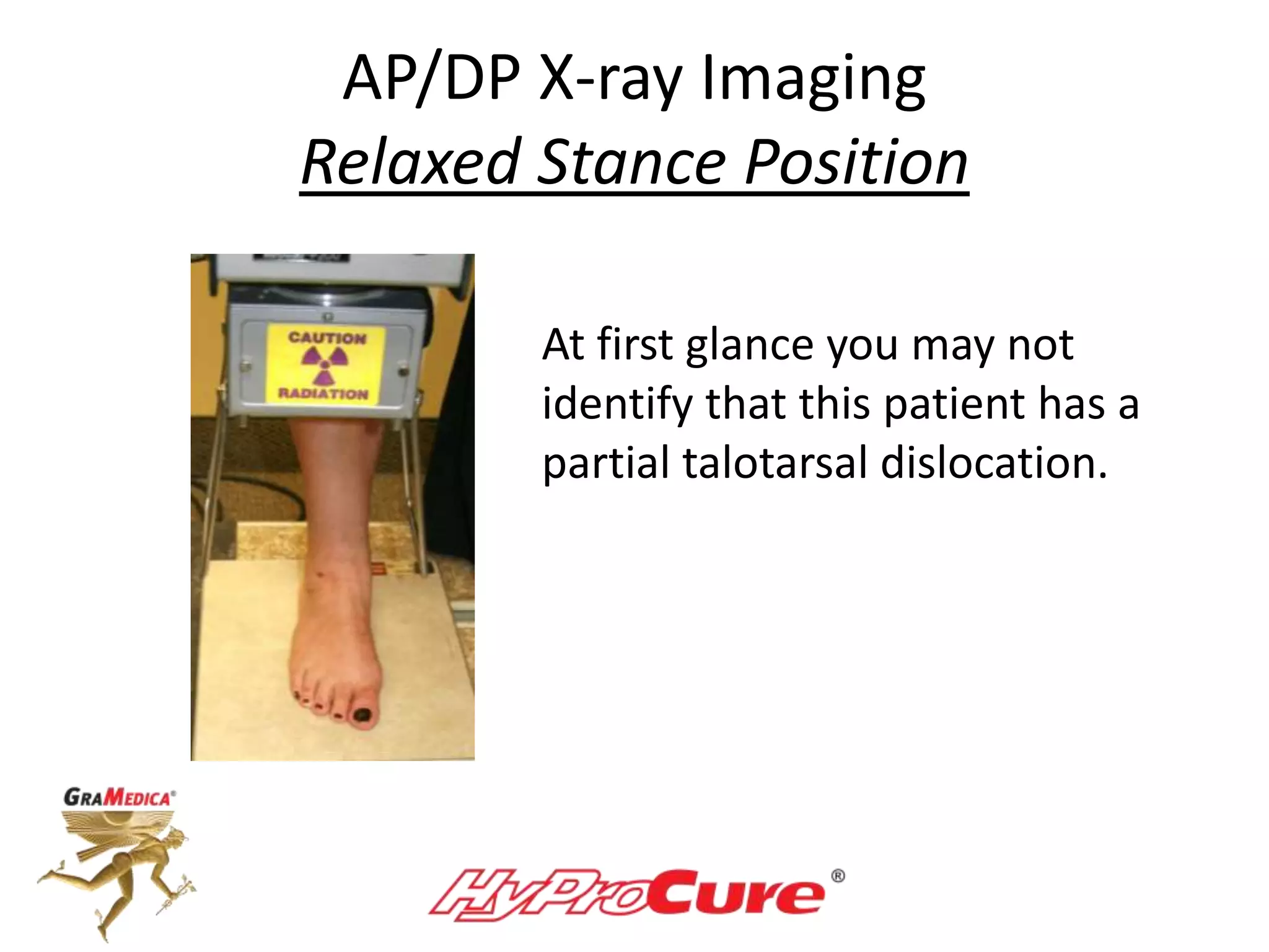
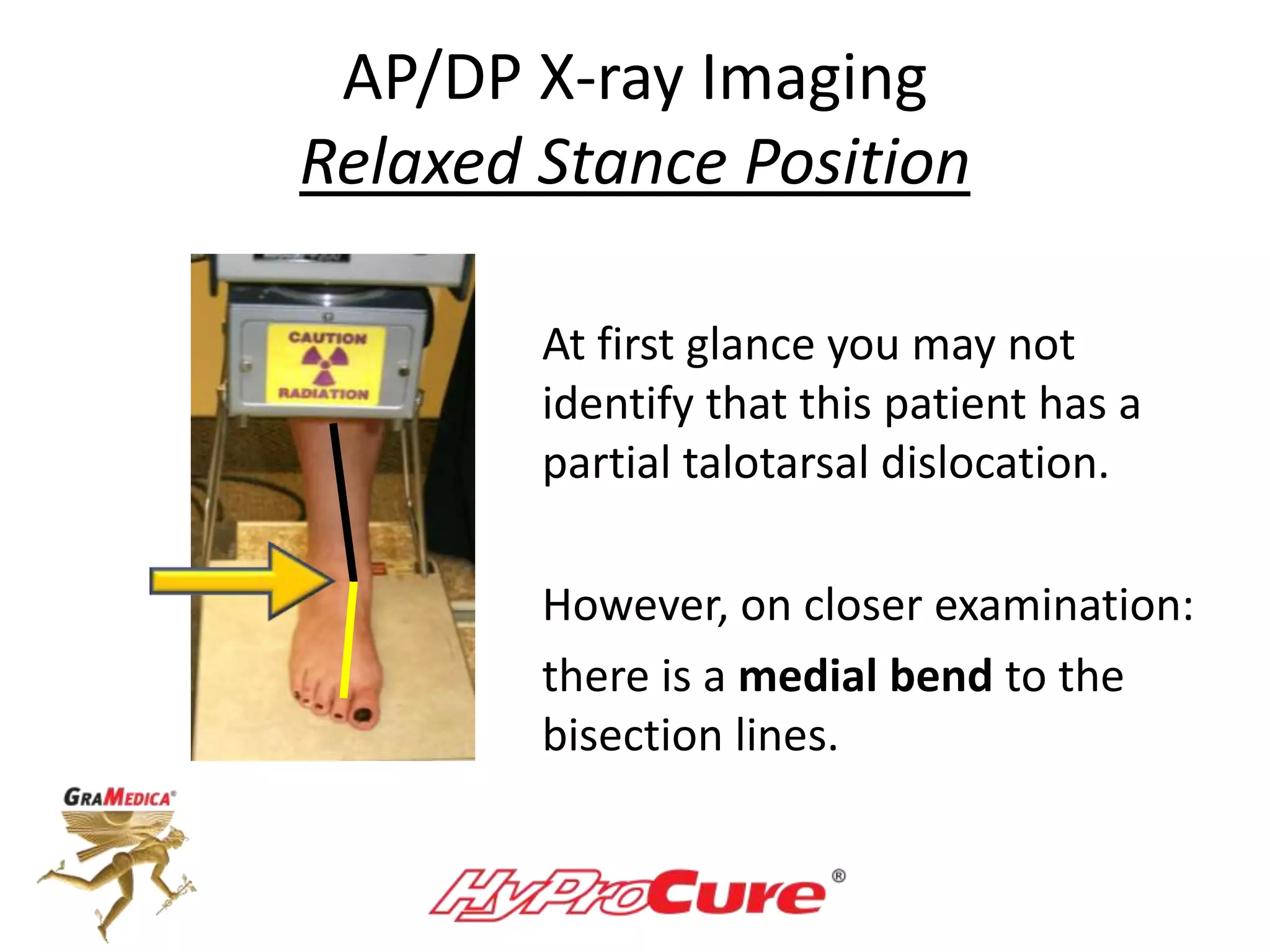
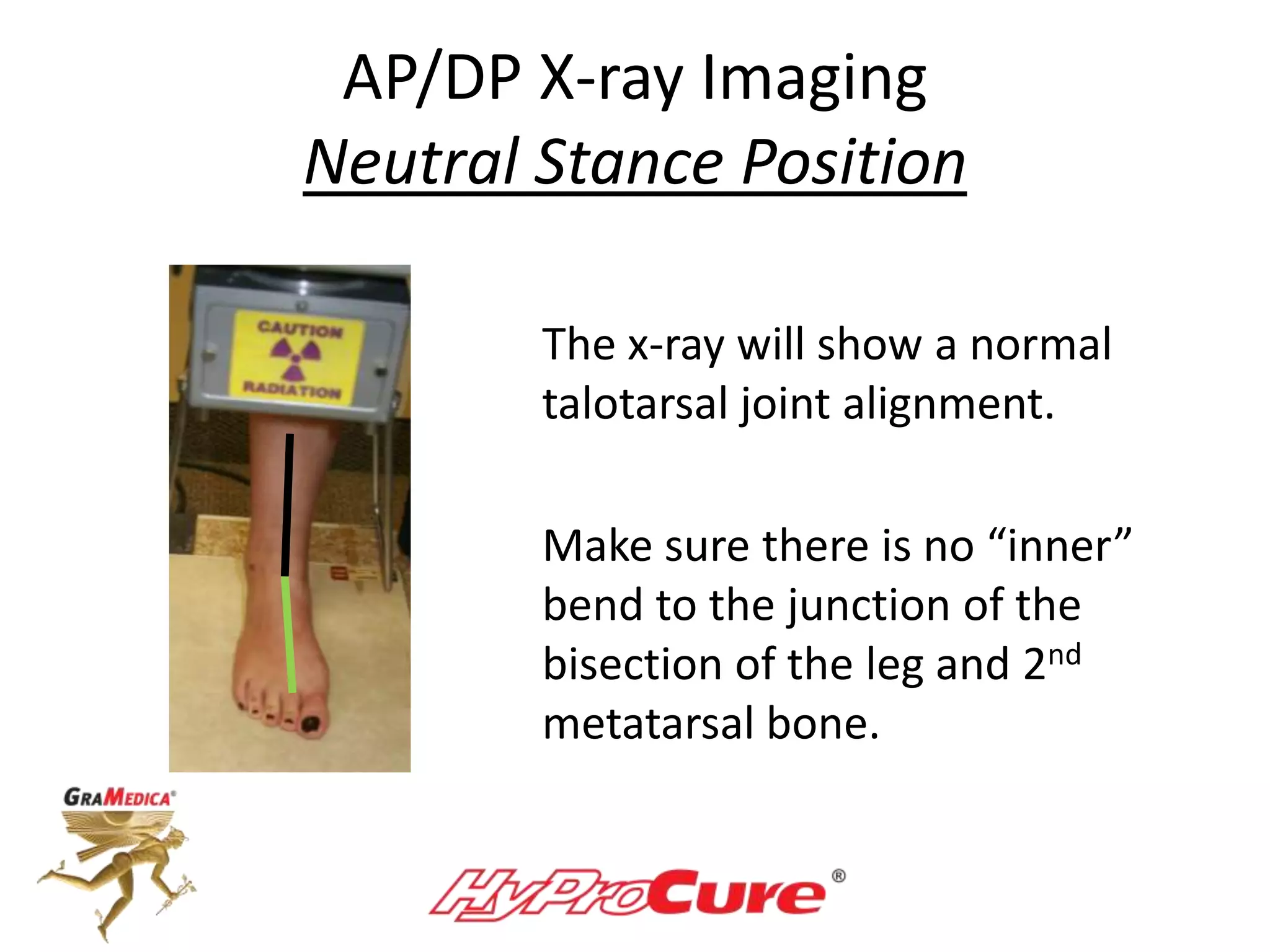
Guide to:
Neutral and Relaxed Stance Position
Radiographs of the TaloTarsal Joint
The best way to diagnose
TaloTarsal Dislocation
is via radiographic examination.
• How flexible/reducible is
this partial talotarsal
dislocation deformity?
• You can’t tell with this static
relaxed position radiograph.
• That’s why there is a need
to take comparison views.
Radiographic Comparison
• Provides objective data (can draw angles to compare
normal versus abnormal accepted angular measurements)
• Excellent educational tool to show the patient
their hindfoot deformity
• Documents the flexibility of the deformity
• Rules out secondary pathologies
Can you pick which view is
relaxed stance or neutral position?
Relaxed Stance Position Neutral Stance Position
If you are going to recommend an
EOTTS-HyProCure procedure it is
very important to show radiographic
evidence that the dislocation
deformity is flexible.
Also, this will help to rule-out the
possibility of a tarsal coalition.
Foot Positioning
Relaxed Stance
Neutral Stance
Relaxed Stance
That’s the easy position –
patient just stands how they
normally stand.
Neutral Position
This is where the TaloTarsal
Joint should naturally be
positioned during stance:
neither
supinated nor pronated.
Normal TaloTarsal Joint Alignment
• The talus should be
balanced on the
calcaneus and navicular.
• Sinus tarsi is “open”
• TTJ articular facets are
in Constant
Congruent
Contact
Foot Positioning
Neutral Stance Position
The bisection of the leg (black line)
should line up with the bisection
of the 2nd metatarsal (green line).
Foot Positioning
Supinated-Overcorrected Position
The juncture of the 2 lines bends
outward/laterally.
This indicates a supinated hindfoot.
It is better to place the foot in a
more supinated verse a more
pronated position. The goal is to
show that the talus can resupinate
on the calcaneus.
Sign of TaloTarsal Dislocation
There is an inner/medial bend
of the bisection of the leg and
2nd metatarsal.
“Pronated” position of the
hindfoot.
Only a very “slight” amount of
pronation is acceptable.
Technique:
Take AP and Lateral Views
Can also take posterior calcaneal
view and AP of the ankle – optional.
Lateral X-ray Imaging
Relaxed Stance Position
• Tube is angle 90 degrees.
Lateral X-ray Imaging
Neutral Stance Position
• The patient’s hindfoot is
repositioned so that it is
neither supinated nor
pronated.
• The talus is repositioned the
talus back on top of the
calcaneus.
Dare to Compare
Relaxed Stance Position Neutral Stance Position
Please make sure your x-ray technician
understands the difference
Relaxed Stance Position Neutral Stance Position
AP/DP X-ray Imaging
Relaxed Stance Position
• Tube head is 15 degrees aimed
at the mid-foot.
• Patient is standing on the x-ray
plate.
AP/DP X-ray Imaging
Relaxed Stance Position
At first glance you may not
identify that this patient has a
partial talotarsal dislocation.
AP/DP X-ray Imaging
Relaxed Stance Position
At first glance you may not
identify that this patient has a
partial talotarsal dislocation.
However, on closer examination:
there is a medial bend to the
bisection lines.
AP/DP X-ray Imaging
Neutral Stance Position
The x-ray will show a normal
talotarsal joint alignment.
Make sure there is no “inner”
bend to the junction of the
bisection of the leg and 2nd
metatarsal bone.
See the difference?
Relaxed
Stance
Position
Neutral
Stance
Position
Good Luck!
There is another module for the interpretation of these views.
www.HyProCureDoctors.com

Neutral and Relaxed Stance X-ray Positioning for TaloTarsal Joint Dislocation

  • 1.
    Guide to: Neutral andRelaxed Stance Position Radiographs of the TaloTarsal Joint
  • 2.
    The best wayto diagnose TaloTarsal Dislocation is via radiographic examination.
  • 3.
    • How flexible/reducibleis this partial talotarsal dislocation deformity? • You can’t tell with this static relaxed position radiograph. • That’s why there is a need to take comparison views.
  • 4.
    Radiographic Comparison • Providesobjective data (can draw angles to compare normal versus abnormal accepted angular measurements) • Excellent educational tool to show the patient their hindfoot deformity • Documents the flexibility of the deformity • Rules out secondary pathologies
  • 5.
    Can you pickwhich view is relaxed stance or neutral position?
  • 6.
    Relaxed Stance PositionNeutral Stance Position
  • 7.
    If you aregoing to recommend an EOTTS-HyProCure procedure it is very important to show radiographic evidence that the dislocation deformity is flexible.
  • 8.
    Also, this willhelp to rule-out the possibility of a tarsal coalition.
  • 9.
  • 10.
    Relaxed Stance That’s theeasy position – patient just stands how they normally stand.
  • 11.
    Neutral Position This iswhere the TaloTarsal Joint should naturally be positioned during stance: neither supinated nor pronated.
  • 12.
    Normal TaloTarsal JointAlignment • The talus should be balanced on the calcaneus and navicular. • Sinus tarsi is “open” • TTJ articular facets are in Constant Congruent Contact
  • 13.
    Foot Positioning Neutral StancePosition The bisection of the leg (black line) should line up with the bisection of the 2nd metatarsal (green line).
  • 14.
    Foot Positioning Supinated-Overcorrected Position Thejuncture of the 2 lines bends outward/laterally. This indicates a supinated hindfoot. It is better to place the foot in a more supinated verse a more pronated position. The goal is to show that the talus can resupinate on the calcaneus.
  • 15.
    Sign of TaloTarsalDislocation There is an inner/medial bend of the bisection of the leg and 2nd metatarsal. “Pronated” position of the hindfoot. Only a very “slight” amount of pronation is acceptable.
  • 16.
    Technique: Take AP andLateral Views Can also take posterior calcaneal view and AP of the ankle – optional.
  • 17.
    Lateral X-ray Imaging RelaxedStance Position • Tube is angle 90 degrees.
  • 18.
    Lateral X-ray Imaging NeutralStance Position • The patient’s hindfoot is repositioned so that it is neither supinated nor pronated. • The talus is repositioned the talus back on top of the calcaneus.
  • 19.
    Dare to Compare RelaxedStance Position Neutral Stance Position
  • 20.
    Please make sureyour x-ray technician understands the difference Relaxed Stance Position Neutral Stance Position
  • 21.
    AP/DP X-ray Imaging RelaxedStance Position • Tube head is 15 degrees aimed at the mid-foot. • Patient is standing on the x-ray plate.
  • 22.
    AP/DP X-ray Imaging RelaxedStance Position At first glance you may not identify that this patient has a partial talotarsal dislocation.
  • 23.
    AP/DP X-ray Imaging RelaxedStance Position At first glance you may not identify that this patient has a partial talotarsal dislocation. However, on closer examination: there is a medial bend to the bisection lines.
  • 24.
    AP/DP X-ray Imaging NeutralStance Position The x-ray will show a normal talotarsal joint alignment. Make sure there is no “inner” bend to the junction of the bisection of the leg and 2nd metatarsal bone.
  • 25.
  • 26.
    Good Luck! There isanother module for the interpretation of these views. www.HyProCureDoctors.com