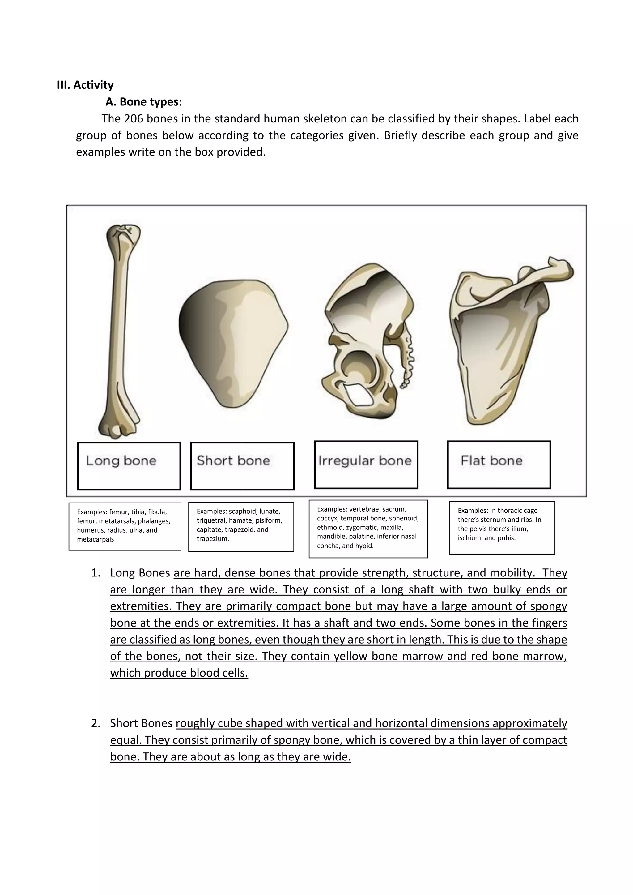
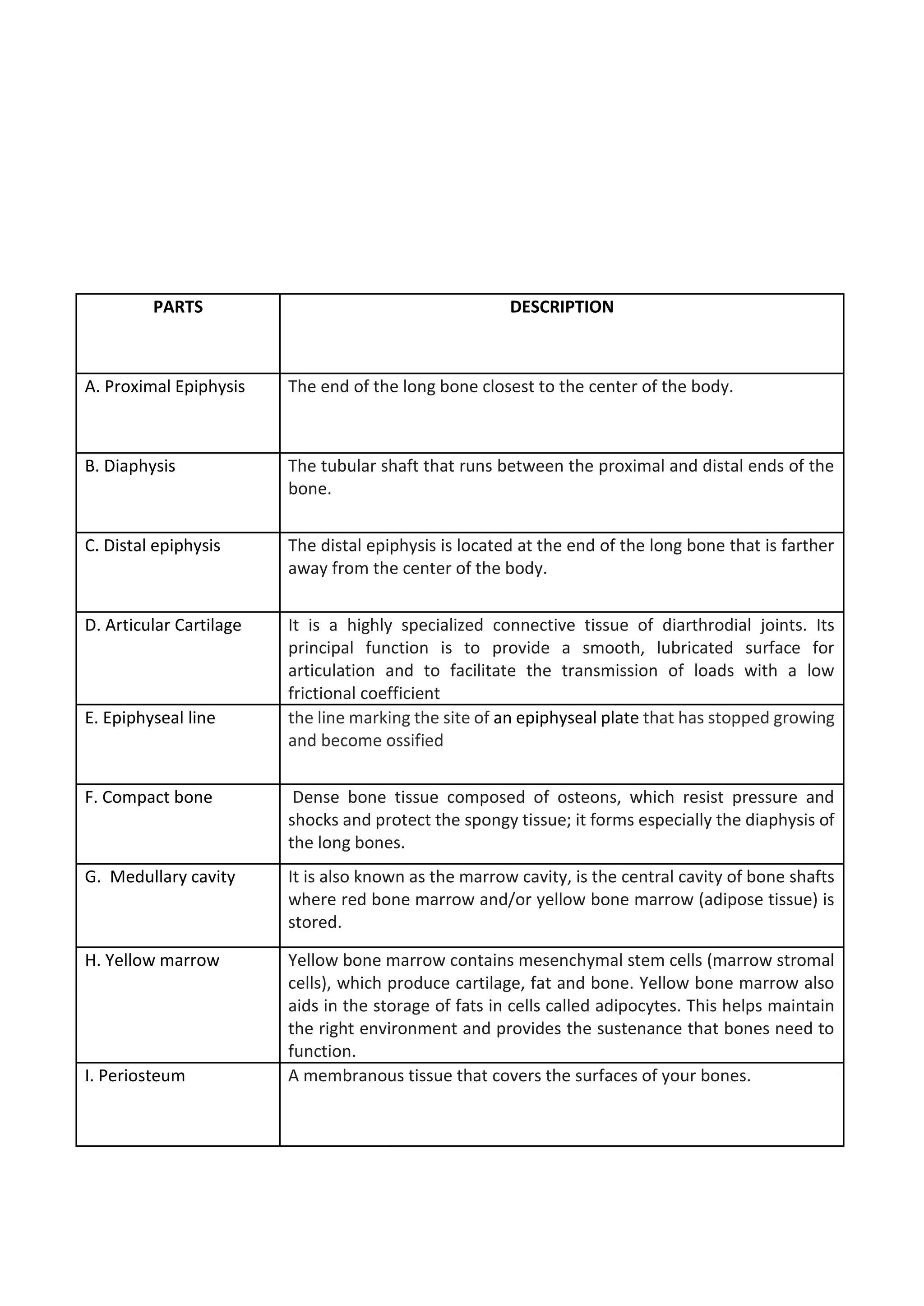
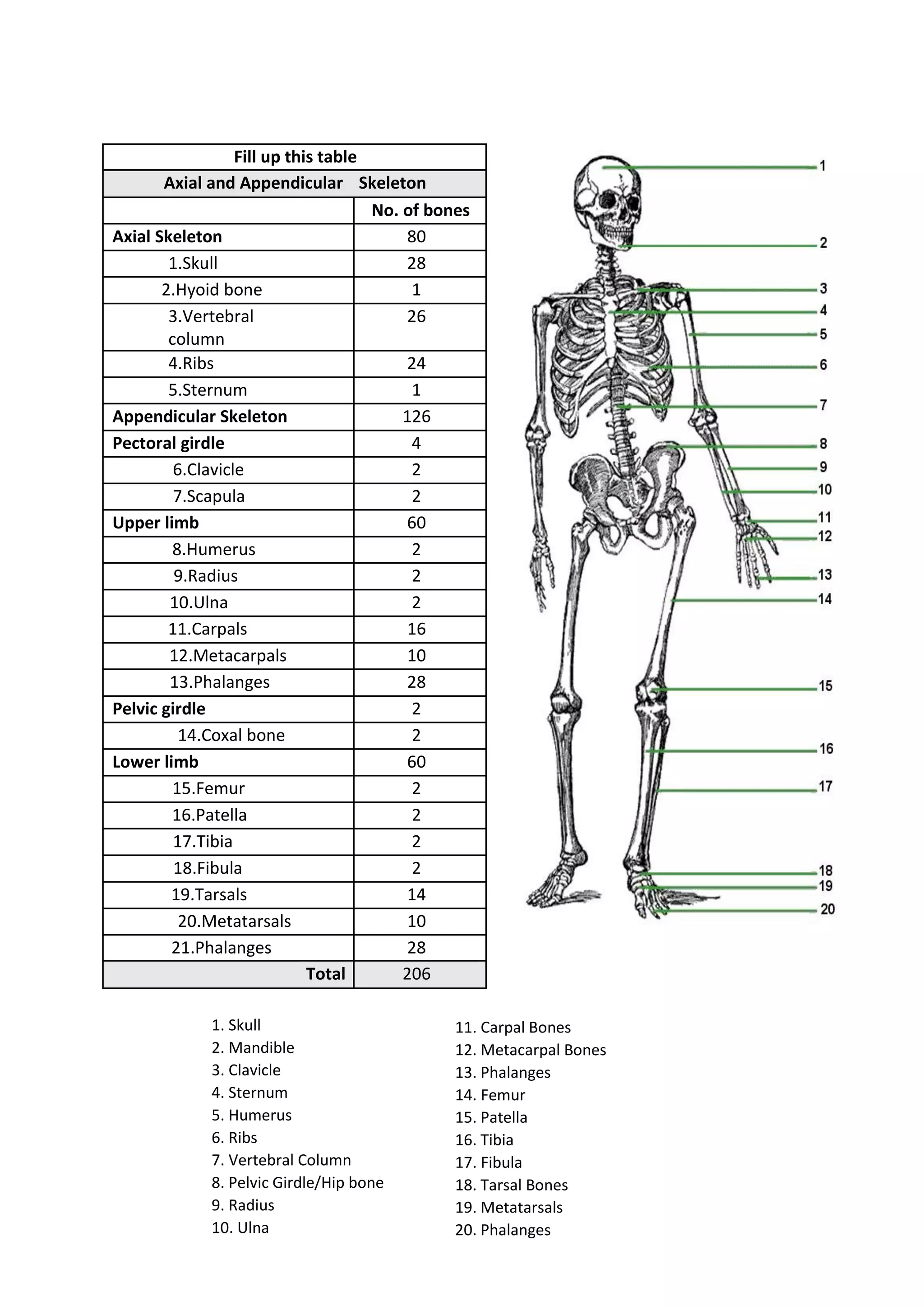
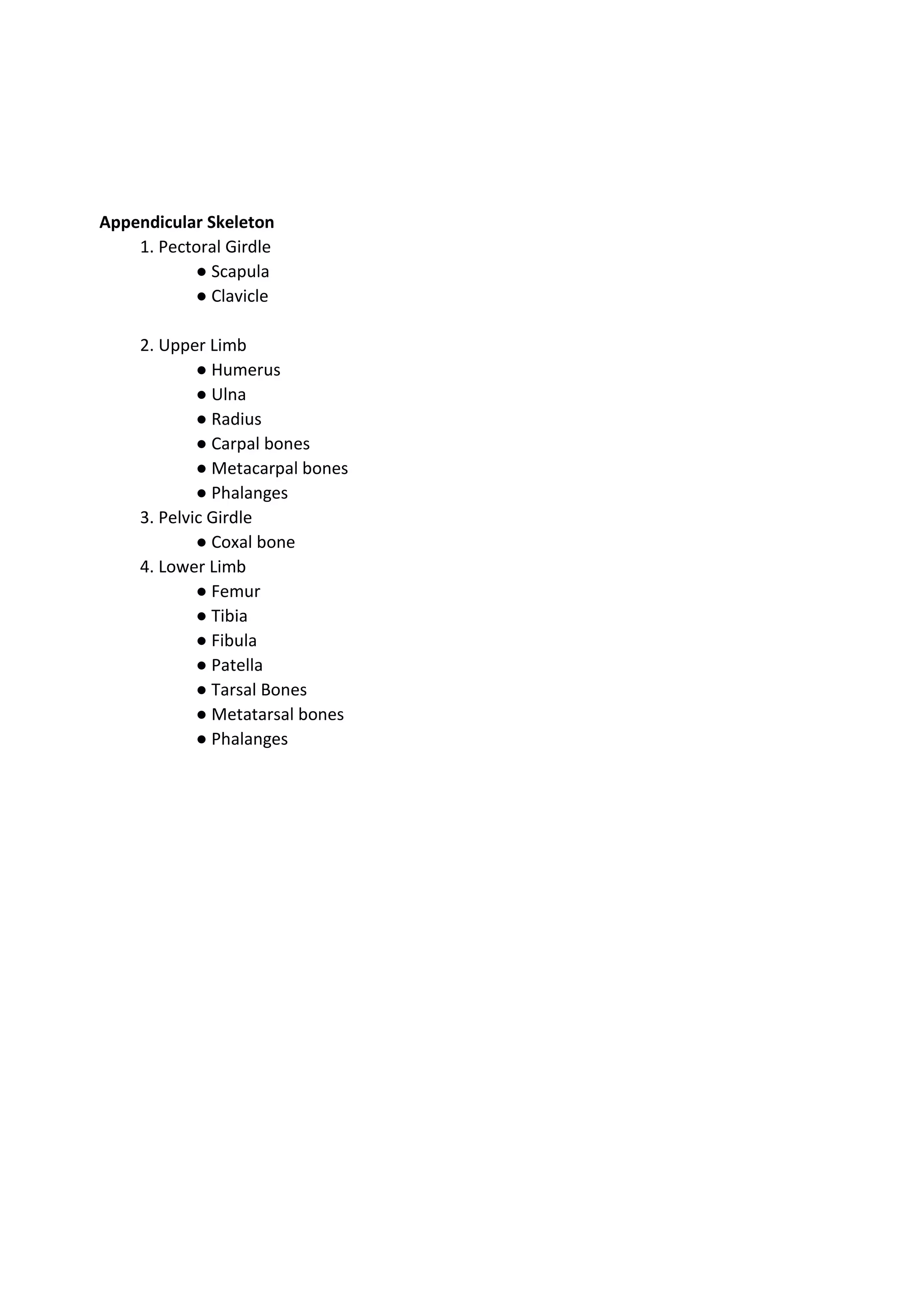
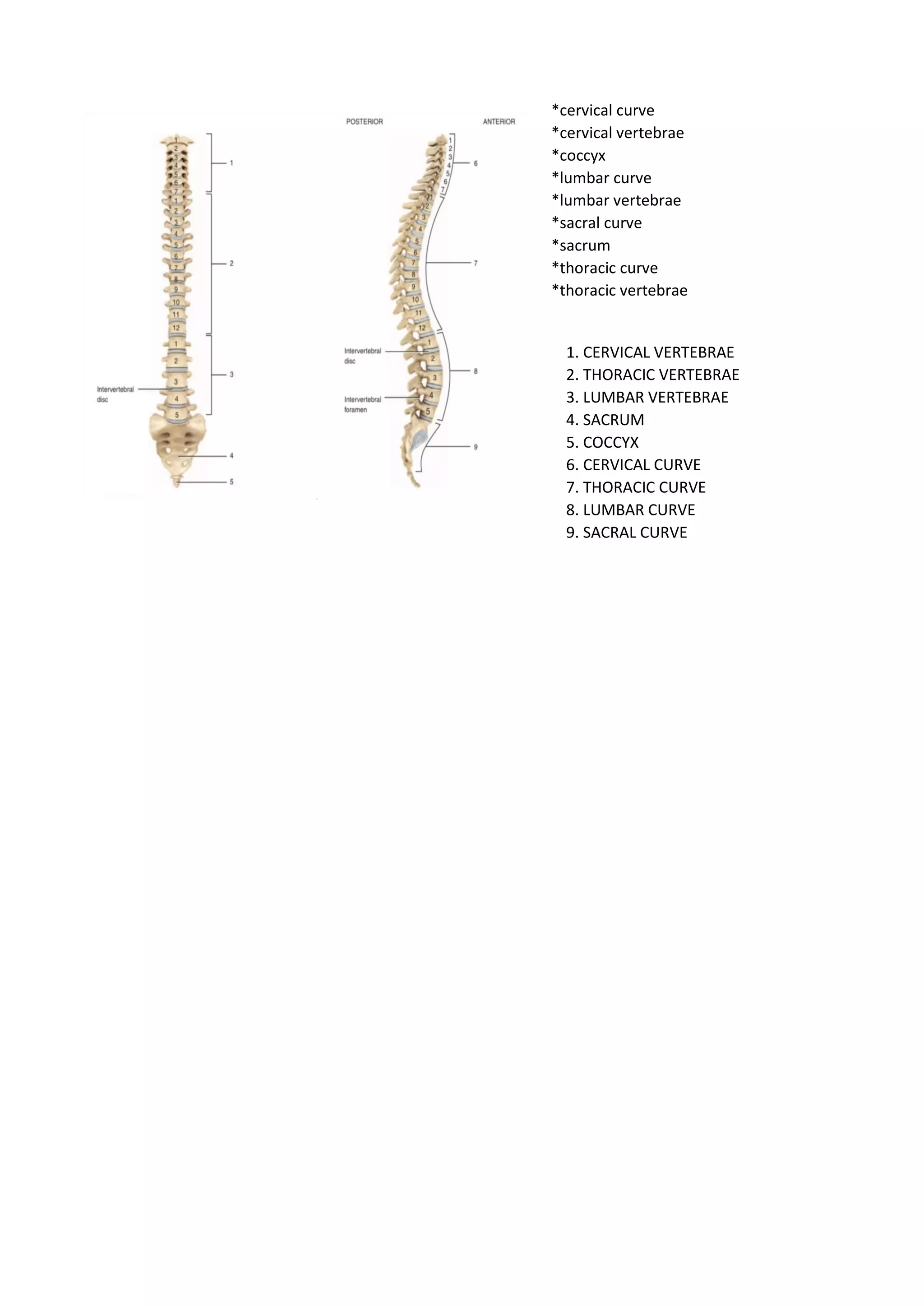
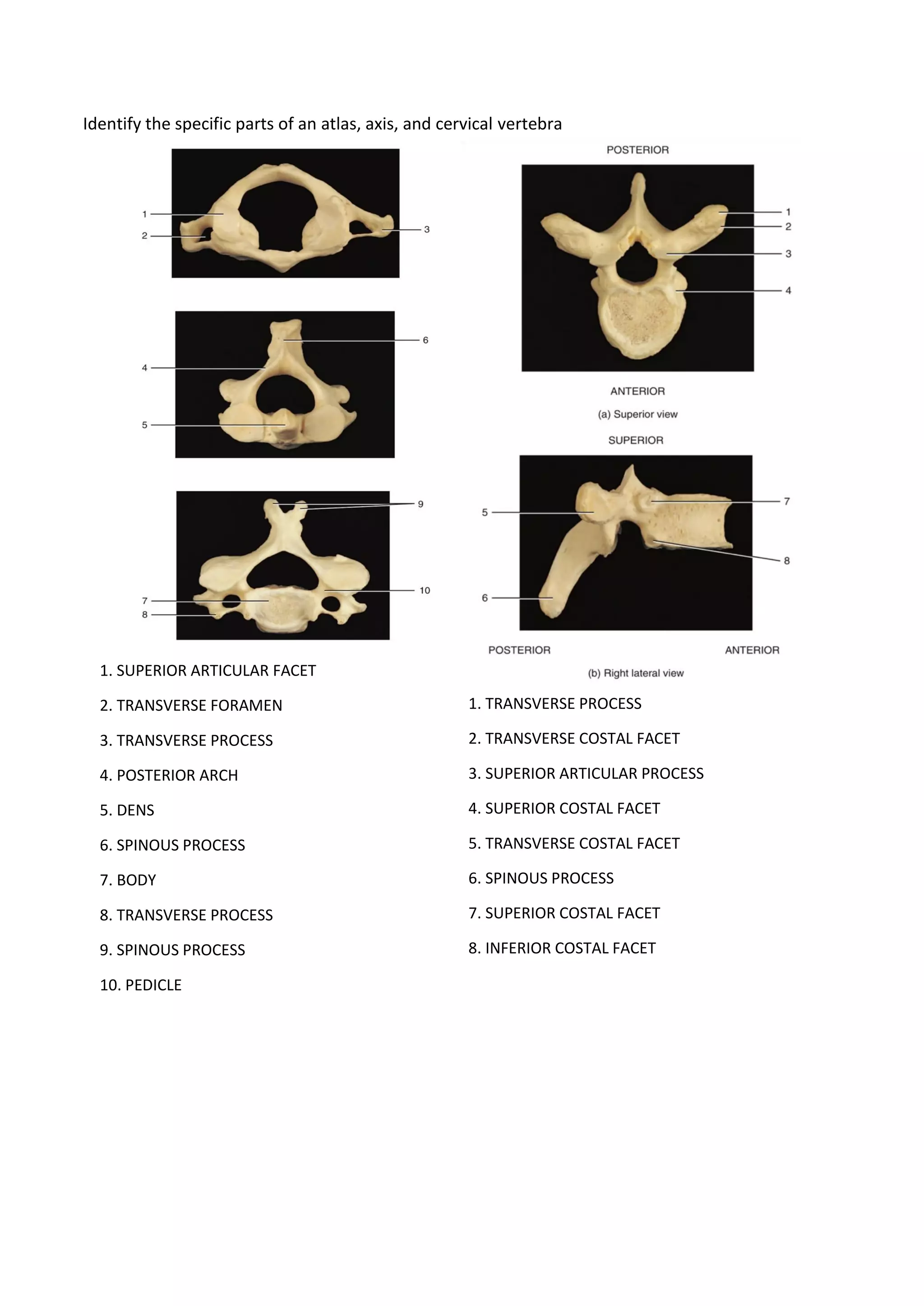
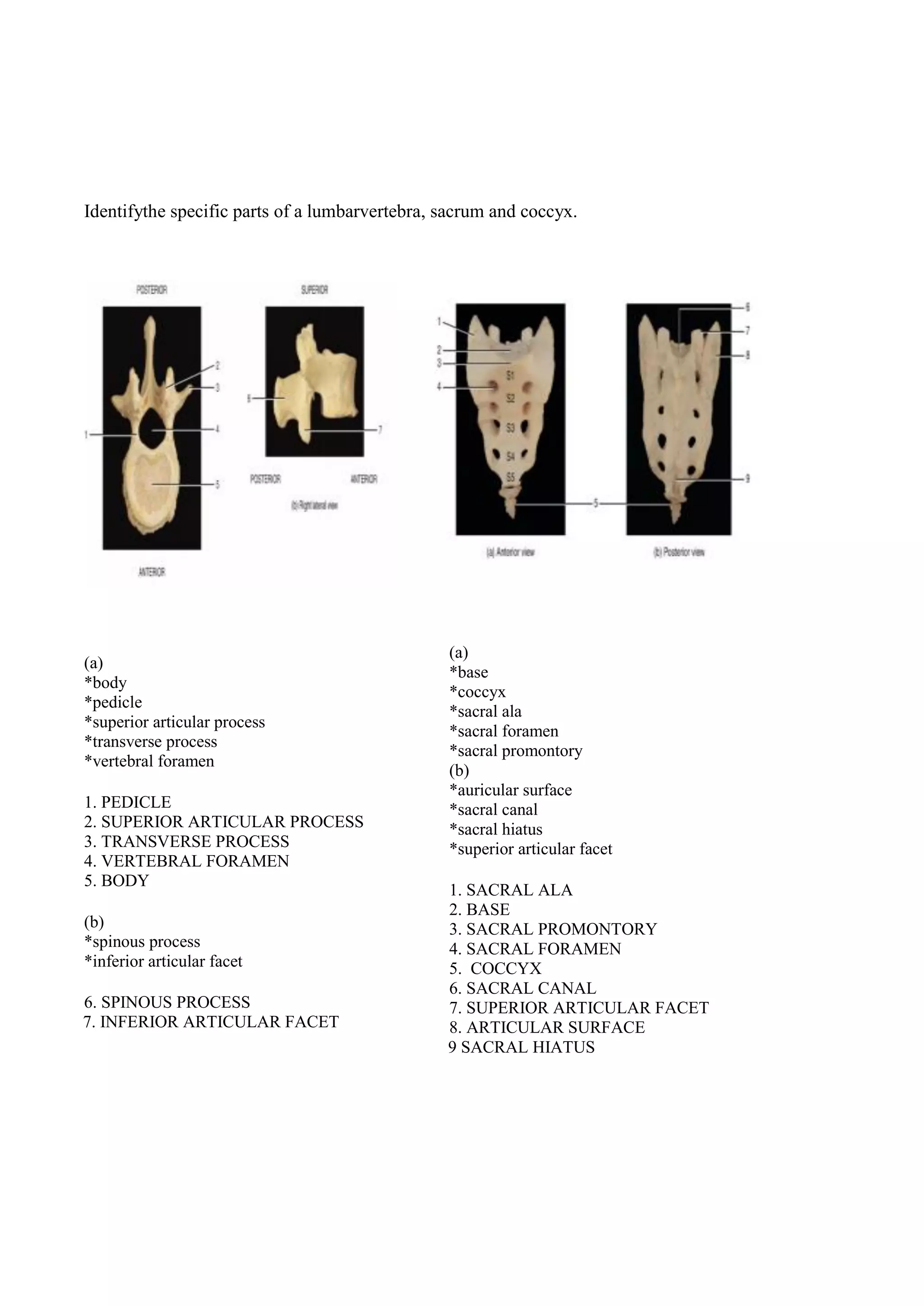
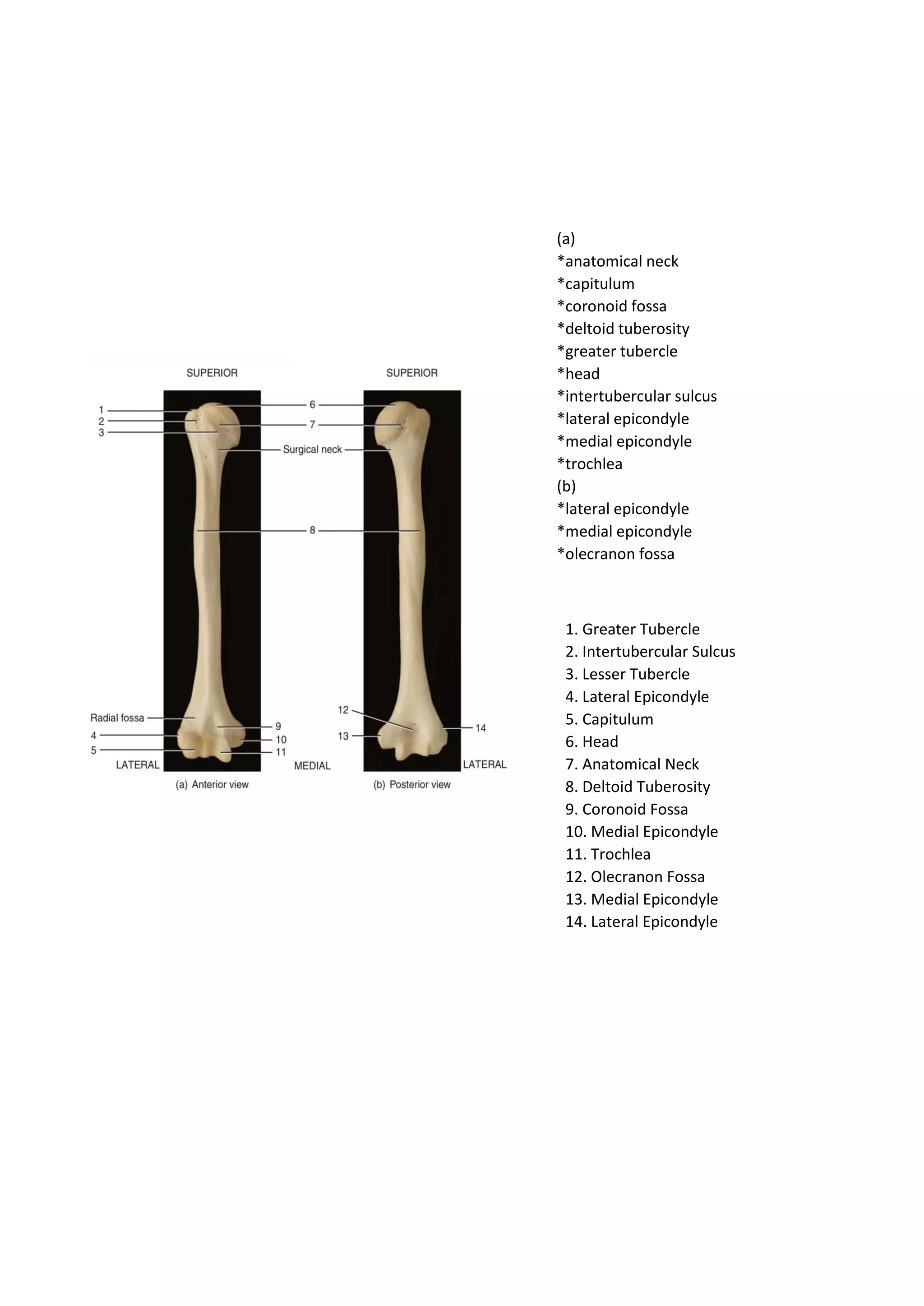
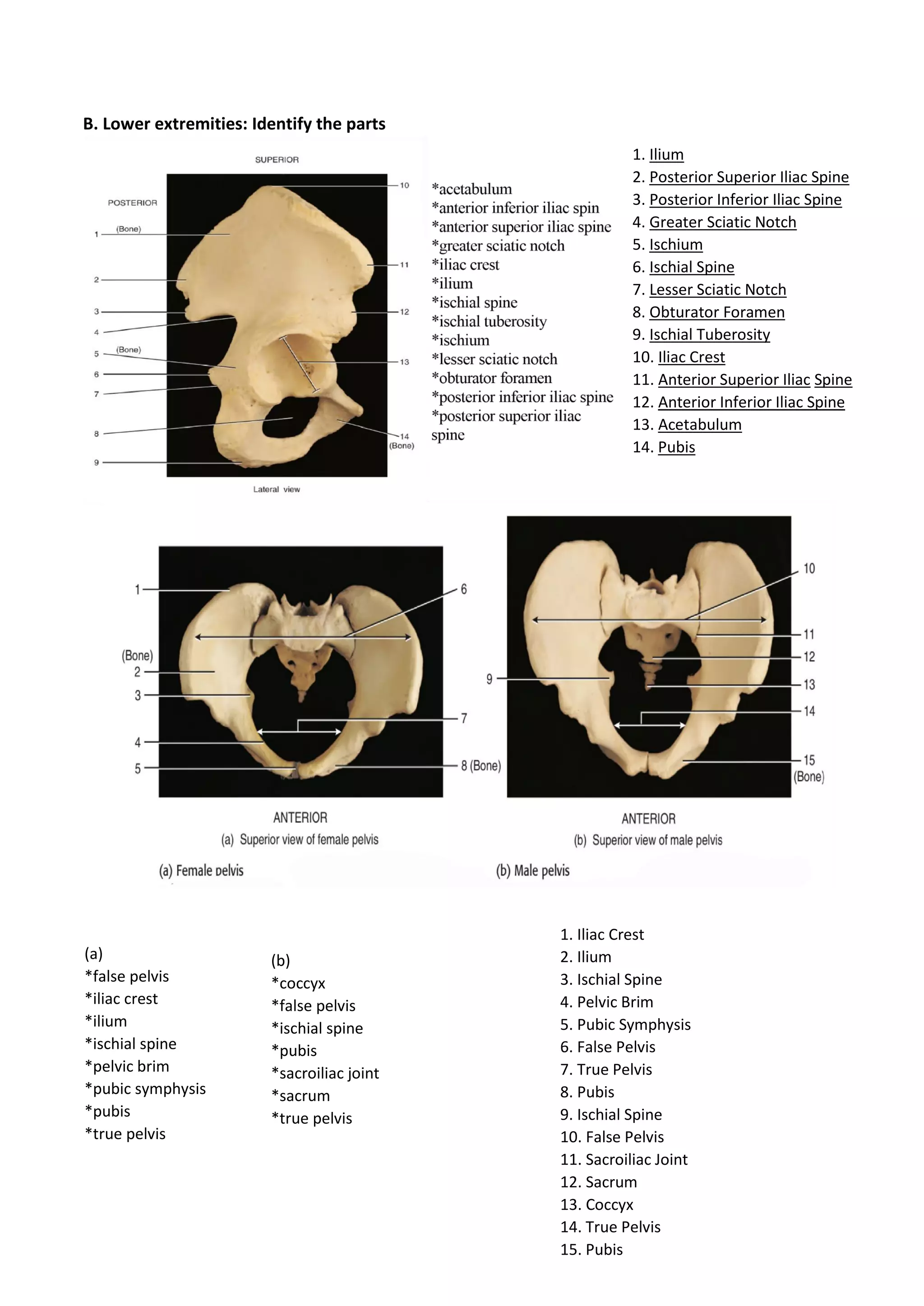
COLLEGE OF MEDICALLABORATORYSCIENCE
CENTRALPHILIPPINE UNIVERSITY
MLS 1102a
HUMANANATOMYAND PHYSIOLOGYWITH PATHOPHYSIOLOGY
Exercise No. 6a Score:
OVERVIEW OF THE SKELETAL SYSTEM
Group No. 2
Names: Natasha Liliane D. Loot (Leader)
Eurika Reign R. Guihama
Edzel T. Gustilo
Date Performed: November 16, 2021
I. Introduction
The human skeletal system is composed of bones, as well as cartilage, ligaments, and other
connective tissues that stabilize or connect bones to each other. Bones are composed primarily of
bone tissue, which serves as the rigid framework that supports the body. It is a highly specialized
connective tissue that is both rigid and flexible.
II. Learning Outcomes
At the end of this exercise, the learners should be able to:
1.Describe the skeletal organs
2.List the primary function/s of the skeletal system
3.Outline the organization of the skeletal system
III. Activity
A. Bone types:
The 206 bones in the standard human skeleton can be classified by their shapes. Label each
group of bones below according to the categories given. Briefly describe each group and give
examples write on the box provided.
1. Long Bones are hard, dense bones that provide strength, structure, and mobility. They
are longer than they are wide. They consist of a long shaft with two bulky ends or
extremities. They are primarily compact bone but may have a large amount of spongy
bone at the ends or extremities. It has a shaft and two ends. Some bones in the fingers
are classified as long bones, even though they are short in length. This is due to the shape
of the bones, not their size. They contain yellow bone marrow and red bone marrow,
which produce blood cells.
2. Short Bones roughly cube shaped with vertical and horizontal dimensions approximately
equal. They consist primarily of spongy bone, which is covered by a thin layer of compact
bone. They are about as long as they are wide.
Examples: femur, tibia, fibula,
femur, metatarsals, phalanges,
humerus, radius, ulna, and
metacarpals
Examples: scaphoid, lunate,
triquetral, hamate, pisiform,
capitate, trapezoid, and
trapezium.
Examples: In thoracic cage
there’s sternum and ribs. In
the pelvis there’s ilium,
ischium, and pubis.
Examples: vertebrae, sacrum,
coccyx, temporal bone, sphenoid,
ethmoid, zygomatic, maxilla,
mandible, palatine, inferior nasal
concha, and hyoid.
3. Flat Bones are made up of a layer of spongy bone between two thin layers of compact
bone. They are thin, flattened, and usually curved. They have marrow, but they do not
have a bone marrow cavity.
4. Irregular Bones are primarily spongy bone that is covered with a thin layer of compact
bone. They often have a fairly complex shape.
B. Gross Structure of a Bone
All bones have the same general structural pattern. Some bones have more “optional
features" than other bones, some less. The long bone is often used as a general specimen for study
because it has all the features that any bone can have.
Instruction: Examine the external aspect/sectioned long bone specimen in the Figure (6b) below.
Label the different features and briefly describe each part.
*articular cartilage
*compact bone
*diaphysis
*distal epiphysis
*endosteum
*epiphyseal line
*medullary cavity
*nutrient artery
*periosteum
*proximal epiphysis
*spongy bone
*yellow marrow
1. proximal epiphysis
2. diaphysis
3. distal epiphysis
4. articular cartilage
5. epiphyseal line
6. compact bone
7. medullary cavity
8. yellow marrow
9. periosteum
10. nutrient artery
11. endosteum
12. spongy bone
PARTS DESCRIPTION
A. Proximal Epiphysis The end of the long bone closest to the center of the body.
B. Diaphysis The tubular shaft that runs between the proximal and distal ends of the
bone.
C. Distal epiphysis The distal epiphysis is located at the end of the long bone that is farther
away from the center of the body.
D. Articular Cartilage It is a highly specialized connective tissue of diarthrodial joints. Its
principal function is to provide a smooth, lubricated surface for
articulation and to facilitate the transmission of loads with a low
frictional coefficient
E. Epiphyseal line the line marking the site of an epiphyseal plate that has stopped growing
and become ossified
F. Compact bone Dense bone tissue composed of osteons, which resist pressure and
shocks and protect the spongy tissue; it forms especially the diaphysis of
the long bones.
G. Medullary cavity It is also known as the marrow cavity, is the central cavity of bone shafts
where red bone marrow and/or yellow bone marrow (adipose tissue) is
stored.
H. Yellow marrow Yellow bone marrow contains mesenchymal stem cells (marrow stromal
cells), which produce cartilage, fat and bone. Yellow bone marrow also
aids in the storage of fats in cells called adipocytes. This helps maintain
the right environment and provides the sustenance that bones need to
function.
I. Periosteum A membranous tissue that covers the surfaces of your bones.
J. Nutrient artery An artery of variable origin that supplies the medullary cavity of a long
bone.
K. Endosteum A membrane lining the inner surface of the bony wall also identified as
the lining membrane of the Bone marrow cavity is endosteum; The
endosteum lines the Haversian canal and all the internal cavities of the
bone.
L. Spongy bone Spongy (cancellous) bone is lighter and less dense than compact bone.
Spongy bone consists of plates (trabeculae) and bars of bone adjacent to
small, irregular cavities that contain red bone marrow.
C. Divisions of the Skeletal System
The usual number given for bones in the human skeleton is 206. This is by no means the
absolute normal number, however. Most people have more bones, but each person has
different types,locations, and numbers of "extra" bones. In this activity, you will examine the
standard 206bones that may be present.
Examine the diagram below and locate the division of the skeletal system (A) axial skeleton;
and (B) appendicular skeleton by coloring each division using colored pens/pencils.
Label the illustration of the skeletal system.
Fill up this table
Axial and Appendicular Skeleton
No. of bones
Axial Skeleton 80
1.Skull 28
2.Hyoid bone 1
3.Vertebral
column
26
4.Ribs 24
5.Sternum 1
Appendicular Skeleton 126
Pectoral girdle 4
6.Clavicle 2
7.Scapula 2
Upper limb 60
8.Humerus 2
9.Radius 2
10.Ulna 2
11.Carpals 16
12.Metacarpals 10
13.Phalanges 28
Pelvic girdle 2
14.Coxal bone 2
Lower limb 60
15.Femur 2
16.Patella 2
17.Tibia 2
18.Fibula 2
19.Tarsals 14
20.Metatarsals 10
21.Phalanges 28
Total 206
1. Skull
2. Mandible
3. Clavicle
4. Sternum
5. Humerus
6. Ribs
7. Vertebral Column
8. Pelvic Girdle/Hip bone
9. Radius
10. Ulna
11. Carpal Bones
12. Metacarpal Bones
13. Phalanges
14. Femur
15. Patella
16. Tibia
17. Fibula
18. Tarsal Bones
19. Metatarsals
20. Phalanges
D. Discussion: Discuss or answer the following items concisely.
1. Describe the skeletal organs
The skeletal organs are the body’s core framework and it serves as a structural support system. It is
composed of 4 main fibrous and mineralized connective tissue, bones, ligaments, tendon, cartilage
and joint. They are the ones who gives shape to our body, allows movement, produces blood cells
and protects the organs. In addition, bone is a rigid form of connective tissue that is a part of the
skeletal system of vertebrates and is made up of calcium and the ligaments is a small band of dense,
white fibrous elastic tissue and it connects the end of the bones to create a joint and they also assist
in holding the organs in place. While tendon is a tough, flexible, and inelastic band of fibrous
connective tissue that connects muscles to bones and lastly, the joints hold the skeleton and
supports body movement.
2. List the primary functions of the skeletal system.
• Supports the body
• Gives body shape
• Body movements
• Stores minerals and adipose tissue
• Blood cell production
• Protects body organs
3. Outline the organization of skeletal system.
Axial Skeleton
1. Skull
● Braincase
● Face
2. Auditory ossicles
3. Hyoid
4. Vertebral Column
● Cervical Vertebrae
● Thoracic Vertebrae
● Lumbar Vertebrae
● Sacrum
● Coccyx
5. Thoracic Cage
● Ribs
● Sternum
Appendicular Skeleton
1. Pectoral Girdle
● Scapula
● Clavicle
2. Upper Limb
● Humerus
● Ulna
● Radius
● Carpal bones
● Metacarpal bones
● Phalanges
3. Pelvic Girdle
● Coxal bone
4. Lower Limb
● Femur
● Tibia
● Fibula
● Patella
● Tarsal Bones
● Metatarsal bones
● Phalanges
COLLEGE OF MEDICALLABORATORYSCIENCE
CENTRALPHILIPPINE UNIVERSITY
MLS 1102a
HUMANANATOMYAND PHYSIOLOGYWITH
PATHOPHYSIOLOGY
Exercise No. 6b Score:
THE SKULL
Group No. 2
Names: Natasha Liliane D. Loot (Leader)
Eurika Reign R. Guihama
Edzel T. Gustilo
Date Performed: November 16, 2021
I. Introduction
The skull is the superior portion of the axial skeleton. For ease of study, the
28bones of the skull are divided into three categories. The cranial bones form a roughly
spherical case for the brain called the cranium. The facial bones include most of the
remaining skull bones, except the 6auditory ossicles of the middle ear.
II. Learning Outcomes
At the end of this exercise, the learners should be able to:
1. Distinguish between the cranial bones and facial bones of the skull;
2. Identify all the bones of the skull and their important markings in a specimen or
figure;
3. Describe the important features of a fetal skull.
III. Activity
A. Bones of the Face and Brain case
Eight of the 28 skull bones form the cranium, or brain case of the skull. Fourteen bones
are classified as facial bones. Identify the bones of the skull in the figures below by writing
your answer to the corresponding number.
BONES OF THE SKULL
ANTERIOR VIEW
*ethmoid bone
*frontal bone
*inferior nasal concha
or turbinate
*lacrimal
*mandible
*maxilla
*nasal bone
*parietal bone
*sphenoid bone
*temporal bone
*vomer
*zygomatic bone
1. parietal bone
2. sphenoid bone
3. ethmoid bone
4. lacrimal bone
5. inferior nasal concha or turbinate
6. vomer
7. frontal bone
8. temporal bone
9. nasal bone
10 zygomatic bone
11. maxilla
12. mandible
BONES OF THE SKULL
LATERALVIEW
BONES OF THE SKULL
INFERIORVIEW
*coronal
*ethmoid
*frontal bone
*lacrimal bone
*lambdoid suture
*mandible
*maxilla
*nasal bone
*occipital bone
*parietal bone
*sphenoid bone
*squamous suture
*temporal bone
*zygomatic bone
1. parietal bone
2. coronal
3. squamous suture
4. temporal bone
5. lambdoid suture
6. occipital bone
7. frontal bone
8. sphenoid bone
9. ethmoid
10. lacrimal bone
11. nasal bone
12. zygomatic bone
13. maxilla
14. mandible
* maxilla
*occipital bone
*palatine bone
*sphenoid bone
*temporal bone
*vomer
*zygomatic bone
1. maxilla
2. vomer
3. sphenoid bone
4. occipital bone
5. zygomatic bone
6. palatine bone
7. temporal bone
B. Anatomy of the External and Middle Ear
The external ear, consisting of the auricle, external auditory canal, and tympanic
membrane, extends from the auricle to the tympanic membrane. Each middle ear, within
the petrous portion of the temporal bone, contains three auditory ossicles. Label the
structures of the external and middle ear in the figure below.
* auditory tube
*auricle
*external auditory
canal
*external ear
*helix
*incus
*internal ear
*lobule
*malleus
*middle ear
*stapes attached to
oval window
*tympanic
membrane
1. lobule
2. auricle
3. helix
4. external ear
5. malleus
6. incus
7. stapes attached to oval window
8. middle ear
9. internal ear
10. auditory tube
11. tympanic membrane
12. external auditory canal
C. The Fetal Skull
The fetal skull features partly ossified skull bones and a large proportion of fibrous and
cartilaginous tissue. Because the flat bones of the cranium have not met to form sutures,
there are fibrous areas called fontanels. Identify each in a fetal skull diagram below.
D. Bone markings. Give the name of the bone marking or feature described.
1. A large process of the temporal bone, just posterior to the external auditory
meatus; it contains sinuses.
Mastoid process
2. A smaller, needle- shaped process just medial to the process described in item
no.1
Styloid process
3. A hole in the sphenoid bone that allows the optic nerve to exit the eye orbit.
Optic foramen
*anterior fontanel
*anterolateral fontanel
*posterior fontanel
*posterolateral fontanel
1. anterolateral fontanel
2. posterolateral fontanel
3. anterior fontanel
4. posterior fontanel
4. Same name for ridge like process on both the maxilla and mandible in which the
teeth are embedded.
Alveolar process
5. A crest or projection on the superior surface of the ethmoid bone.
Crista Galli
6. A curved plate of bone projecting from the lateral wall of the nasal cavity, just
above the inferior nasal conchae (part of the ethmoid bone).
Middle nasal conchae
7. One of two holes on the anterior portion of the mandibular body.
Mantle foramen
8. A hole in the maxilla just below the orbit of the eye.
Infra orbital foramen
9. Part of the ethmoid that forms the upper portion of the nasal septum.
Perpendicular plate
10. Structure formed by both the zygomatic process of the temporal bone and the
temporal process of the zygomatic bone.
Zygomatic arch
E. Sutures. Determine the name of suture joint described.
1. Joins the parietal bones together. Sagittal suture
2. Joins the superior margin of the temporal bone to the frontal, parietal, and
occipital bone. Squamosal suture
3. Joins the palatine bone to the maxilla. Transverse palatine suture
4. Joins the frontal bone to the two parietal bones. Coronal suture
COLLEGE OF MEDICAL LABORATORY SCIENCE
CENTRALPHILIPPINE UNIVERSITY
MLS 1102a
HUMANAN ATOMY AND PHYSIOLOGY WITH PATHOPHYSIOLOGY
Exercise No. 6c Score:
THE VERTEBRAL COLUMN AND THORACIC CAGE
Group No. 2
Names: Natasha Liliane D. Loot (Leader)
Eurika Reign R. Guihama
Edzel T. Gustilo
Date Performed: November 16, 2021
I. Introduction
The vertebral column is a set of 26 bones stacked one on another to form a slightly curved,
flexible support rod. The 25 bones of the rib cage, or thoracic cage, protect the lungs and heart
within the thorax. The hyoid bone is not attached to any other bone but is very close to the skull
and vertebral column and lies along the central axis of the body.
II. Learning outcomes
At the end of this exercise, the learners should be able to:
1. Name the component bones of the vertebral column and thoracic cage;
2. Identify the bones and markings of the vertebral column and thorax in figure.
III. Activity
A. The Vertebral Column
The 26 bones of the vertebral column are divided among the bones listed here. Identify
the 5 regions and 4normal curvatures of the vertebral column in the figure below and write
your answer on the corresponding number.
*cervical curve
*cervical vertebrae
*coccyx
*lumbar curve
*lumbar vertebrae
*sacral curve
*sacrum
*thoracic curve
*thoracic vertebrae
1. CERVICAL VERTEBRAE
2. THORACIC VERTEBRAE
3. LUMBAR VERTEBRAE
4. SACRUM
5. COCCYX
6. CERVICAL CURVE
7. THORACIC CURVE
8. LUMBAR CURVE
9. SACRAL CURVE
Identify the specific parts of an atlas, axis, and cervical vertebra
1. TRANSVERSE PROCESS
2. TRANSVERSE COSTAL FACET
3. SUPERIOR ARTICULAR PROCESS
4. SUPERIOR COSTAL FACET
5. TRANSVERSE COSTAL FACET
6. SPINOUS PROCESS
7. SUPERIOR COSTAL FACET
8. INFERIOR COSTAL FACET
1. SUPERIOR ARTICULAR FACET
2. TRANSVERSE FORAMEN
3. TRANSVERSE PROCESS
4. POSTERIOR ARCH
5. DENS
6. SPINOUS PROCESS
7. BODY
8. TRANSVERSE PROCESS
9. SPINOUS PROCESS
10. PEDICLE
Identifythe specific parts of a lumbarvertebra, sacrum and coccyx.
(a)
*body
*pedicle
*superior articular process
*transverse process
*vertebral foramen
1. PEDICLE
2. SUPERIOR ARTICULAR PROCESS
3. TRANSVERSE PROCESS
4. VERTEBRAL FORAMEN
5. BODY
(b)
*spinous process
*inferior articular facet
6. SPINOUS PROCESS
7. INFERIOR ARTICULAR FACET
(a)
*base
*coccyx
*sacral ala
*sacral foramen
*sacral promontory
(b)
*auricular surface
*sacral canal
*sacral hiatus
*superior articular facet
1. SACRAL ALA
2. BASE
3. SACRAL PROMONTORY
4. SACRAL FORAMEN
5. COCCYX
6. SACRAL CANAL
7. SUPERIOR ARTICULAR FACET
8. ARTICULAR SURFACE
9 SACRAL HIATUS
B. The Thoracic Cage
The 25bones of the rib cage, or thoracic cage, form a partially flexible, protective shield
for the heart, lungs and other thoracic organs. The thoracic cage also helps protect some
organs of the upper abdomen, such as the liver and spleen. Label the parts of the thorax.
*body of sternum
*costal cartilage
*false ribs
*floating ribs
*manubrium
*sternal angle
*sternum
*jugular notch
*true ribs
*xiphoid process
1. JUGULAR NOTCH
2. MANUBRIUM
3. BODY OF STERNUM
4. XIPHOID PROCESS
5. STERNUM
6. COSTAL CARTILAGE
7. STERNAL ANGLE
8. TRUE RIBS
9. FLOATING RIBS
10. FALSE RIBS
C. Fill in the numbers as indicated.
Number Structures
26 Bones in the vertebral column
24 Unfused vertebrae
7 Cervical vertebrae
12 Thoracic vertebrae
5 Lumbar vertebrae
4 Spinal curves
24 Bones in the thoracic cage
12 Pairs of ribs
7 Pairs of true ribs
5 Pairs of false ribs
2 Pairs of floating ribs
COLLEGE OF MEDICALLABORATORYSCIENCE
CENTRALPHILIPPINE UNIVERSITY
MLS 1102a
HUMANANATOMYAND PHYSIOLOGYWITH PATHOPHYSIOLOGY
Exercise No. 6d Score:
THE APPENDICULAR SKELETON
Group No. 2
Names: Natasha Liliane D. Loot (Leader)
Eurika Reign R. Guihama
Edzel T. Gustilo
Date Performed: November 16, 2021
I. Introduction
The appendicular skeleton consists of all 126 bones that form the upper and lower
extremities. The shoulder girdle and arms (upper extremities) have 64 bones altogether,
whereas the pelvic girdle and the legs (lower extremities) have a total of 62bones.
II. Learning outcomes
At the end of this exercise, the learners should be able to:
1. name correctly the bones of the appendicular skeleton;
2. identify appendicular bones and markings in figures.
III. Activity
A. Upper extremities: Identify the parts
(b)
*acromion
*coracoid
*glenoid fossa
*lateral border
*medial border
*subscapular fossa
(c)
*acromion
*glenoid cavity
* infraspinous fossa
*spine of scapula
*supraspinous fossa
1. (no label)
2. (no label)
3. Acromion
4. Coracoid
5. Glenoid Fossa
6. Lateral Border
7. Subscapular Fossa
8. Medial Border
9. Supraspinous Fossa
10. Infraspinous Fossa
11. Acromion
12. Spine of Scapula
13. Glenoid Cavity
(a)
*anatomical neck
*capitulum
*coronoid fossa
*deltoid tuberosity
*greater tubercle
*head
*intertubercular sulcus
*lateral epicondyle
*medial epicondyle
*trochlea
(b)
*lateral epicondyle
*medial epicondyle
*olecranon fossa
1. Greater Tubercle
2. Intertubercular Sulcus
3. Lesser Tubercle
4. Lateral Epicondyle
5. Capitulum
6. Head
7. Anatomical Neck
8. Deltoid Tuberosity
9. Coronoid Fossa
10. Medial Epicondyle
11. Trochlea
12. Olecranon Fossa
13. Medial Epicondyle
14. Lateral Epicondyle
1. Head of Radius
2. Radial Tuberosity
3. Styloid Process of Radius
4. Olecranon Process
5. Trochlear Notch
6. Coronoid Process
7. Radial Notch
8. Styloid process of Ulna
9. Radius
10. Ulna
(a)
*coronoid process
*head of radius
*olecranon process
*radial notch
*radial tuberosity
*styloid process of radius
*styloid process of ulna
*trochlear notch
(b)
*radius
*ulna
Palm Anterior
1. Radius
2. Ulna
3. Capitate
4. Scaphoid
5. Trapezium
6. Trapeziod
7. Lunate
8. Triquetrum
9. Pisiform
10. Hamate
B. Lower extremities: Identify the parts
1. Ilium
2. Posterior Superior Iliac Spine
3. Posterior Inferior Iliac Spine
4. Greater Sciatic Notch
5. Ischium
6. Ischial Spine
7. Lesser Sciatic Notch
8. Obturator Foramen
9. Ischial Tuberosity
10. Iliac Crest
11. Anterior Superior Iliac Spine
12. Anterior Inferior Iliac Spine
13. Acetabulum
14. Pubis
(a)
*false pelvis
*iliac crest
*ilium
*ischial spine
*pelvic brim
*pubic symphysis
*pubis
*true pelvis
(b)
*coccyx
*false pelvis
*ischial spine
*pubis
*sacroiliac joint
*sacrum
*true pelvis
1. Iliac Crest
2. Ilium
3. Ischial Spine
4. Pelvic Brim
5. Pubic Symphysis
6. False Pelvis
7. True Pelvis
8. Pubis
9. Ischial Spine
10. False Pelvis
11. Sacroiliac Joint
12. Sacrum
13. Coccyx
14. True Pelvis
15. Pubis
1. Head of Femur
2. Greater Trochanter
3. Neck
4. Lesser Trochanter
5. Medial Epicondyle
6. Medial Condyle
7. Linea Aspera
8. Lateral Epicondyle
9. Lateral Condyle
1. Lateral Condyle
2. Head of Fibula
3. Fibula
4. Lateral Malleolus
5. Medial Condyle
6. Tibial Tuberosity
7. Tibia
8. Medial Malleolus
C.
Table. Fill- in the numbers as
indicated.
Number Structures
126 Bones in the appendicular skeleton
64 Bones in the upper extremities
62 Bones in the lower extremities
16 Wrist bones (total)
14 Ankle bones (total)
27 Hand/finger bones (total)
26 Foot/toe bones (total)
1. Calcaneus
2. Talus
3. Navicular
4. Lateral Cuneiform
5. Intermediate Cuneiform
6. Medial Cuneiform
7. Cuboid
8. Metatarsals
9. Proximal Phalanx
10. Middle Phalanx
11. Distal Phalanx

Group 2 activity6

  • 1.
    COLLEGE OF MEDICALLABORATORYSCIENCE CENTRALPHILIPPINEUNIVERSITY MLS 1102a HUMANANATOMYAND PHYSIOLOGYWITH PATHOPHYSIOLOGY Exercise No. 6a Score: OVERVIEW OF THE SKELETAL SYSTEM Group No. 2 Names: Natasha Liliane D. Loot (Leader) Eurika Reign R. Guihama Edzel T. Gustilo Date Performed: November 16, 2021 I. Introduction The human skeletal system is composed of bones, as well as cartilage, ligaments, and other connective tissues that stabilize or connect bones to each other. Bones are composed primarily of bone tissue, which serves as the rigid framework that supports the body. It is a highly specialized connective tissue that is both rigid and flexible. II. Learning Outcomes At the end of this exercise, the learners should be able to: 1.Describe the skeletal organs 2.List the primary function/s of the skeletal system 3.Outline the organization of the skeletal system
  • 2.
    III. Activity A. Bonetypes: The 206 bones in the standard human skeleton can be classified by their shapes. Label each group of bones below according to the categories given. Briefly describe each group and give examples write on the box provided. 1. Long Bones are hard, dense bones that provide strength, structure, and mobility. They are longer than they are wide. They consist of a long shaft with two bulky ends or extremities. They are primarily compact bone but may have a large amount of spongy bone at the ends or extremities. It has a shaft and two ends. Some bones in the fingers are classified as long bones, even though they are short in length. This is due to the shape of the bones, not their size. They contain yellow bone marrow and red bone marrow, which produce blood cells. 2. Short Bones roughly cube shaped with vertical and horizontal dimensions approximately equal. They consist primarily of spongy bone, which is covered by a thin layer of compact bone. They are about as long as they are wide. Examples: femur, tibia, fibula, femur, metatarsals, phalanges, humerus, radius, ulna, and metacarpals Examples: scaphoid, lunate, triquetral, hamate, pisiform, capitate, trapezoid, and trapezium. Examples: In thoracic cage there’s sternum and ribs. In the pelvis there’s ilium, ischium, and pubis. Examples: vertebrae, sacrum, coccyx, temporal bone, sphenoid, ethmoid, zygomatic, maxilla, mandible, palatine, inferior nasal concha, and hyoid.
  • 3.
    3. Flat Bonesare made up of a layer of spongy bone between two thin layers of compact bone. They are thin, flattened, and usually curved. They have marrow, but they do not have a bone marrow cavity. 4. Irregular Bones are primarily spongy bone that is covered with a thin layer of compact bone. They often have a fairly complex shape. B. Gross Structure of a Bone All bones have the same general structural pattern. Some bones have more “optional features" than other bones, some less. The long bone is often used as a general specimen for study because it has all the features that any bone can have. Instruction: Examine the external aspect/sectioned long bone specimen in the Figure (6b) below. Label the different features and briefly describe each part. *articular cartilage *compact bone *diaphysis *distal epiphysis *endosteum *epiphyseal line *medullary cavity *nutrient artery *periosteum *proximal epiphysis *spongy bone *yellow marrow 1. proximal epiphysis 2. diaphysis 3. distal epiphysis 4. articular cartilage 5. epiphyseal line 6. compact bone 7. medullary cavity 8. yellow marrow 9. periosteum 10. nutrient artery 11. endosteum 12. spongy bone
  • 4.
    PARTS DESCRIPTION A. ProximalEpiphysis The end of the long bone closest to the center of the body. B. Diaphysis The tubular shaft that runs between the proximal and distal ends of the bone. C. Distal epiphysis The distal epiphysis is located at the end of the long bone that is farther away from the center of the body. D. Articular Cartilage It is a highly specialized connective tissue of diarthrodial joints. Its principal function is to provide a smooth, lubricated surface for articulation and to facilitate the transmission of loads with a low frictional coefficient E. Epiphyseal line the line marking the site of an epiphyseal plate that has stopped growing and become ossified F. Compact bone Dense bone tissue composed of osteons, which resist pressure and shocks and protect the spongy tissue; it forms especially the diaphysis of the long bones. G. Medullary cavity It is also known as the marrow cavity, is the central cavity of bone shafts where red bone marrow and/or yellow bone marrow (adipose tissue) is stored. H. Yellow marrow Yellow bone marrow contains mesenchymal stem cells (marrow stromal cells), which produce cartilage, fat and bone. Yellow bone marrow also aids in the storage of fats in cells called adipocytes. This helps maintain the right environment and provides the sustenance that bones need to function. I. Periosteum A membranous tissue that covers the surfaces of your bones.
  • 5.
    J. Nutrient arteryAn artery of variable origin that supplies the medullary cavity of a long bone. K. Endosteum A membrane lining the inner surface of the bony wall also identified as the lining membrane of the Bone marrow cavity is endosteum; The endosteum lines the Haversian canal and all the internal cavities of the bone. L. Spongy bone Spongy (cancellous) bone is lighter and less dense than compact bone. Spongy bone consists of plates (trabeculae) and bars of bone adjacent to small, irregular cavities that contain red bone marrow. C. Divisions of the Skeletal System The usual number given for bones in the human skeleton is 206. This is by no means the absolute normal number, however. Most people have more bones, but each person has different types,locations, and numbers of "extra" bones. In this activity, you will examine the standard 206bones that may be present. Examine the diagram below and locate the division of the skeletal system (A) axial skeleton; and (B) appendicular skeleton by coloring each division using colored pens/pencils. Label the illustration of the skeletal system.
  • 6.
    Fill up thistable Axial and Appendicular Skeleton No. of bones Axial Skeleton 80 1.Skull 28 2.Hyoid bone 1 3.Vertebral column 26 4.Ribs 24 5.Sternum 1 Appendicular Skeleton 126 Pectoral girdle 4 6.Clavicle 2 7.Scapula 2 Upper limb 60 8.Humerus 2 9.Radius 2 10.Ulna 2 11.Carpals 16 12.Metacarpals 10 13.Phalanges 28 Pelvic girdle 2 14.Coxal bone 2 Lower limb 60 15.Femur 2 16.Patella 2 17.Tibia 2 18.Fibula 2 19.Tarsals 14 20.Metatarsals 10 21.Phalanges 28 Total 206 1. Skull 2. Mandible 3. Clavicle 4. Sternum 5. Humerus 6. Ribs 7. Vertebral Column 8. Pelvic Girdle/Hip bone 9. Radius 10. Ulna 11. Carpal Bones 12. Metacarpal Bones 13. Phalanges 14. Femur 15. Patella 16. Tibia 17. Fibula 18. Tarsal Bones 19. Metatarsals 20. Phalanges
  • 7.
    D. Discussion: Discussor answer the following items concisely. 1. Describe the skeletal organs The skeletal organs are the body’s core framework and it serves as a structural support system. It is composed of 4 main fibrous and mineralized connective tissue, bones, ligaments, tendon, cartilage and joint. They are the ones who gives shape to our body, allows movement, produces blood cells and protects the organs. In addition, bone is a rigid form of connective tissue that is a part of the skeletal system of vertebrates and is made up of calcium and the ligaments is a small band of dense, white fibrous elastic tissue and it connects the end of the bones to create a joint and they also assist in holding the organs in place. While tendon is a tough, flexible, and inelastic band of fibrous connective tissue that connects muscles to bones and lastly, the joints hold the skeleton and supports body movement. 2. List the primary functions of the skeletal system. • Supports the body • Gives body shape • Body movements • Stores minerals and adipose tissue • Blood cell production • Protects body organs 3. Outline the organization of skeletal system. Axial Skeleton 1. Skull ● Braincase ● Face 2. Auditory ossicles 3. Hyoid 4. Vertebral Column ● Cervical Vertebrae ● Thoracic Vertebrae ● Lumbar Vertebrae ● Sacrum ● Coccyx 5. Thoracic Cage ● Ribs ● Sternum
  • 8.
    Appendicular Skeleton 1. PectoralGirdle ● Scapula ● Clavicle 2. Upper Limb ● Humerus ● Ulna ● Radius ● Carpal bones ● Metacarpal bones ● Phalanges 3. Pelvic Girdle ● Coxal bone 4. Lower Limb ● Femur ● Tibia ● Fibula ● Patella ● Tarsal Bones ● Metatarsal bones ● Phalanges
  • 9.
    COLLEGE OF MEDICALLABORATORYSCIENCE CENTRALPHILIPPINEUNIVERSITY MLS 1102a HUMANANATOMYAND PHYSIOLOGYWITH PATHOPHYSIOLOGY Exercise No. 6b Score: THE SKULL Group No. 2 Names: Natasha Liliane D. Loot (Leader) Eurika Reign R. Guihama Edzel T. Gustilo Date Performed: November 16, 2021 I. Introduction The skull is the superior portion of the axial skeleton. For ease of study, the 28bones of the skull are divided into three categories. The cranial bones form a roughly spherical case for the brain called the cranium. The facial bones include most of the remaining skull bones, except the 6auditory ossicles of the middle ear. II. Learning Outcomes At the end of this exercise, the learners should be able to: 1. Distinguish between the cranial bones and facial bones of the skull; 2. Identify all the bones of the skull and their important markings in a specimen or figure; 3. Describe the important features of a fetal skull.
  • 10.
    III. Activity A. Bonesof the Face and Brain case Eight of the 28 skull bones form the cranium, or brain case of the skull. Fourteen bones are classified as facial bones. Identify the bones of the skull in the figures below by writing your answer to the corresponding number. BONES OF THE SKULL ANTERIOR VIEW *ethmoid bone *frontal bone *inferior nasal concha or turbinate *lacrimal *mandible *maxilla *nasal bone *parietal bone *sphenoid bone *temporal bone *vomer *zygomatic bone 1. parietal bone 2. sphenoid bone 3. ethmoid bone 4. lacrimal bone 5. inferior nasal concha or turbinate 6. vomer 7. frontal bone 8. temporal bone 9. nasal bone 10 zygomatic bone 11. maxilla 12. mandible
  • 11.
    BONES OF THESKULL LATERALVIEW BONES OF THE SKULL INFERIORVIEW *coronal *ethmoid *frontal bone *lacrimal bone *lambdoid suture *mandible *maxilla *nasal bone *occipital bone *parietal bone *sphenoid bone *squamous suture *temporal bone *zygomatic bone 1. parietal bone 2. coronal 3. squamous suture 4. temporal bone 5. lambdoid suture 6. occipital bone 7. frontal bone 8. sphenoid bone 9. ethmoid 10. lacrimal bone 11. nasal bone 12. zygomatic bone 13. maxilla 14. mandible * maxilla *occipital bone *palatine bone *sphenoid bone *temporal bone *vomer *zygomatic bone 1. maxilla 2. vomer 3. sphenoid bone 4. occipital bone 5. zygomatic bone 6. palatine bone 7. temporal bone
  • 12.
    B. Anatomy ofthe External and Middle Ear The external ear, consisting of the auricle, external auditory canal, and tympanic membrane, extends from the auricle to the tympanic membrane. Each middle ear, within the petrous portion of the temporal bone, contains three auditory ossicles. Label the structures of the external and middle ear in the figure below. * auditory tube *auricle *external auditory canal *external ear *helix *incus *internal ear *lobule *malleus *middle ear *stapes attached to oval window *tympanic membrane 1. lobule 2. auricle 3. helix 4. external ear 5. malleus 6. incus 7. stapes attached to oval window 8. middle ear 9. internal ear 10. auditory tube 11. tympanic membrane 12. external auditory canal
  • 13.
    C. The FetalSkull The fetal skull features partly ossified skull bones and a large proportion of fibrous and cartilaginous tissue. Because the flat bones of the cranium have not met to form sutures, there are fibrous areas called fontanels. Identify each in a fetal skull diagram below. D. Bone markings. Give the name of the bone marking or feature described. 1. A large process of the temporal bone, just posterior to the external auditory meatus; it contains sinuses. Mastoid process 2. A smaller, needle- shaped process just medial to the process described in item no.1 Styloid process 3. A hole in the sphenoid bone that allows the optic nerve to exit the eye orbit. Optic foramen *anterior fontanel *anterolateral fontanel *posterior fontanel *posterolateral fontanel 1. anterolateral fontanel 2. posterolateral fontanel 3. anterior fontanel 4. posterior fontanel
  • 14.
    4. Same namefor ridge like process on both the maxilla and mandible in which the teeth are embedded. Alveolar process 5. A crest or projection on the superior surface of the ethmoid bone. Crista Galli 6. A curved plate of bone projecting from the lateral wall of the nasal cavity, just above the inferior nasal conchae (part of the ethmoid bone). Middle nasal conchae 7. One of two holes on the anterior portion of the mandibular body. Mantle foramen 8. A hole in the maxilla just below the orbit of the eye. Infra orbital foramen 9. Part of the ethmoid that forms the upper portion of the nasal septum. Perpendicular plate 10. Structure formed by both the zygomatic process of the temporal bone and the temporal process of the zygomatic bone. Zygomatic arch E. Sutures. Determine the name of suture joint described. 1. Joins the parietal bones together. Sagittal suture 2. Joins the superior margin of the temporal bone to the frontal, parietal, and occipital bone. Squamosal suture 3. Joins the palatine bone to the maxilla. Transverse palatine suture 4. Joins the frontal bone to the two parietal bones. Coronal suture
  • 15.
    COLLEGE OF MEDICALLABORATORY SCIENCE CENTRALPHILIPPINE UNIVERSITY MLS 1102a HUMANAN ATOMY AND PHYSIOLOGY WITH PATHOPHYSIOLOGY Exercise No. 6c Score: THE VERTEBRAL COLUMN AND THORACIC CAGE Group No. 2 Names: Natasha Liliane D. Loot (Leader) Eurika Reign R. Guihama Edzel T. Gustilo Date Performed: November 16, 2021 I. Introduction The vertebral column is a set of 26 bones stacked one on another to form a slightly curved, flexible support rod. The 25 bones of the rib cage, or thoracic cage, protect the lungs and heart within the thorax. The hyoid bone is not attached to any other bone but is very close to the skull and vertebral column and lies along the central axis of the body. II. Learning outcomes At the end of this exercise, the learners should be able to: 1. Name the component bones of the vertebral column and thoracic cage; 2. Identify the bones and markings of the vertebral column and thorax in figure. III. Activity A. The Vertebral Column The 26 bones of the vertebral column are divided among the bones listed here. Identify the 5 regions and 4normal curvatures of the vertebral column in the figure below and write your answer on the corresponding number.
  • 16.
    *cervical curve *cervical vertebrae *coccyx *lumbarcurve *lumbar vertebrae *sacral curve *sacrum *thoracic curve *thoracic vertebrae 1. CERVICAL VERTEBRAE 2. THORACIC VERTEBRAE 3. LUMBAR VERTEBRAE 4. SACRUM 5. COCCYX 6. CERVICAL CURVE 7. THORACIC CURVE 8. LUMBAR CURVE 9. SACRAL CURVE
  • 17.
    Identify the specificparts of an atlas, axis, and cervical vertebra 1. TRANSVERSE PROCESS 2. TRANSVERSE COSTAL FACET 3. SUPERIOR ARTICULAR PROCESS 4. SUPERIOR COSTAL FACET 5. TRANSVERSE COSTAL FACET 6. SPINOUS PROCESS 7. SUPERIOR COSTAL FACET 8. INFERIOR COSTAL FACET 1. SUPERIOR ARTICULAR FACET 2. TRANSVERSE FORAMEN 3. TRANSVERSE PROCESS 4. POSTERIOR ARCH 5. DENS 6. SPINOUS PROCESS 7. BODY 8. TRANSVERSE PROCESS 9. SPINOUS PROCESS 10. PEDICLE
  • 18.
    Identifythe specific partsof a lumbarvertebra, sacrum and coccyx. (a) *body *pedicle *superior articular process *transverse process *vertebral foramen 1. PEDICLE 2. SUPERIOR ARTICULAR PROCESS 3. TRANSVERSE PROCESS 4. VERTEBRAL FORAMEN 5. BODY (b) *spinous process *inferior articular facet 6. SPINOUS PROCESS 7. INFERIOR ARTICULAR FACET (a) *base *coccyx *sacral ala *sacral foramen *sacral promontory (b) *auricular surface *sacral canal *sacral hiatus *superior articular facet 1. SACRAL ALA 2. BASE 3. SACRAL PROMONTORY 4. SACRAL FORAMEN 5. COCCYX 6. SACRAL CANAL 7. SUPERIOR ARTICULAR FACET 8. ARTICULAR SURFACE 9 SACRAL HIATUS
  • 19.
    B. The ThoracicCage The 25bones of the rib cage, or thoracic cage, form a partially flexible, protective shield for the heart, lungs and other thoracic organs. The thoracic cage also helps protect some organs of the upper abdomen, such as the liver and spleen. Label the parts of the thorax. *body of sternum *costal cartilage *false ribs *floating ribs *manubrium *sternal angle *sternum *jugular notch *true ribs *xiphoid process 1. JUGULAR NOTCH 2. MANUBRIUM 3. BODY OF STERNUM 4. XIPHOID PROCESS 5. STERNUM 6. COSTAL CARTILAGE 7. STERNAL ANGLE 8. TRUE RIBS 9. FLOATING RIBS 10. FALSE RIBS
  • 20.
    C. Fill inthe numbers as indicated. Number Structures 26 Bones in the vertebral column 24 Unfused vertebrae 7 Cervical vertebrae 12 Thoracic vertebrae 5 Lumbar vertebrae 4 Spinal curves 24 Bones in the thoracic cage 12 Pairs of ribs 7 Pairs of true ribs 5 Pairs of false ribs 2 Pairs of floating ribs
  • 21.
    COLLEGE OF MEDICALLABORATORYSCIENCE CENTRALPHILIPPINEUNIVERSITY MLS 1102a HUMANANATOMYAND PHYSIOLOGYWITH PATHOPHYSIOLOGY Exercise No. 6d Score: THE APPENDICULAR SKELETON Group No. 2 Names: Natasha Liliane D. Loot (Leader) Eurika Reign R. Guihama Edzel T. Gustilo Date Performed: November 16, 2021 I. Introduction The appendicular skeleton consists of all 126 bones that form the upper and lower extremities. The shoulder girdle and arms (upper extremities) have 64 bones altogether, whereas the pelvic girdle and the legs (lower extremities) have a total of 62bones. II. Learning outcomes At the end of this exercise, the learners should be able to: 1. name correctly the bones of the appendicular skeleton; 2. identify appendicular bones and markings in figures. III. Activity A. Upper extremities: Identify the parts (b) *acromion *coracoid *glenoid fossa *lateral border *medial border *subscapular fossa (c) *acromion *glenoid cavity * infraspinous fossa *spine of scapula *supraspinous fossa 1. (no label) 2. (no label) 3. Acromion 4. Coracoid 5. Glenoid Fossa 6. Lateral Border 7. Subscapular Fossa 8. Medial Border 9. Supraspinous Fossa 10. Infraspinous Fossa 11. Acromion 12. Spine of Scapula 13. Glenoid Cavity
  • 22.
    (a) *anatomical neck *capitulum *coronoid fossa *deltoidtuberosity *greater tubercle *head *intertubercular sulcus *lateral epicondyle *medial epicondyle *trochlea (b) *lateral epicondyle *medial epicondyle *olecranon fossa 1. Greater Tubercle 2. Intertubercular Sulcus 3. Lesser Tubercle 4. Lateral Epicondyle 5. Capitulum 6. Head 7. Anatomical Neck 8. Deltoid Tuberosity 9. Coronoid Fossa 10. Medial Epicondyle 11. Trochlea 12. Olecranon Fossa 13. Medial Epicondyle 14. Lateral Epicondyle
  • 23.
    1. Head ofRadius 2. Radial Tuberosity 3. Styloid Process of Radius 4. Olecranon Process 5. Trochlear Notch 6. Coronoid Process 7. Radial Notch 8. Styloid process of Ulna 9. Radius 10. Ulna (a) *coronoid process *head of radius *olecranon process *radial notch *radial tuberosity *styloid process of radius *styloid process of ulna *trochlear notch (b) *radius *ulna Palm Anterior 1. Radius 2. Ulna 3. Capitate 4. Scaphoid 5. Trapezium 6. Trapeziod 7. Lunate 8. Triquetrum 9. Pisiform 10. Hamate
  • 24.
    B. Lower extremities:Identify the parts 1. Ilium 2. Posterior Superior Iliac Spine 3. Posterior Inferior Iliac Spine 4. Greater Sciatic Notch 5. Ischium 6. Ischial Spine 7. Lesser Sciatic Notch 8. Obturator Foramen 9. Ischial Tuberosity 10. Iliac Crest 11. Anterior Superior Iliac Spine 12. Anterior Inferior Iliac Spine 13. Acetabulum 14. Pubis (a) *false pelvis *iliac crest *ilium *ischial spine *pelvic brim *pubic symphysis *pubis *true pelvis (b) *coccyx *false pelvis *ischial spine *pubis *sacroiliac joint *sacrum *true pelvis 1. Iliac Crest 2. Ilium 3. Ischial Spine 4. Pelvic Brim 5. Pubic Symphysis 6. False Pelvis 7. True Pelvis 8. Pubis 9. Ischial Spine 10. False Pelvis 11. Sacroiliac Joint 12. Sacrum 13. Coccyx 14. True Pelvis 15. Pubis
  • 25.
    1. Head ofFemur 2. Greater Trochanter 3. Neck 4. Lesser Trochanter 5. Medial Epicondyle 6. Medial Condyle 7. Linea Aspera 8. Lateral Epicondyle 9. Lateral Condyle 1. Lateral Condyle 2. Head of Fibula 3. Fibula 4. Lateral Malleolus 5. Medial Condyle 6. Tibial Tuberosity 7. Tibia 8. Medial Malleolus
  • 26.
    C. Table. Fill- inthe numbers as indicated. Number Structures 126 Bones in the appendicular skeleton 64 Bones in the upper extremities 62 Bones in the lower extremities 16 Wrist bones (total) 14 Ankle bones (total) 27 Hand/finger bones (total) 26 Foot/toe bones (total) 1. Calcaneus 2. Talus 3. Navicular 4. Lateral Cuneiform 5. Intermediate Cuneiform 6. Medial Cuneiform 7. Cuboid 8. Metatarsals 9. Proximal Phalanx 10. Middle Phalanx 11. Distal Phalanx