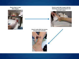
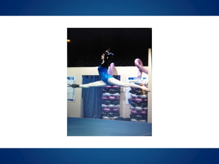
Case Study: Grace
Varus Ankle & Medial Physeal Bar
12 years to 13 years
David S. Feldman, MD
Chief of Pediatric Orthopedic Surgery
Professor of Orthopedic Surgery & Pediatrics
NYU Langone Medical Center & NYU Hospital for Joint Diseases
BACKGROUND
At the age of 10, Grace dislocated and
fractured her right ankle during gymnastics.
She underwent surgery at another facility to
repair the injury which had also affected her
growth plate (physis) and heel. After surgery,
she was placed in short leg casts for six
weeks, made a full recovery, and was able
to return to gymnastics.
A few months before her 12th birthday,
Grace’s mother noticed her right foot had
begun to turn outward. Her pediatrician
ordered CT scans and MRIs for evaluation
and found that a bony bridge had developed
in her physis (physeal bar).
Another surgery was performed to remove a
portion of her fibula along with the bony
bridge as it would have likely delayed or
prevented further growth of the bone. She
was able to return to gymnastics after
surgery but began to feel pain in her instep
along with ongoing numbness and tingling in
her heel.
A few months after her second surgery,
Grace visited me for a second opinion on
her right ankle.
EVALUATION
During our first appointment, I examined
Grace and reviewed her medical films. I
found that her physis seemed to have
stopped growing and as a result of her
previous surgeries, there was only a small
distal fibula remaining near her ankle and it
was not attached to the rest of her fibula.
An x-ray of Grace’s lower limbs. Note the right limb’s lack of space
around the physes and the disconnected parts of the fibula.
I was concerned that this small amount of
bone would be insufficient to buttress and
protect her ankle. In addition, the ankle was
bowing (varus) and surgery would become a
necessity if it continued to drift into a more
varus position.
TREATMENT
Grace began physical therapy for treatment
of muscle atrophy in her ankle which helped
to decrease her pain and stopped the
tingling in her heel. However, over the next
few months her ankle varus increased and
the range of motion in her ankle mildly
decreased. The physeal bar also
reappeared in her ankle and required
surgical intervention to prevent bone growth
issues in her leg.
Grace’s ankle before surgery
Distal Tibia Epiphysiodesis
A small incision was made at the distal tibia
and the entire lateral distal tibial physis as
well as the physis of the medial and lateral
fibula were removed. The surgical site was
irrigated, closed in layers, and dry sterile
dressings were applied.
Post-op Care
Grace’s surgical wounds healed within a few
weeks but she had to wear a cam walker
fracture boot and was not allowed to
participate in any sports for six weeks.
Grace’s ankle after the sutures
and pins were removed.
Grace’s leg in a cast
after surgery.
Grace’s ankle after surgery with the
dressings removed but sutures and
pins still in place.
CONCLUSION
Four months after surgery, x-rays showed
that Grace’s right leg and ankle were normal
and within a year her ankle had full range of
motion with no pain or tenderness.
Since then, Grace has been doing very well
and has returned to gymnastics and track.
Grace still receives ongoing strengthening
therapy and remains under observation for
lower limb issues that might arise as a result
of her previous injuries.
As this case shows, in some instances big
problems can be resolved with minimally
invasive procedures that allow kids to
quickly recover and return to their sports and
activities.
David S. Feldman, MD
Pediatric Orthopedic Surgeon
www.davidsfeldmanmd.com

Grace: Varus Ankle & Medial Physeal Bar Case Study

  • 1.
    Case Study: Grace VarusAnkle & Medial Physeal Bar 12 years to 13 years David S. Feldman, MD Chief of Pediatric Orthopedic Surgery Professor of Orthopedic Surgery & Pediatrics NYU Langone Medical Center & NYU Hospital for Joint Diseases
  • 2.
  • 3.
    At the ageof 10, Grace dislocated and fractured her right ankle during gymnastics. She underwent surgery at another facility to repair the injury which had also affected her growth plate (physis) and heel. After surgery, she was placed in short leg casts for six weeks, made a full recovery, and was able to return to gymnastics.
  • 4.
    A few monthsbefore her 12th birthday, Grace’s mother noticed her right foot had begun to turn outward. Her pediatrician ordered CT scans and MRIs for evaluation and found that a bony bridge had developed in her physis (physeal bar).
  • 5.
    Another surgery wasperformed to remove a portion of her fibula along with the bony bridge as it would have likely delayed or prevented further growth of the bone. She was able to return to gymnastics after surgery but began to feel pain in her instep along with ongoing numbness and tingling in her heel.
  • 6.
    A few monthsafter her second surgery, Grace visited me for a second opinion on her right ankle.
  • 7.
  • 8.
    During our firstappointment, I examined Grace and reviewed her medical films. I found that her physis seemed to have stopped growing and as a result of her previous surgeries, there was only a small distal fibula remaining near her ankle and it was not attached to the rest of her fibula.
  • 9.
    An x-ray ofGrace’s lower limbs. Note the right limb’s lack of space around the physes and the disconnected parts of the fibula.
  • 10.
    I was concernedthat this small amount of bone would be insufficient to buttress and protect her ankle. In addition, the ankle was bowing (varus) and surgery would become a necessity if it continued to drift into a more varus position.
  • 11.
  • 12.
    Grace began physicaltherapy for treatment of muscle atrophy in her ankle which helped to decrease her pain and stopped the tingling in her heel. However, over the next few months her ankle varus increased and the range of motion in her ankle mildly decreased. The physeal bar also reappeared in her ankle and required surgical intervention to prevent bone growth issues in her leg.
  • 13.
  • 14.
    Distal Tibia Epiphysiodesis Asmall incision was made at the distal tibia and the entire lateral distal tibial physis as well as the physis of the medial and lateral fibula were removed. The surgical site was irrigated, closed in layers, and dry sterile dressings were applied.
  • 15.
    Post-op Care Grace’s surgicalwounds healed within a few weeks but she had to wear a cam walker fracture boot and was not allowed to participate in any sports for six weeks.
  • 16.
    Grace’s ankle afterthe sutures and pins were removed. Grace’s leg in a cast after surgery. Grace’s ankle after surgery with the dressings removed but sutures and pins still in place.
  • 17.
  • 18.
    Four months aftersurgery, x-rays showed that Grace’s right leg and ankle were normal and within a year her ankle had full range of motion with no pain or tenderness.
  • 19.
    Since then, Gracehas been doing very well and has returned to gymnastics and track. Grace still receives ongoing strengthening therapy and remains under observation for lower limb issues that might arise as a result of her previous injuries.
  • 20.
    As this caseshows, in some instances big problems can be resolved with minimally invasive procedures that allow kids to quickly recover and return to their sports and activities.
  • 22.
    David S. Feldman,MD Pediatric Orthopedic Surgeon www.davidsfeldmanmd.com