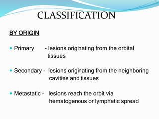
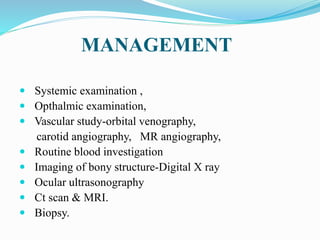
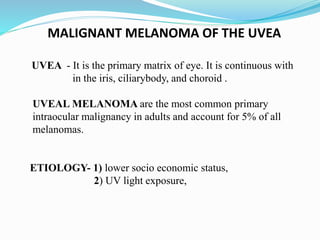
2. INCIDENCE & EPIDEMOLOGY
ο Tumors of the eye and orbit are rare.
ο Male to female incidence is similar.
ο In adults, Melanoma is the most common
primary intraocular cancer, followed by
Lymphoma
ο In children Retinoblastoma is the most common
tumor, followed by Medulloepithelioma
ο Metastases, or secondary intraocular tumors, are
more common than primary tumors and typically
come from breast or lung cancers.
3. ORBIT
ο The orbit is the cavity or socket of the skull in which
the eye and its appendages are situated.
ο In the adult human, the volume of the orbit is 30 mL,
of which the eye occupies 6.5 ml.
ο The orbits are conical or four-sided pyramidal cavities,
which open into the midline of the face and point back
into the head.
4.
5. CLASSIFICATION
BY ORIGIN
ο Primary - lesions originating from the orbital
tissues
ο Secondary - lesions originating from the neighboring
cavities and tissues
ο Metastatic - lesions reach the orbit via
hematogenous or lymphatic spread
6. BENIGN ORBITAL TUMORS
a. Pterygium
b. Chorodial Hemangiomas
c. Orbital Pseudotumors
d. Thyroid associated orbitopathy
8. MALIGNANT ORBITAL TUMORS
The most common malignant orbital tumors in adults
include:
LYMPHOMA - It is the most common type of malignant
orbital tumor in the adults.
ο Occurs mainly in the lacrimal gland, but can occur in any
other orbital structure.
OPTIC NERVE GLIOMA - It is extremely rare tumor that
begins in the optic nerve and spreads to the orbit
9. ο SARCOMA β
almost any type of sarcoma can involve
the orbit
ο Angiosarcoma
ο Fibrosarcoma
ο Osteosarcoma
ο Chondrosarcoma
ο Liposarcoma
ο Malignant fibrous histiocytoma
10. β’ Cancers from other parts of the body, such as the
breast, lung, prostate, brain and kidney, can also
spread (metastasize) to the orbit.
β’ Metastasis from melanoma can also occur but it is
uncommon.
β’ SECONDARY CARCINOMAS - they can also be
secondary cancers that have spread to the orbit from
nearby structures, such as the eyeball (intraocular
tumors), eyelid, conjunctiva, sinuses or nasal cavity.
11. TNM STAGING
ο TX - Primary tumor cannot be assessed
ο T0 -No evidence of primary tumor
ο T1 - Tumor is 15 mm (0.6 inch) or less in size.
ο T2 - Tumor is more than 15 mm in size. It has not spread into
the globe of the eye or the bony wall of the orbit.
ο T3 -Tumor is any size and has spread into the orbital tissues or
bony walls of the orbit.
12. T4 - Tumour has spread into one or more of the following:
ο the globe
ο periorbital structures, such as the eyelids or temporal fossa
ο nasal cavity and paranasal sinuses
ο central nervous system (the brain and spinal cord)
14. SIGNS & SYMPTOMS
ο Proptosis β the most important sign
ο Blurred vision,
ο Diplopia,
ο Strabismus ,
ο Whitish or yellowish glow through the pupil,
ο Decreasing/loss of vision,
ο Red and painful Eye
ο Pain
16. TREATMENT
Orbital tumor may be treated by:
ο Surgery
ο Chemotherapy
ο External beam radiotherapy
ο Plaque brachytherapy
17. LACRIMAL GLAND TUMORS
ο Lacrimal gland tumors are seen more frequently in the third
decade of life, and the second bimodal peak is in the teenage
years.
ο The lacrimal gland is a bilobed eccrine secretory gland, which
is situated in the Superotemporal orbit.
ο The 2 lobes of the lacrimal gland
- Orbital lobe
- Palpebral lobe,
19. ο Lacrimal gland swelling can be classified broadly into
- Inflammatory
- Neoplastic subtypes.
ο Inflammatory etiologies, include
- Dacryoadenitis,
- Sarcoidosis .
ο Most of the neoplastic lesions in the lacrimal gland are
epithelial in origin,
ο 50% classified as
- BENIGN and 50% as MALIGNANT PSEUDO TUMOR.
20. ο BENIGN LESIONS -
- Pleomorphic adenomas ,
- Reactive lymphoid hyperplasia,
- Oncocytomas.
These lesions are slowly growing masses more
commonly found in adults in their forth to fifth decades of
life.
21. ο MALIGNANT LESIONS :
- Adenoid cystic carcinoma- comprising 50% of
malignant tumors of lacrimal gland and 25% of all
lacrimal gland tumors
- Adenocarcinoma,
- Squamous cell carcinoma,
- Mucoepidermoid carcinoma,
- Lymphomas
22. TNM STAGING
ο TX: The primary tumor cannot be assesed.
ο T0 : no evidence of primary tumor.
ο T1: The tumor is 2 centimeters (cm) or smaller and may or may
not extend outside of the lacrimal gland to the orbital soft tissue.
ο T2: The tumor is between 2 cm and 4 cm and likely extends to
the orbital soft tissue.
ο T3: The tumor is greater than 4 cm and likely extends to the
orbital soft tissue.
ο T4: The tumor has invaded the periosteum (the membrane of
connective tissue that covers the bone) or the orbital bone.
ο T4a: The tumor has invaded the periosteum.
ο T4b: The tumor has invaded the orbital bone.
ο T4c: The tumor has extended beyond the orbit to adjacent
structures, including the brain and sinuses.
23. TNM STAGING
ο NX: The regional lymph nodes cannot be evaluated.
ο N0 : There is no regional lymph node metastasis.
ο N1: There is regional lymph node metastasis.
ο MX: Distant metastasis cannot be evaluated.
ο M0 : There is no distant metastasis.
ο M1: There is metastasis to other parts of the bod
24. SIGNS AND SYMPTOMS
ο Malignant lesions characteristically present with a
subacute course of proptosis and temporal sensory
loss in the distribution of the lacrimal nerve in one
third of patients.
ο Diplopia and diminished visual acuity can be seen in
rapid progressive lesions.
ο Benign lesions commonly present with painless
inferonasal globe displacement and fullness of the
superotemporal lid and orbit.
25. DIAGNOSIS
ο CT scan of BENIGN epithelial lesions, such as
pleomorphic adenomas, reveals a well-circumscribed,
pseudoencapsulated lesion in the superotemporal
fossa.
ο In contrast, MALIGNANT epithelial lesions, such as
adenoid cystic carcinoma, usually present as an
irregular mass, producing bony erosion (70%) and
occasional calcification (20%).
26. TREATMENT
ο SURGERY : Surgery is the mainstay treatment of lacrimal
gland tumors.
ο RADIOTHERAPY:
ο It is most often used for lacrimal gland lymphoma.
ο The dosage of radiation used and the site and type of the
tumor significantly affect the risks of side effects.
27. ο Cataracts are a very common side effect of radiation
therapy to the eye area.β
ο Also, loss of eyelashes and/or a dry eye can occur with
external-beam radiation therapy.
ο Other side effects include radiation retinopathy.
ο Radiation optic neuropathy involves nerve damage in the
eye.
SIDE EFFECTS
28. ο CHEMOTHERAPY -
ο IMMUNOTHERAPY - Rituximab (Rituxan) is the
most common targeted therapy used in the treatment
of a lacrimal gland tumor
29. MALIGNANT MELANOMA OF THE UVEA
UVEA - It is the primary matrix of eye. It is continuous with
in the iris, ciliarybody, and choroid .
UVEAL MELANOMA are the most common primary
intraocular malignancy in adults and account for 5% of all
melanomas.
ETIOLOGY- 1) lower socio economic status,
2) UV light exposure,
30. ο CHOROIDAL MELANOMAS are located posterior to
the ciliary body ,
ο IRIS MELANOMA are the most easily visible,They
tend to be smallest at diagnosis,and lest likely to
metastasis.
ο CILIARYBODY MELANOMA are least visible as they
are hidden behind iris ,and more likely to
metastasized.
32. SYMPTOMS
ο Decreased or blurry vision,
ο Flashes of light,
ο Distortion or loss of vision.
ο Dark spot on the colored part of the eye or a distorted
pupil.
33. DIAGNOSIS
ο Ocular ultrasonography
ο Fine needle aspiration biopsy,
ο Fluorescein angiography and photography(by slit lamp,
gonioscopy for iris and ciliary body tumors)
34. MANAGEMENT
ο ENUCLEATION -
Enucleation is required in a subset of patients, because
of complications of conservative therapy.
ο ENDORESECTION -
Transretinal endoresection is controversial, mainly
because of fear of seeding of tumor cells.
35. ο TRANSSCLERAL RESECTION -
Transscleral local resection has been promoted for
tumors >6-mm thick in patients highly motivated to
retain vision.
ο TRANSPUPILLARY THERMOTHERAPY -
In this technique 1-minute applications of 3-mm
spots of low-energy diode laser are administered to the
tumor and the surrounding choroid.
36. PLAQUE BRACHYTHERAPY
ο Plaque brachytherapy is the mainstay of treatment in
many centers, with iodine-125 and ruthenium-106
being the most common isotopes, although
palladium-103 has also been used effectively.
ο 91 Iodine emits Ξ³-rays, which have a range sufficient for
tumors up to 8- to 10-mm thick, while ruthenium
delivers beta particles that have a more limited range,
which is suitable for tumors upto 5 mm.
37. ο The general objective with all plaques is to deliver
approximately 80 Gy to the tumor apex by fixing the
plaque in the exact location of the tumor.
ο Best results were obtained in eyes with small tumors
outside a radius of 5 mm from the optic disc and foveola.
39. PROTON BEAM THERAPY
ο Proton beam therapy may be used to treat uveal
melanoma and is usually indicated for tumors that
extend close to the optic disc,
ο ADVANTAGE - Proton beam delivers a homogeneous
dose to tumor and has a sharp edge, a high tumor dose
can be delivered with relative sparing of the optic
nerve.
40. STEREOTACTIC RADIOTHERAPY
ο Stereotactic radiation can be delivered by
LINEAR ACCELERATOR (LINAC)
ο By specialized devices -
GAMMA KNIFE which provides focused radiation with a
multitude of sources.
41. COMPLICATIONS
The most frequently encountered complications are
ο Exudative retinopathy
ο Neovascular glaucoma
ο Vitreous hemorrhage
ο Radiation retinopathy
ο Cataract