Minimally Invasive Glaucoma Surgery (MIGS)

Minimally invasive glaucoma surgery (MIGS) is a group of procedures that minimizes the invasive rate of glaucoma with five characteristics: ab interno microincision, minimal trauma, more effective, high safety profile, and quick recovery. MIGS is a surgery that uses an incision in a clear cornea and is indicated in patients with mild to moderate open angle glaucoma. The technique of MIGS is based on several mechanisms, namely trabecular meshwork bypass stents including iStent, trabectome, and Hydrus microstent; Suprachoroidal implant using Cypass microstent; And subconjungtiva filtration using XEN gel stent. MIGS technology has potential advantages in glaucoma management by reducing the burden of treatment, improving patients quality of life, and cutting or delaying more invasive surgeries.

Recommended
More Related Content
What's hot
What's hot (20)
Similar to Minimally Invasive Glaucoma Surgery (MIGS)
Similar to Minimally Invasive Glaucoma Surgery (MIGS) (20)
More from Meironi Waimir
More from Meironi Waimir (20)
Recently uploaded
Recently uploaded (20)
Minimally Invasive Glaucoma Surgery (MIGS)
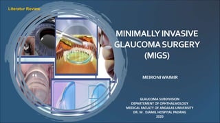
- 1. MINIMALLYINVASIVE GLAUCOMASURGERY (MIGS) GLAUCOMA SUBDIVISION DEPARTEMENT OF OPHTHALMOLOGY MEDICAL FACULTY OF ANDALAS UNIVERSITY DR. M . DJAMIL HOSPITAL PADANG 2020 MEIRONIWAIMIR Literatur Review
- 2. INTRODUCTION • Leading cause of irreversible blindness worldwide. • The management medical therapy, laser to surgery. The golden standard Trabeculectomy Several potential complications. This has triggered a lot of research and development of new procedures that effective in reducing IOP. Glaucoma Characterized by glaucomatous optic neuropathy, visual field defect and can be accompanied by increased intraocular pressure (IOP).
- 3. INTRODUCTION Ab interno microincision Minimal trauma More effective High safety profile Quick recovery and relatively easy to do Minimally invasive glaucoma surgery or micro invasive glaucoma surgery (MIGS) group of procedures that minimizes the invasive rate of glaucoma. The term MIGS is based on five characteristics:
- 4. Trabecular meshwork bypass stents Trabectome iStent Suprachoroidal implants CyPass Microstent Subconjungtiva filtration XEN gel stents INTRODUCTION Technique operation of MIGS has developed with different mechanisms. Hydrus microstent * MIGS modality that has been approved by the Food and Drugs Administration (US-FDA)
- 5. AnteriorChamberAngle Roombetweencorneaandiris Anterior chamber is limited by • Anterior: endothelium cornea • Peripheral: trabecular meshwork, part of the ciliary corpus, and iris roots • Posterior: iris surface and pupil Structure: • Schwalbe line • Trabecular meshwork • Scleral spur • Ciliary body band • Schlemm canal • Peripheral iris
- 6. AnteriorChamberAngle Schwalbe line located in the transition between cornea and trabecular meshwork. Trabecular meshwork is a connective tissue formed by trabeculocytes. Uveal Meshwork Corneoscleral Meshwork Juxtacanalicular Meshwork Scleral Spur is a fibrous ring which cross sectionally shaped like a wedge. • Consists of collagen types I, III and elastic tissue. Ciliary body band is a part from ciliary corpus. Processus Iris
- 7. SchlemmCanal Inner wall consists of irregular endothelial cell that have giant vacuoles. • Giant vacuoles formed in response to pressure gradients from aqueous humor flow when the endothelial canal wall is stretched due to increased IOP. Circular tubes resemble lymphatic vessels, located at the border of the cornea and sclera. Outer wall consists of smooth horizontal cells and channel that connect with episcleral vein.
- 8. CollectorChannel • Schlemm canal collector channel (intrascleral, episcleral, and sub-conjunctival venous complexes). • smooth muscle able to constrict to regulate aqueous humor flow. • Collector channel divided into: • Direct channel: Greater canal, (4-6 channels) with a diameter 70 micron flows directly into the episcleral vein. • Indirect channel: Smaller canal (15-20 channels) with a diameter 50 microns forms intrasclera plexus before it flows into the episclera vein.
- 9. AqueousHumorOutflow Available 2 lane aqueous humor outflow: o Conventional pathway (Trabecular outflow) involves the trabecular meshwork, Schlemm's canal, and episcleral veins. o Unconventional pathway (Uveoscleral outflow) through the anterior ciliary muscle and iris stroma to reach the supracillary and supracoroidal spaces.
- 10. There are three mechanisms which glaucoma implants can reduce IOP: Trabecular Meshwork Bypass Stents Increased outflow to the Schlemm canal achieved by creating a large direct pathway between the AC and Schlemm canal. • iStent, trabectome, and Hydrus microstent. Increasing the uveoskleral outflow into the suprachoroidal space. • CyPass microstent. Suprachoroidal Implant MODALITYANDPROCEDURE MINIMALLYINVASIVEGLAUCOMASURGERY Subkonjungtiva Filtration Increase outflow by creating new channels into the subconjunctival space by forming an external bleb. • Xen gel stents.
- 12. IStent and IStent Inject Mild to moderate OAG that using one to three ocular hypotension drugs. In patients with very narrow angle it should be avoided more difficult and risk of iris or endothelial damage. Usually performed on patients who have stable disease and well-controlled IOP.
- 13. IStent • Coated heparin titanium implant, size 1 x 0.3 mm implanted through TM into the Schlemm canal. • An "L" shaped device with a pointed tip that can penetrate TM. • the "snorkel" section facing the AC allows aqueous drainage from the AC to the Schlemm canal • Retention arches facing the TM keeping the stent in place. • The half-cylinder pipe prevents obstruction. IStent
- 14. • Smaller than 1st generation device. • Made of titanium coated heparin. • Bullet shaped with a length only 360 microns. • Easier to use no displacement of the stents needed for positioning. • IStent inject can be implanted with only one inserter. IStentInject 2nd generation and has been certified by FDA
- 15. • Aqueous humor flow into the Schlemm canal by passing through the juxtacanalikular. • The advantage Patency of bypass outflow because it has a heparin layer. • Complications mild hyphema of the Schlemm canal, transient IOP elevation, corneal edema, stenting malposition, lumen obstruction by clots or iris. • Decrease in IOP ≥20% in patients with open angle glaucoma. IStentandIStentInject
- 18. HydrusMicrostent • A device that is implanted through a clear corneal incision into the Schlemm canal. • Made from a material with very elastic biocompatibility called nitinol (metal alloy of nickel and titanium). • Crescent-shaped with 8 mm length. • There are three windows along the surface.
- 19. • The ideal patients Mild to moderate open angle glaucoma and have moderate to dense pigments in TM. • Inserted into the Schlemm canal across the TM using a manual inserter gonioscopy guided. HydrusMicrostent • Reduces the resistance of aquous humor outflow by two mechanisms. 1. Passing the trabecular meshwork which is the place with the highest resistance. 2. Expand and install three windows to the Schlemm canal.
- 21. Complications subconjunctival bleeding, hyphema, and focal peripheral anterior synechia • Pfeiffer et al Hydrus implantation can reduce IOP 20% in 80% of OAG patients (from 26.3 ± 4.4 mmHg to 16.9 ± 3.3 mmHg). HydrusMicrostent
- 23. Trabectome • Procedure using high frequency electrocautery performed under gonioscopic to erode TM and the inner walls of the Schlemm canal. • Consists of a disposable hand piece used for aspiration, irrigation and electrocautery
- 24. • This procedure can be done 90 or 120 degrees thermal damage to the inner walls of the Schlemm canal make a direct connection between the AC and the Schlemm canal. • The advantage Removes the area of greatest resistance of aquous humor outflow and removes tissue that can reduce inflammatory stimulation so that potential scar tissue is formed. • Performed in open-angle glaucoma Requires adequate visualization of TM. Trabectome
- 25. • Trabectome generally achieves postoperative IOP in the low to moderate range, with average reduction of IOP around 30% after 6 months. Trabectome • Complications a sudden decrease in IOP on the first day, intraoperative blood reflux from the Schlemm canal, goniosynechiae and membrane growth.
- 27. • Made with polyamides biocompatible and not biodegradable. • It is 6.35 mm long and has a single lumen of 300 µm. • Used in patients who want to reduce their dependence on drugs for controlled IOP. • Aqueous humor enters the supracillary space through some of the fenestrations that exist along the tool. CyPassMicrostent
- 29. • Reduce IOP 30-35%. • The CyPass Clinical Experiance Study Reported a reduction in IOP of 26-37%. • Complications transient early hypotension (13.8%), transient IOP elevation (10.5%), and transient hyphema (6%). CyPassMicrostent
- 31. • Gelatin stents allowing aqueous outflow from AC to the subconjunctival space by a clear corneal incision without conjunctival dissection. Xen Gel Stent • Soft flexible hydrophilic tube composed of gelatin with glutaraldehyde. • The length is 6 mm and the width varies by model.
- 32. • Soft, biocompatible and non-inflammatory. • Flexible when hydrated softens within 1-2 minutes after implantation and can adjust to the surrounding tissue. • Implanted using an injector. Selection criteria: Schaffer grade 2 or wider and the conjunctiva can accommodate bleb formation. Xen Gel Stent
- 34. Xen Gel Stent A study of XEN implants combined with cataract surgery Reduction in IOP from 22.4 (+/- 4.2) mmHg to 15.4 (+/- 3.0) mmHg at 12 months postoperative and there was a reduction in drug use glaucoma from 2.5 +/- 1.4 to 0.9 +/- 1.0. In another study using XEN implantation alone (n = 49 eyes) 40% succeeded in reducing IOP at 12 months post implantation (IOP </ = 18mmHg and> / = 20% reduction in IOP).
- 35. CONCLUSION • Minimally invasive glaucoma surgery (MIGS) is a group of procedures that minimizes the invasive rate of glaucoma with five characteristics: ab interno microincision, minimal trauma, more effective, high safety profile, and quick recovery. • MIGS is a surgery that uses an incision in a clear cornea and is indicated in patients with mild to moderate open angle glaucoma.
- 36. • The technique of MIGS is based on several mechanisms, namely trabecular meshwork bypass stents including iStent, trabectome, and Hydrus microstent; Suprachoroidal implant using Cypass microstent; And subconjungtiva filtration using XEN gel stent. • MIGS technology has potential advantages in glaucoma management by reducing the burden of treatment, improving patients quality of life, and cutting or delaying more invasive surgeries. CONCLUSION
- 37. THANK YOU Kerinci Mountain 3805 mdpl
Editor's Notes
- The golden standard for glaucoma surgical therapy Trabeculectomy
- An incision is made on the clear cornea with a micro incision causing a slight anatomic distortion
- Uveal meshwork has fewer elastic fibers than corneoscleral meshwork. hole size about 25µm-75µm. Provide little resistance to aqueous humor outflow Corneoscleral meshwork forms the largest center of trabecular meshwork. smaller than uveal meshwork (5µ - 50µ) Juxtacanalicular form the outermost part of the canal Schlemm. Plays a major role in the normal resistance of the aqueous humor due to its narrow and winding path.
- Schlemm canal empties into a number of collector channel
- implantation become more difficult
- Allows aqueous humor flow into the Schlemm canal by passing through the juxtacanalikular which is the highest outflow resistance area.
- TM pigmentation will improve the surgeon's view of the target tissue at the AC angle easier to place Hydrus into the Schlemm canal.
- membrane growth which can cause IOP elevations.
- Based on the principle There is a pressure gradient 1-5 mmHg between AC and suprachoroidal space. The pressure in the suprachoroidal space is lower creates a directional flow towards the suprachoroidal space.