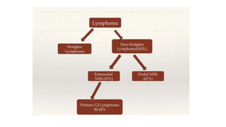
- Gastrointestinal lymphomas originate from lymphoid cells in the gastrointestinal tract. The two main types are non-Hodgkin lymphoma and Hodgkin lymphoma.
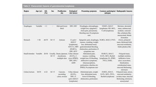
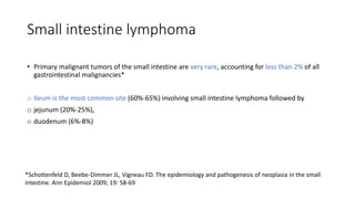
- Primary gastrointestinal lymphomas are rare, accounting for about 1-4% of gastrointestinal malignancies. The stomach is the most commonly involved site. Histopathologically, almost 90% are B cell lymphomas.
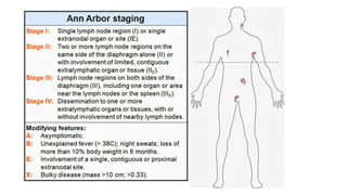
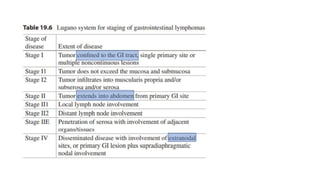
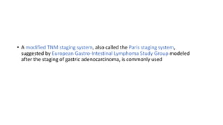
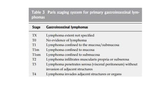
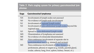
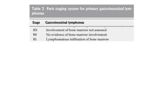
- Proper staging of primary gastrointestinal lymphomas includes physical exam, endoscopic ultrasound, CT scan, PET scan, and bone marrow biopsy according to the Lugano or Paris staging systems. Treatment involves chemotherapy, radiation, surgery or a combination depending on the histological subtype and extent of disease.
Primary GIT lymphoma typically refers to a lymphoma that predominantly involves any section of the GIT from the oropharynx to the rectum. The GIT is the predominant site of extra nodal lymphoma involvement, mostly are non-Hodgkin lymphomas (NHLs).
Recent Update on Management of Ulcerative ColitisDr Amit Dangi
Recent update on the surgical and medical management of ulcerative colitis, including various controversies regarding IPAA and recent medical management incorporating the role of biologicals
A power point presentation on classification, types and investigations of gastric/stomach cancer presented by students to the faculty of private medical teaching hospital and approved by the surgery department of the college. The resources taken are guyton and hall book of physiology and bailey and Love's short practice if surgery.
REFERENCES
cancer.org | 1.800.227.2345
Advances in the diagnosis and management
of lymphoma
Zachary H Word1
Matthew J Matasar1,2
1
Lymphoma Service, Department of
Medicine, Memorial Sloan–Kettering
Cancer Center, 2
Department of
Medicine, New York Presbyterian
Hospital, New York, NY, USA
Correspondence: Matthew J Matasar
Memorial Sloan–Kettering Cancer Center,
1275 York Avenue, New York,
NY 10065, USA
Tel +1 212 639 8889
Fax +1 646 422 2291
Email matasarm@mskcc.org
Lymphoma and CLL Forms
Parameswaran Hari, MD, MS
CLymphoma 101: The Basics
Neha Mehta-Shah, MD, MSCI
Assistant Professor
Department of Medicine
Division of Oncology
IBMTR , Milwaukee
FDG PET/CT plays an important role in staging, restaging, prognostication, planning treatment strategies, monitoring therapy, and detecting relapse. In this lecture I try my best to explain it for our fellows .
esophageal carcinoma is one of the common gastrointestinal malignancy. Its usually present at advanced stage. Its management requires diagnosis as early as possible and staging followed by proper planning of treatment. Its treatment include endoscopic, surgical, adjuvant chemotherapy and palliative management.
Acute and Late Radiation Related Side Effects and their Management in Pelvic ...Dr Kartik Kadia
Acute and Late Radiation Related Side Effects and their Management in Pelvic Malignancies
Carcinoma Cervix - Radiation Related Toxicities and Management
Detailed Seminar on Carcinoma Pancreas with -
Anatomy, Epidemiology, Enteropathogenesis, Pathology, Staging , Diagnostic workup and different modalities of Treatment
Basavarajeeyam is an important text for ayurvedic physician belonging to andhra pradehs. It is a popular compendium in various parts of our country as well as in andhra pradesh. The content of the text was presented in sanskrit and telugu language (Bilingual). One of the most famous book in ayurvedic pharmaceutics and therapeutics. This book contains 25 chapters called as prakaranas. Many rasaoushadis were explained, pioneer of dhatu druti, nadi pareeksha, mutra pareeksha etc. Belongs to the period of 15-16 century. New diseases like upadamsha, phiranga rogas are explained.
Ethanol (CH3CH2OH), or beverage alcohol, is a two-carbon alcohol
that is rapidly distributed in the body and brain. Ethanol alters many
neurochemical systems and has rewarding and addictive properties. It
is the oldest recreational drug and likely contributes to more morbidity,
mortality, and public health costs than all illicit drugs combined. The
5th edition of the Diagnostic and Statistical Manual of Mental Disorders
(DSM-5) integrates alcohol abuse and alcohol dependence into a single
disorder called alcohol use disorder (AUD), with mild, moderate,
and severe subclassifications (American Psychiatric Association, 2013).
In the DSM-5, all types of substance abuse and dependence have been
combined into a single substance use disorder (SUD) on a continuum
from mild to severe. A diagnosis of AUD requires that at least two of
the 11 DSM-5 behaviors be present within a 12-month period (mild
AUD: 2–3 criteria; moderate AUD: 4–5 criteria; severe AUD: 6–11 criteria).
The four main behavioral effects of AUD are impaired control over
drinking, negative social consequences, risky use, and altered physiological
effects (tolerance, withdrawal). This chapter presents an overview
of the prevalence and harmful consequences of AUD in the U.S.,
the systemic nature of the disease, neurocircuitry and stages of AUD,
comorbidities, fetal alcohol spectrum disorders, genetic risk factors, and
pharmacotherapies for AUD.
New Drug Discovery and Development .....NEHA GUPTA
The "New Drug Discovery and Development" process involves the identification, design, testing, and manufacturing of novel pharmaceutical compounds with the aim of introducing new and improved treatments for various medical conditions. This comprehensive endeavor encompasses various stages, including target identification, preclinical studies, clinical trials, regulatory approval, and post-market surveillance. It involves multidisciplinary collaboration among scientists, researchers, clinicians, regulatory experts, and pharmaceutical companies to bring innovative therapies to market and address unmet medical needs.
TEST BANK for Operations Management, 14th Edition by William J. Stevenson, Ve...kevinkariuki227
TEST BANK for Operations Management, 14th Edition by William J. Stevenson, Verified Chapters 1 - 19, Complete Newest Version.pdf
263778731218 Abortion Clinic /Pills In Harare ,sisternakatoto
263778731218 Abortion Clinic /Pills In Harare ,ABORTION WOMEN’S CLINIC +27730423979 IN women clinic we believe that every woman should be able to make choices in her pregnancy. Our job is to provide compassionate care, safety,affordable and confidential services. That’s why we have won the trust from all generations of women all over the world. we use non surgical method(Abortion pills) to terminate…Dr.LISA +27730423979women Clinic is committed to providing the highest quality of obstetrical and gynecological care to women of all ages. Our dedicated staff aim to treat each patient and her health concerns with compassion and respect.Our dedicated group ABORTION WOMEN’S CLINIC +27730423979 IN women clinic we believe that every woman should be able to make choices in her pregnancy. Our job is to provide compassionate care, safety,affordable and confidential services. That’s why we have won the trust from all generations of women all over the world. we use non surgical method(Abortion pills) to terminate…Dr.LISA +27730423979women Clinic is committed to providing the highest quality of obstetrical and gynecological care to women of all ages. Our dedicated staff aim to treat each patient and her health concerns with compassion and respect.Our dedicated group of receptionists, nurses, and physicians have worked together as a teamof receptionists, nurses, and physicians have worked together as a team wwww.lisywomensclinic.co.za/
Knee anatomy and clinical tests 2024.pdfvimalpl1234
This includes all relevant anatomy and clinical tests compiled from standard textbooks, Campbell,netter etc..It is comprehensive and best suited for orthopaedicians and orthopaedic residents.
Title: Sense of Smell
Presenter: Dr. Faiza, Assistant Professor of Physiology
Qualifications:
MBBS (Best Graduate, AIMC Lahore)
FCPS Physiology
ICMT, CHPE, DHPE (STMU)
MPH (GC University, Faisalabad)
MBA (Virtual University of Pakistan)
Learning Objectives:
Describe the primary categories of smells and the concept of odor blindness.
Explain the structure and location of the olfactory membrane and mucosa, including the types and roles of cells involved in olfaction.
Describe the pathway and mechanisms of olfactory signal transmission from the olfactory receptors to the brain.
Illustrate the biochemical cascade triggered by odorant binding to olfactory receptors, including the role of G-proteins and second messengers in generating an action potential.
Identify different types of olfactory disorders such as anosmia, hyposmia, hyperosmia, and dysosmia, including their potential causes.
Key Topics:
Olfactory Genes:
3% of the human genome accounts for olfactory genes.
400 genes for odorant receptors.
Olfactory Membrane:
Located in the superior part of the nasal cavity.
Medially: Folds downward along the superior septum.
Laterally: Folds over the superior turbinate and upper surface of the middle turbinate.
Total surface area: 5-10 square centimeters.
Olfactory Mucosa:
Olfactory Cells: Bipolar nerve cells derived from the CNS (100 million), with 4-25 olfactory cilia per cell.
Sustentacular Cells: Produce mucus and maintain ionic and molecular environment.
Basal Cells: Replace worn-out olfactory cells with an average lifespan of 1-2 months.
Bowman’s Gland: Secretes mucus.
Stimulation of Olfactory Cells:
Odorant dissolves in mucus and attaches to receptors on olfactory cilia.
Involves a cascade effect through G-proteins and second messengers, leading to depolarization and action potential generation in the olfactory nerve.
Quality of a Good Odorant:
Small (3-20 Carbon atoms), volatile, water-soluble, and lipid-soluble.
Facilitated by odorant-binding proteins in mucus.
Membrane Potential and Action Potential:
Resting membrane potential: -55mV.
Action potential frequency in the olfactory nerve increases with odorant strength.
Adaptation Towards the Sense of Smell:
Rapid adaptation within the first second, with further slow adaptation.
Psychological adaptation greater than receptor adaptation, involving feedback inhibition from the central nervous system.
Primary Sensations of Smell:
Camphoraceous, Musky, Floral, Pepperminty, Ethereal, Pungent, Putrid.
Odor Detection Threshold:
Examples: Hydrogen sulfide (0.0005 ppm), Methyl-mercaptan (0.002 ppm).
Some toxic substances are odorless at lethal concentrations.
Characteristics of Smell:
Odor blindness for single substances due to lack of appropriate receptor protein.
Behavioral and emotional influences of smell.
Transmission of Olfactory Signals:
From olfactory cells to glomeruli in the olfactory bulb, involving lateral inhibition.
Primitive, less old, and new olfactory systems with different path
Report Back from SGO 2024: What’s the Latest in Cervical Cancer?bkling
Are you curious about what’s new in cervical cancer research or unsure what the findings mean? Join Dr. Emily Ko, a gynecologic oncologist at Penn Medicine, to learn about the latest updates from the Society of Gynecologic Oncology (SGO) 2024 Annual Meeting on Women’s Cancer. Dr. Ko will discuss what the research presented at the conference means for you and answer your questions about the new developments.
HOT NEW PRODUCT! BIG SALES FAST SHIPPING NOW FROM CHINA!! EU KU DB BK substit...GL Anaacs
Contact us if you are interested:
Email / Skype : kefaya1771@gmail.com
Threema: PXHY5PDH
New BATCH Ku !!! MUCH IN DEMAND FAST SALE EVERY BATCH HAPPY GOOD EFFECT BIG BATCH !
Contact me on Threema or skype to start big business!!
Hot-sale products:
NEW HOT EUTYLONE WHITE CRYSTAL!!
5cl-adba precursor (semi finished )
5cl-adba raw materials
ADBB precursor (semi finished )
ADBB raw materials
APVP powder
5fadb/4f-adb
Jwh018 / Jwh210
Eutylone crystal
Protonitazene (hydrochloride) CAS: 119276-01-6
Flubrotizolam CAS: 57801-95-3
Metonitazene CAS: 14680-51-4
Payment terms: Western Union,MoneyGram,Bitcoin or USDT.
Deliver Time: Usually 7-15days
Shipping method: FedEx, TNT, DHL,UPS etc.Our deliveries are 100% safe, fast, reliable and discreet.
Samples will be sent for your evaluation!If you are interested in, please contact me, let's talk details.
We specializes in exporting high quality Research chemical, medical intermediate, Pharmaceutical chemicals and so on. Products are exported to USA, Canada, France, Korea, Japan,Russia, Southeast Asia and other countries.
New Directions in Targeted Therapeutic Approaches for Older Adults With Mantl...i3 Health
i3 Health is pleased to make the speaker slides from this activity available for use as a non-accredited self-study or teaching resource.
This slide deck presented by Dr. Kami Maddocks, Professor-Clinical in the Division of Hematology and
Associate Division Director for Ambulatory Operations
The Ohio State University Comprehensive Cancer Center, will provide insight into new directions in targeted therapeutic approaches for older adults with mantle cell lymphoma.
STATEMENT OF NEED
Mantle cell lymphoma (MCL) is a rare, aggressive B-cell non-Hodgkin lymphoma (NHL) accounting for 5% to 7% of all lymphomas. Its prognosis ranges from indolent disease that does not require treatment for years to very aggressive disease, which is associated with poor survival (Silkenstedt et al, 2021). Typically, MCL is diagnosed at advanced stage and in older patients who cannot tolerate intensive therapy (NCCN, 2022). Although recent advances have slightly increased remission rates, recurrence and relapse remain very common, leading to a median overall survival between 3 and 6 years (LLS, 2021). Though there are several effective options, progress is still needed towards establishing an accepted frontline approach for MCL (Castellino et al, 2022). Treatment selection and management of MCL are complicated by the heterogeneity of prognosis, advanced age and comorbidities of patients, and lack of an established standard approach for treatment, making it vital that clinicians be familiar with the latest research and advances in this area. In this activity chaired by Michael Wang, MD, Professor in the Department of Lymphoma & Myeloma at MD Anderson Cancer Center, expert faculty will discuss prognostic factors informing treatment, the promising results of recent trials in new therapeutic approaches, and the implications of treatment resistance in therapeutic selection for MCL.
Target Audience
Hematology/oncology fellows, attending faculty, and other health care professionals involved in the treatment of patients with mantle cell lymphoma (MCL).
Learning Objectives
1.) Identify clinical and biological prognostic factors that can guide treatment decision making for older adults with MCL
2.) Evaluate emerging data on targeted therapeutic approaches for treatment-naive and relapsed/refractory MCL and their applicability to older adults
3.) Assess mechanisms of resistance to targeted therapies for MCL and their implications for treatment selection
2. • The term LYMPHOMA identifies a heterogeneous group of
biologically and clinically distinct neoplasms that originate from cells
in the lymphoid organs and have been historically divided into :
• Non-Hodgkin lymphoma (NHL) and
• Hodgkin lymphoma (HL)
3.
4. Introduction to Lymphoma
• Although NHLs and Hodgkin lymphoma (HL) both frequently involve
lymphohematopoietic tissues, their biologic and clinical behaviors are
distinct.
5. Hodgkin’s Lymphoma NHL
Cells of origin B cells B cells, T cells, NK cells
Classification Nodular sclerosing,
Mixed celluarity,
Lymphocyte predominant,
Lymphocyte depleted
DLBC,
Follicular mixed,
Burkitt, Small Lymphocytic
Epidemiology Bimodal :
20-25 years
>60 years
<65 years, HIV,EBV,
Congenital
immunodeficiencies
H/P Painless Cervical
Lymphadenopathy, B
symptoms (Fever, Night
sweats, weight loss)
Painless Disseminated
Lymphadenopathy, B
symptoms
LN More often localized to
single axial group of nodes
More frequent
involvement of multiple
peripheral nodes
Cure rates High (80%) Low (<50%)
6. • Current lymphoma classification systems divide the lymphomas into different
entities based on their perceived cell of origin
• During embryogenesis, hematopoietic stem cells from the liver and the placenta
give rise to progenitor cells that migrate to the Bone marrow and the Thymus
where they undergo a program of antigen-independent differentiation into B-
and T-cell lineage precursor cells, respectively
Introduction to Lymphoma
7.
8. Immunophenotyping of Lymphoid Cells
• Lymphocytes at various stages in development can be defined and differentiated
by the detection of certain antigens on the cell surface (CD)
• This antigen fingerprint is referred to as the immunophenotype of the cell.
9. • It can be detected by flow cytometric analysis of single cell suspensions of
o whole blood,
o bone marrow,
o body fluids, or
o disaggregated tissue
• using fluorescently labeled antibodies against these antigens or by
immunohistochemistry
Immunophenotyping of Lymphoid Cells
10.
11.
12. Primary gastrointestinal lymphoma
• Gastrointestinal tract is the most common extranodal site involved by lymphoma
accounting for 5%-20% of all cases*
• Primary gastrointestinal lymphoma, however, is very rare, constituting only about
1%-4% of all gastrointestinal malignancies.
• Gastrointestinal lymphoma is usually secondary to the widespread nodal
diseases.
*Freeman C, Berg JW, Cutler SJ. Occurrence and prognosis of extranodal lymphomas. Cancer 1972; 29:
252-260
13. • The most commonly involved sites are as follows*:
ostomach (60–75%),
osmall intestine (9%),
oileocecal region (7%), and
ocolorectal region (<1%).
*Herrmann R, Panahon AM, Barcos MP, Walsh D, Stutzman L. Gastrointestinal involvement in non-
Hodgkin's lymphoma. Cancer 1980; 46: 215-222
14. • Histopathologically, almost 90% of the primary gastrointestinal
lymphomas are of B cell type with very few T-cell lymphomas and
Hodgkin lymphoma.
*Herrmann R, Panahon AM, Barcos MP, Walsh D, Stutzman L. Gastrointestinal involvement in non-
Hodgkin's lymphoma. Cancer 1980; 46: 215-222
15. • Certain histological subtypes have been noted to have a relative
predilection site as –
*Rizvi MA, Evens AM, Tallman MS, Nelson BP, Rosen ST. T-cell non-Hodgkin lymphoma. Blood 2006; 107: 1255-1264
16. • Certain risk factors have been implicated in the pathogenesis of
gastrointestinal lymphoma including –
oViral Infection
oBacterial Infection
oImmunosuppression
oCeliac disease
oInflammatory bowel disease
17. Bacteria -
• Helicobacter pylori (H. pylori)
• Campylobacter jejuni (C. jejuni),
H. pylori
C. jejuni
18. Virus -
• Human immunodeficiency virus (HIV),
• Epstein-Barr virus (EBV),
• hepatitis B virus (HBV),
• human T-cell lymphotropic virus-1 (HTLV-1)
19. • Dawson’s criteria are used for labeling primary gastrointestinal lymphoma –
(1) absence of peripheral lymphadenopathy at the time of presentation;
(2) lack of enlarged mediastinal lymph nodes;
(3) normal total and differential white blood cell count;
(4) predominance of bowel lesion at the time of laparotomy with only lymph
nodes obviously affected in the immediate vicinity; and
(5) no lymphomatous involvement of liver and spleen
*Dawson IM, Cornes JS, Morson BC. Primary malignant lymphoid tumours of the intestinal tract. Report of 37 cases with a study
of factors influencing prognosis. Br J Surg 1961; 49: 80-89
22. A proper staging for GI lymphomas will include
• Physical examination:
oevaluation of superficial lymph nodes;
oabdomen palpation to detect liver enlargement,
osplenomegaly, and
oabdominal masses.
23. • Endoscopic ultrasonography,
which is the gold standard in defining the locoregional GI involvement
A proper staging for GI lymphomas will include
24. • Computed tomography
of the neck, chest, and abdomen in order to detect involvement of nodes above
and below the diaphragm and also other extranodal involvement not pertaining to
the GI tract.
A proper staging for GI lymphomas will include
25. • Positron emission tomography
is not generally indicated as a staging procedure, but it retains a role in defining
the pretreatment lymphomatous involvement and response to treatment
A proper staging for GI lymphomas will include
26. • Bone marrow biopsy:
bone marrow biopsy should be performed in order to exclude a marrow
involvement that could influence treatment and follow-up management
A proper staging for GI lymphomas will include
27. • Ann Arbor staging is commonly employed to stage lymphoma and the
international prognostic index has been used to define the prognostic
subgroups, but Paris staging has increasingly gained its significance.
28.
29. • The spreading patterns of extranodal lymphomas are different from primary
nodal lymphomas.
• Because of this, the use of Ann Arbor staging is not suitable, especially for
primary gastric lymphomas.
30. Several adaptations and modifications have been done for
gastrointestinal lymphomas by Musshoff
31. • The Lugano staging system is a modification of the original Ann Arbor staging
system designed for the staging of primary gastrointestinal lymphomas.
• It was developed to incorporate measures of depth of invasion and distant nodal
involvement
32.
33. • A modified TNM staging system, also called the Paris staging system,
suggested by European Gastro-Intestinal Lymphoma Study Group modeled
after the staging of gastric adenocarcinoma, is commonly used
34.
35.
36.
37. Esophageal lymphoma
• The esophagus is a rarely involved site, accounting for < 1% of all gastrointestinal
lymphomas.
• Esophageal involvement usually results from metastasis from cervical or
mediastinal lymph nodes or extension from gastric lymphoma.
• Primary esophageal lymphoma is extremely rare, with less than 30 cases
reported in the literature*
* Coppens E, El Nakadi I, Nagy N, Zalcman M. Primary Hodgkin's lymphoma of the esophagus. AJR Am J
Roentgenol 2003; 180: 1335-1337
38. • The majority are the DLBCL type of NHL.
• Only few cases of MALT lymphoma, MCL, T-cell lymphoma and HL involving the esophagus
have been reported
• The etiology of esophageal lymphoma is unknown and the role of EBV in its pathogenesis is
controversial.
• It has been shown that esophageal lymphoma is most common in immunocompromised
patients, with HIV infection as a probable risk factor*
*Weeratunge CN, Bolivar HH, Anstead GM, Lu DH. Primary esophageal lymphoma: a diagnostic challenge in
acquired immunodeficiency syndrome--two case reports and review. South Med J 2004; 97: 383-387
39. • The age of presentation is variable
The common symptoms of patients with esophageal lymphoma include –
• dysphagia,
• odynophagia,
• weight loss,
• chest pain or
• present as a result of complications such as hemorrhage, obstruction or
perforation with a tracheoesophageal fistula.
• Constitutional B symptoms (fever, night sweats) are not typically present.
40. • The morphological features seen by
endoscopy* are
onodular,
opolypoidal,
oulcerated or
ostenosis
*Zhu Q, Xu B, Xu K, Li J, Jin XL. Primary non-Hodgkin's lymphoma in the esophagus. J Dig Dis 2008; 9: 241-244
41. • EUS has gained clinical acceptance for the assessment of lymphoma and
preoperative staging, because it can accurately depict the structural
abnormalities and depth of invasion of the lesions.
• EUS findings, however, are not pathognomonic, with presentation varied as
anechoic, hypoechoic or even hyperechoic masses
42. • CT findings in esophageal lymphoma are nonspecific and not diagnostic, with features
such as thickening of the wall mimicking other common tumors, such as esophageal
carcinoma.
• CT, however, they are valuable for the evaluation of –
o extraluminal component of esophageal mass,
o its mediastinal extension, and
o status of lymph nodes,
• thus playing a role in staging disease, assisting in stratification of available treatment
modalities, evaluating treatment responses, monitoring disease progression, and
detecting relapses
*Paes FM, Kalkanis DG, Sideras PA, Serafini AN. FDG PET/ CT of extranodal involvement in non-Hodgkin lymphoma
and Hodgkin disease. Radiographics 2010; 30: 269-291
43. • Radiological findings in esophageal lymphoma vary greatly and are nonspecific, which
poses diagnostic challenges when it is differentiated from other benign and malignant
lesions.
• Radiographic patterns* of esophageal lymphoma include –
o stricture,
o ulcerated mass,
o multiple submucosal nodules,
o varicoid pattern,
o progressive aneurysmal dilatation, and
o tracheoesophageal fistula formation, but none of them is diagnostic.
*Carnovale RL, Goldstein HM, Zornoza J, Dodd GD. Radiologic manifestations of esophageal lymphoma. AJR Am J
Roentgenol 1977; 128: 751-754
44. • Incorporation of PET/CT has emerged as an indispensable tool in staging the
disease and following up the patients with extranodal involvement of Hodgkin’s
and non-Hodgkin’s lymphoma*
• Intensity of FDG uptake in lymphoma is influenced by various intrinsic tumor
factors such as histological features and grade
• FDG PET/CT can also detect the indolent lesions that are undetectable on
conventional cross-sectional imaging
*Paes FM, Kalkanis DG, Sideras PA, Serafini AN. FDG PET/ CT of extranodal involvement in non-Hodgkin lymphoma
and Hodgkin disease. Radiographics 2010; 30: 269-291
45. Treatment of Esophageal lymphoma
• Due to the rarity of esophageal lymphoma, no standardized approaches to its
management have been formulated.
• Secondary lymphoma involving the esophagus can be treated with
chemotherapy, while primary esophageal lymphoma can be managed with
surgery, chemotherapy and radiotherapy or their combination.
• Treatment protocols vary depending on its histological subtypes and extent.
46. • Although surgery is the initial treatment modality, it has been recently reserved
for cases with their diagnosis not possibly made at endoscopic biopsy or for those
who warrant surgical intervention due to complications such as perforations
47. • Chemotherapy or radiotherapy alone can be used as its initial therapy.
• The commonly employed chemotherapy regimen is CHOP in combination with
Rituximab.
• It was reported that external beam radiation at the dose of 40 Gy can also be
used*
*Chadha KS, Hernandez-Ilizaliturri FJ, Javle M. Primary Ghimire P et al. Primary GI lymphoma
WJG|www.wjgnet.com 707 February 14, 2011|Volume 17|Issue 6| esophageal lymphoma: case series and
review of the literature. Dig Dis Sci 2006; 51: 77-83
50. Epidemiology
• Stomach is the most commonly involved site (60%-75%) in
gastrointestinal tract followed by small bowel, ileocecal region and
rectum*
• Gastric lymphoma accounts for 3%-5% of all malignant tumors of the
stomach
*Ferrucci PF, Zucca E. Primary gastric lymphoma pathogenesis and treatment: what has changed over the past
10 years? Br J Haematol 2007; 136: 521-538
51. • Primary gastric lymphoma can arise from mainly 2 different sources:
o extranodal marginal B-cell lymphoma of mucosa-associated lymphoid tissue
(MALT) type and
o diffuse large B-cell lymphoma (DLBCL)
The majority of primary lymphomas of the stomach are MALT type (40–50%), of
which 70–80% are confined to the stomach (stage IE)*
*Cogliatti SB, Schmid U, Schumacher U, Eckert F, Hansmann ML, Hedderich J, Takahashi H, Lennert K.
Primary B-cell gastric lymphoma: a clinicopathological study of 145 patients. Gastroenterology 1991; 101:
1159-1170
52. • MALT lymphoma can be divided into
• H. pylori positive or negative
based on the presence of Helicobacter pylori.
• Most patients (>90%) with gastric MALT lymphoma are H. pylori positive
• H. pylori negative MALT lymphoma tends to have a higher frequency for t(11;18)
(q21;q21) translocation compared to H. pylori positive MALT lymphoma
53. The development of MALT lymphomas is linked to the clonal expansion of B cells that accompanies
chronic gastritis in the presence of H. pylori
H. pylori-induced gastritis first leads to the accumulation of CD4+ lymphocytes and mature B cells in the
gastric lamina propria
Antigens derived from H. pylori drive the activation of T cells, B-cell proliferation, and lymphoid follicle
formation, which if persistent
can evolve into a monoclonal lymphoma
54. • Extranodal marginal zone B-cell lymphoma (MZL), also called low-grade B-cell lymphoma
of mucosa-associated lymphoid tissue, is an extranodal lymphoma that arises in a
number of epithelial tissues, including –
• stomach,
• salivary gland,
• lung,
• small bowel,
• thyroid, and elsewhere.
• It was originally referred to as a “pseudo-lymphoma” because of its tendency to remain
localized, but it is now recognized that it is a clonal B-cell neoplasm that frequently
recurs locally and has the potential for systemic spread and transformation to a high-
grade B-cell lymphoma.
55. • It has been shown that individuals with positive HBsAg have an increased risk of
developing Gastric Lymphoma*
• It was reported that HBV plays a role in the development of B-cell NHL*
*Engels EA, Cho ER, Jee SH. Hepatitis B virus infection and risk of non-Hodgkin lymphoma in South Korea: a cohort study.
Lancet Oncol 2010; 11: 827-834
56. • In contrast, primary gastric lymphoma with a T-cell phenotype is relatively rare,
accounting for only 7% of primary gastric lymphomas in HTLV-1 infected endemic
areas
• Primary gastric T-cell lymphoma without HTLV-1 infection is rare, and sporadic
cases have been reported*
*Sugita S, Iijima T, Furuya S, Kano J, Yanaka A, Ohta K, Kojima H, Noguchi M. Gastric T-cell lymphoma with cytotoxic
phenotype. Pathol Int 2007; 57: 108-114
58. • Clinical symptoms of gastric lymphoma are nonspecific and indistinguishable from other
benign and malignant conditions.
• The most common complaints of gastric lymphoma patients are
o epigastric pain,
o weight loss,
o nausea and vomiting.
• Occasionally, an abdominal mass is palpable.
• Lymphadenopathy is rare and its patients often have no physical signs.
• Unlike nodal lymphoma, B constitutional symptom is not common.
59. • Endoscopy cannot distinguish* gastric lymphoma
from the more common gastric carcinoma.
• The three main patterns that can be recognized
at endoscopy include –
o ulceration,
o diffuse infiltration, and
o polypoid mass
• Endoscopy, however, is an indispensable tool for
the initial diagnosis and follow-up of cases as
well as for obtaining biopsy specimens.
*Taal BG, Burgers JM. Primary non-Hodgkin's lymphoma of the stomach: endoscopic diagnosis and the role of surgery.
Scand J Gastroenterol Suppl 1991; 188: 33-37
60. • EUS can assess the extent of lesion and its invasion.
• Lesions are usually hypoechoic although few hyperechoic cases have
been reported*
*Taal BG, Burgers JM. Primary non-Hodgkin's lymphoma of the stomach: endoscopic diagnosis and the role of surgery.
Scand J Gastroenterol Suppl 1991; 188: 33-37
61. • Infiltrative carcinoma tends to have a vertical growth in gastric wall, while
lymphoma tends to show mainly a horizontal extension and more involvement of
perigastric lymph nodes*
• EUS is highly accurate in detecting the
o depth of lymphomatous infiltration and the
o presence of perigastric lymph nodes,
thus providing additional information for treatment planning, and can
differentiate lymphoma from carcinoma
*Püspök A, Raderer M, Chott A, Dragosics B, Gangl A, Schöfl R. Endoscopic ultrasound in the follow up and response
assessment of patients with primary gastric lymphoma. Gut 2002; 51: 691-694
62. • Radiographic patterns of gastric lymphoma observed in double-contrast UGI
studies include –
o ulcers,
o polypoid mass,
o thickened fold,
o mucosal nodularities or
o infiltrating lesions, which are not conclusive,
thus posing a diagnostic challenge while differentiating from other malignant and
benign lesions, hence requiring pathological confirmation
63. • Preservation of gastric distensibility and pliability, with gastric fold thickening, is a
finding more suggestive of lymphoma.
• Gastric wall thickening is much less severe in low-grade lymphoma than in high-
grade lymphoma on CT images, and abdominal lymphadenopathy is less common
in low-grade lymphoma.
64. The MRI features include –
• irregularly thickened mucosal folds,
• irregular submucosal infiltration,
• annular constricting lesion,
• exophytic tumor growth,
• mesenteric masses and
• mesenteric/retroperitoneal lymphadenopathy.
MRI – Gastric Lymphoma
65. • Application of 18F-FDG PET/CT in diagnosis of gastric lymphoma is challenging
due to the physiologic FDG activity in the stomach and variability in the degree of
uptake in various histologic subtypes
• It was reported that aggressive gastric lymphoma has more intense uptake than
low grade MALT lymphoma*
*Radan L, Fischer D, Bar-Shalom R, Dann EJ, Epelbaum R, Haim N, Gaitini D, Israel O. FDG avidity and
PET/CT patterns in primary gastric lymphoma. Eur J Nucl Med Mol Imaging 2008; 35: 1424-1430
67. Treatment - Gastric lymphoma
• Gastric lymphoma has better prognosis than
o intestinal lymphoma and
o gastric adenocarcinoma, perhaps
because of the tendency of gastric lymphomas to remain localized in the stomach
for a long time
68. • Antibiotic therapy can achieve a long-term remission in 60%-100% patients with
localized H. pylori-positive MALT lymphoma without t (11;18) chromosomal
translocation.
• Histological assessment of treatment response, however, faces the problem of
standardization, thus mandating serial followup.
69. • The GELA histologic evaluation system is commonly employed at certain centers.
It has been shown that monoclonal B-cells still exist in almost half of the patients
despite histological and endoscopic remission following antibiotic therapy*
*Stathis A, Chini C, Bertoni F, Proserpio I, Capella C, Mazzucchelli L, Pedrinis E, Cavalli F, Pinotti G, Zucca E. Long-
term outcome following Helicobacter pylori eradication in a retrospective study of 105 patients with localized
gastric marginal zone B-cell lymphoma of MALT type. Ann Oncol 2009; 20: 1086-1093
70. • No definite guidelines have been advocated for the treatment of advanced or H. pylori
negative MALT-type of gastric lymphoma.
• Although surgery has been used as its initial treatment, recent studies showed that
radiotherapy alone can achieve a complete remission with a 5-year disease free period
• Thus, “involved-field” irradiation at the total dose of 30 Gy has become the treatment of
choice for stagesⅠand Ⅱ MALT lymphoma without H. pylori or with persistent
lymphoma following therapy.
71. • Surgery is, at present, reserved only for those with complications such as
perforation, hemorrhage or obstruction that cannot be treated with other
alternative therapies.
• Systemic therapy similar to that for indolent and advanced lymphoma must be
taken into consideration in patients with their disease spread.
72. • Diffuse large B-cell lymphoma of the stomach is treated with aggressive poly-
chemotherapy, which is usually combined with Rituximab.
• Thus, gastric lymphoma should be treated with the front-line
chemoimmunotherapy with 3-4 cycles of standard R-CHOP (rituximab,
cyclophosphamide, doxorubicin, vincristine and prednisone) followed by
“involved-field” radiotherapy.
73. Follow up – Gastric Lymphoma
oGood clinical judgment,
oa careful history, and
ophysical examination
are the cornerstones of patient follow-up.
74. • Patients initially treated with H. pylori eradication therapy need to be evaluated
4–8 weeks after the completion of treatment to determine
whether the H. pylori was successfully eradicated and
whether there has been a tumor response
75. • Following the documentation of the achieved H. pylori eradication, a strict
endoscopic follow-up is recommended,
with multiple biopsies taken 2–3 months after treatment to rule out tumor
progression, and subsequently (twice per year for 2 years) to monitor the
histological regression of the lymphoma
76. • Because of high recurrence rate in Gastric lymphomas, monitoring is necessary.
• A long-term careful endoscopic and systemic follow-up
(clinical examination, blood counts and adequate radiological or ultrasound
examinations every 12–18 months) is recommended for all patients
78. Small intestine lymphoma
• Primary malignant tumors of the small intestine are very rare, accounting for less than 2% of all
gastrointestinal malignancies*
o Ileum is the most common site (60%-65%) involving small intestine lymphoma followed by
o jejunum (20%-25%),
o duodenum (6%-8%)
*Schottenfeld D, Beebe-Dimmer JL, Vigneau FD. The epidemiology and pathogenesis of neoplasia in the small
intestine. Ann Epidemiol 2009; 19: 58-69
79. The clinical presentation of small intestinal lymphoma is non specific and the
patients have symptoms, such as –
• colicky abdominal pain,
• nausea,
• vomiting,
• weight loss and
• rarely acute obstructive symptoms,
• Intussusception or
• perforation
80. The World Health Organization (WHO) classification system
• Most small intestinal lymphomas are of B-cell origin and Non-Hodgkin’s
lymphomas
• Lymphomas of T-cell origin constitute only 20%; and Hodgkin lymphomas of the
gastrointestinal tract, either primary or secondary, are extremely rare
81. • Small intestinal lymphomas are mainly divided into three groups:
o MALT lymphomas,
o Other B-cell lymphomas except MALT lymphoma (e.g., diffuse large B-cell
lymphoma, mantle cell lymphoma, Burkitt’s lymphoma, follicular lymphoma,
etc.), and
o T-cell lymphomas.
The World Health Organization (WHO) classification system
82. • MALT lymphomas are also subdivided into two groups:
oimmunoproliferative small intestinal disease (IPSID) and
o non-IPSID MALT lymphomas.
• There are two main types of T-cell intestinal lymphomas:
o Enteropathy associated T-cell lymphoma (EATL) and
o extranodal NK-/ T cell lymphoma
The World Health Organization (WHO) classification system
83. • IPSID, also known as alpha chain disease, is a MALT
associated lymphoma due to C. jejuni infection and
characterized by “centrocyte like” mucosal infiltration
with
plasma cells that secrete monotypic and truncated
immunoglobulin, a heavy chain lacking of an
associated light chain.
84. • IPSID mainly affects older children and younger adults with a predominant
involvement of proximal small intestine, the symptoms of its patients are
diarrhea and abdominal pain
85. • MCL primarily affects individuals at the age of over 50 years, and
involves terminal ileum and jejunum appearing as numerous polyps, hence called
multiple lymphomatous polyposis
86. • The prototype MCL is positive for pan B-cell antigens, although few cases of CD5-
MCL have been reported
• Cytogenetic analysis of MCL has shown the rearrangement of bcl-1 locus on
chromosome 11 due to t (11;14) (q13;q32) translocation, accompanying cyclin D1
antigen overexpression.
• Few cases of cyclin D1-negative MCL, however, have been reported with up-
regulated cyclin D2 or D3
87. • Burkitt’s lymphoma mainly affects children and is associated with EBV and
HIV/AIDS*
• T cell lymphoma of the small intestine accounts for approximately 10%-25% of all
primary intestinal lymphomas primarily occurring as enteropathy associated T cell
lymphoma, and most of them are often complicated by Crohn’s disease
* Biko DM, Anupindi SA, Hernandez A, Kersun L, Bellah R. Childhood Burkitt lymphoma: abdominal and
pelvic imaging findings. AJR Am J Roentgenol 2009; 192: 1304-1315
89. • Evaluation of the small intestinal
lymphoma has been revolutionized since
the introduction of
capsule endoscopy (CE) and
double-balloon technique of push-and
pull enteroscopy
which is capable of enabling biopsies as
well as performing interventions, and
limiting major surgical interventions.
90. • Small intestine lymphoma appears as a
mass, polyp and ulcer on CE which
cannot be distinguished from other
lesions
91. • The common features of small intestine
lymphoma seen in CT include-
opolypoid form,
omultiple nodules,
oinfiltrative form
Small intestine lymphoma seen in CT
92. Treatment - Intestinal lymphoma
• The treatment outcome of intestinal lymphoma is relatively poorer than that of
gastric lymphoma depending on their histologic subtypes.
• Lymphoma primarily located in the small intestine usually warrants laparotomy
with the affected segment removed both for its diagnosis and further treatment.
93. • Low-grade B-cell lymphoma of the small intestine (stage IE) only requires surgical
resection.
• Although few studies have reported its benefit for localized intestinal lymphoma
particularly that of the duodenum and rectum,
radiotherapy in particular is not beneficial for intestinal lymphoma due to the
multifocal involvement and its spread.
94. • No therapeutic guidelines are available for MALT lymphoma involving the small
intestine with various modalities depending on the disease burden and other
clinical parameters.
• Local intestinal lymphoma can be managed with surgical or endoscopic resection,
while some cases of colonic MALT lymphoma can benefit from H. pylori therapy
alone
95. • Multi-agent chemotherapeutic strategy is warranted for advanced stage intestinal
lymphoma with multifocal presentation of MALT lymphoma.
• A wait and watch policy for indolent FL at stage IE is advocated by some authors
until they are symptomatic or show evidence of its progression *
*Yamamoto S, Nakase H, Yamashita K, Matsuura M, Takada M, Kawanami C, Chiba T. Gastrointestinal follicular
lymphoma: review of the literature. J Gastroenterol 2010; 45: 370-388
96. • Symptomatic cases, or advanced disease of FL necessitates surgery,
chemotherapy (CHOP) and/or irradiation intervention.
• Although Rituximab is beneficial for FL, its true value has not been well
ascertained*
*Dickson BC, Serra S, Chetty R. Primary gastrointestinal tract lymphoma: diagnosis and management of
common neoplasms. Expert Rev Anticancer Ther 2006; 6: 1609-1628
97. • MCL treatment response and prognosis are poor with a short unmaintained
remission after chemotherapy.
• Treatment is stratified based on the eligibility of patients for stem cell
transplantation
• Those who are eligible for grafting are previously induced with R-CHOP or R-
HyperCVAD (Rituximab, cyclophosphamide, vincristine, doxorubicin and
dexamethasone).
98. • The mammalian target of rapamycin inhibitors, antibodies, bendamustine or
radioimmuno conjugates, can achieve a promising outcome in patients with
relapse or refractory setting single-agent* -
o bortezomib,
o temsirolimus and
o ibritumomab tiuxetan
*Ogura M. Current treatment strategy and new agents in mantle cell lymphoma. Int J Hematol
2010; 92: 25-32
99. • IPSID (immunoproliferative small intestinal disease) in early stage responds to antibiotics
such as tetracycline or combined metronidazole and ampicillin
• IPSID at intermediate or advanced stage responds to anthracycline-based
chemotherapy, with added antibiotics such as tetracycline.
100. • High grade lymphoma frequently presents with complications, thus mandating
surgical intervention.
• No optimized therapeutic protocol is available for Burkitt lymphoma which
usually requires an aggressive approach.
101. • High dose chemoradiotherapy and hematopoietic SCT are beneficial for almost
50% of Burkitt lymphoma patients*
*Song KW, Barnett MJ, Gascoyne RD, Horsman DE, Forrest DL, Hogge DE, Lavoie JC, Nantel SH, Nevill TJ, Shepherd JD, Smith CA,
Sutherland HJ, Voss NJ, Toze CL, Connors JM. Haematopoietic stem cell transplantation as primary therapy of sporadic adult Burkitt
lymphoma. Br J Haematol 2006; 133: 634-637
102. • Radiotherapy is not beneficial for DLBCL involving the small intestine*
• Systemic treatment with anthracycline-based chemotherapy followed by
radiotherapy is proposed for wide spread advanced intestinal lymphoma which
cannot be removed.
*Aleman BM, Haas RL, van der Maazen RW. Role of radiotherapy in the treatment of
lymphomas of the gastrointestinal tract. Best Pract Res Clin Gastroenterol 2010; 24: 27-34
103. Treatment of intestinal T-cell lymphomas
• No guidelines are available for the management of EATL (Enteropathy associated T-cell
lymphoma) although anthracyclin-based chemotherapy is a mainstay treatment modality for
overt EATL with a poor response*
*Babel N, Paragi P, Chamberlain RS. Management of Enteropathy-Associated T-Cell Lymphoma: An
Algorithmic Approach. Case Rep Oncol 2009; 2: 36-43
104. • Treatment of either EATL or extranodal NK-/T-cell lymphoma, must be aggressive.
• Historical data with standard CHOP show poor response in all T-cell lymphomas.
Treatment of intestinal T-cell lymphomas
105. • Based on these unsatisfactory outcomes in extranodal NK-/T-cell lymphomas, a
novel regimen called SMILE (steroid, methotrexate, ifosfamide, L-asparaginase,
and etoposide) was developed.
Treatment of intestinal T-cell lymphomas
106. • This regimen consists of –
o methotrexate 2 g/m2 on day 1;
o ifosfamide 1500 mg/m2 on day 1,
o etoposide 100 mg/m2 on day 1, and
o dexamethasone 40 mg/body from days 2 to 4; and
o L-asparaginase a total of 7 doses 6000 U/m2 every other day from day 8 to day 20.
• SMILE appeared to be more effective than CHOP*
Treatment of intestinal T-cell lymphomas
*Yamaguchi M, Suzuki R, Kwong YL, Kim WS, Hasegawa Y, Izutsu K, et al. Phase I study of dexamethasone, methotrexate,
ifosfamide, L-asparaginase, and etoposide (SMILE) chemotherapy for advanced-stage, relapsed or refractory extranodal
natural killer (NK)/T-cell lymphoma and leukemia. Cancer Sci. 2008;99(5):1016–20
107. • It was reported that 66% of EATL patients undergoing surgical resection followed
by combination chemotherapy and autologous stem cell transplantation can
achieve a sustained complete response*
*Bishton MJ, Haynes AP. Combination chemotherapy followed by autologous stem cell transplant for
enteropathyassociated T cell lymphoma. Br J Haematol 2007; 136: 111-113
109. Colorectal lymphoma
• Colorectal lymphoma constitutes 6%-12% of all gastrointestinal lymphomas.
• Most colorectal lymphomas are secondary involvement of the wide spread
diseases.
110. • Primary colorectal lymphoma is very rare, constituting only 0.2% of all
malignant tumors arising from the colorectal region with caecum, ascending
colon and rectum more often affected
111. • The disease predominantly affects males in the fifth-seventh decade of life with
main complaints of –
o abdominal pain,
o loss of weight,
o palpable abdominal mass or
o lower gastrointestinal bleeding.
112. • Obstruction and perforation are relatively rare in patients with colorectal
lymphoma
• Lymphoma of the colorectal region is mostly the B-cell lineage as other sites of
the gastrointestinal tract.
• Primary colorectal lymphoma comprises low grade B-cell lymphoma arising from
MALT, MCL and T-cell lymphoma besides large B cell lymphoma.
113. • The role of H. pylori in the pathogenesis of colorectal lymphoma has not been
fully established*
• Colorectal MALT-lymphoma is less common in colon and rectum than in small
intestine.
• MCL in the colorectal region presents usually in the setting of diffuse systemic
diseases.
*Niino D, Yamamoto K, Tsuruta O, Maeda T, Yakushijin Y, Aoki R, Kimura Y, Hashikawa K, Kiyasu J, Takeuchi M,
Sugita Y, Ohshima K. Regression of rectal mucosa-associated lymphoid tissue (MALT) lymphoma after antibiotic
treatments. Pathol Int 2010; 60: 438-442
114. • The radiologic appearances of colorectal lymphoma are variable
and significantly overlapped with other benign and malignant
condition of the colorectal region.
115. • Peripheral T-cell lymphoma presents as a diffuse or focal segmental
lesion with extensive mucosal ulceration similar to that observed in
granulomatous conditions as Crohn’s disease or tuberculosis.
117. Treatment – Colorectal Lymphoma
• Although chemotherapy remains the mainstay of management of aggressive lymphomas, the vast
majority of patients undergo surgery
• The regimens containing cyclophosphamide, doxorubicin, vincristine, and prednisone (CHOP) or
addition of rituximab to CHOP are first-line therapies according to the cell of origin
118. • Curative approach to MCL, which is primarily seen in elderly men, consists of
chemotherapy followed by radiation of 30 Gy to the involved site
• Radiotherapy with lower doses may also play a role in a palliative setting,
especially in bleeding in colorectal lymphomas.
120. • 40% of NHL cases will present with a primary or secondary extranodal involvement*
• Secondary involvement of extranodal tissue as part of generalized lymphoma is more common
than primary extranodal disease in which there is a dominant extranodal component without or
with minor nodal involvement.
Extranodal Involvement of Gastrointestinal Tract by
Systemic Lymphomas
*Koniaris LG, Drugas G, Katzman PJ, Salloum R. Management of gastrointestinal lymphoma. J Am Coll Surg.
2003;197(1):127–41.
121. • The GI tract is the most common extranodal site involved, with lymphoma
accounting for 5–20% of all cases*
*Ghimire P, Wu GY, Zhu L. Primary gastrointestinal lymphoma. World J Gastroenterol. 2011;17:697–707.
122. • The clinical manifestations of secondary GI lymphomas depend principally on the
involved site and can be lymphoma-related “B-symptoms”
• Site-related symptoms are mainly -
o dyspepsia,
o abdominal pain,
o nausea or vomiting, and
o anorexia.
• Symptoms such as weight loss, vomiting, hematemesis/ melena, and perforation
are alarming and are more frequent in aggressive lymphomas
123. • Diffuse large B-cell lymphoma (DLBCL),
• follicular lymphoma (FL),
• mantle cell lymphoma (MCL), and
• Burkett’s lymphoma (BL)
are the dominant systemic histological subtypes in extranodal lymphomas,
especially in the gastrointestinal tract.
124. • The diagnosis of systemic lymphomas is best made by examining the excisional
tissue biopsy, most commonly a lymph node.
• This allows assessment of nodal architecture and provides adequate material for
molecular studies.
126. Treatment – Secondary GI Lymphoma
• Secondary GI lymphomas are generally in advanced stage at the time of
presentation
• Rituximab-based chemotherapy is the cornerstone of the treatment in secondary
GI lymphomas
127. • The nodal DLBCL with non-bulky limited stage are treated with combined
modality therapy consisting of abbreviated systemic chemotherapy (i.e.,
three cycles of R-CHOP) and involved field radiation therapy
• An acceptable alternative is the administration of full course (6–8 cycles) systemic
chemotherapy plus rituximab without radiation
128. • Multiple regimens used in other B-cell lymphomas have demonstrated activity in
FL, including
o CHOP and
o CVP (cyclophosphamide, vincristine, and prednisone) as well as
o Fludarabine based regimens (fludarabine and cyclophosphamide [FC];
fludarabine, cyclophosphamide, and mitoxantrone [FCM]),
o Chlorambucil, and
o Bendamustine in combination with rituximab
129. • MCL is usually responsive to a variety of initial therapies, but relatively short-term
remissions are obtained with conventional chemotherapy regimen
• When the patients become symptomatic, first-line therapy choices include
o R-CHOP,
o R-Bendamustine
130. • Chemotherapy for BL has traditionally involved intensive therapy with regimens
such as -
o R-HyperCVAD,
o CODOX-M/ IVAC (cyclophosphamide, doxorubicin, vincristine,
methotrexate/ifosfamide, etoposide, high dose cytarabine)
134. FUTURE PERSPECTIVE
• There has been a tremendous leap in the diagnosis, staging and management of
gastrointestinal lymphoma in the last two decades
• With a better insight into its etiology and molecular aspect, various critical
signaling pathways provide an impetus with greater benefits.
• Identification of the cell surface antigens has led to the introduction of
monoclonal antibodies like Rituximab and radioimmunotherapy that can result in
a more targeted approach with a significant impact for the overall management
of lymphoma.
135. • A deep understanding of the role of monoclonal antibodies* in the pathogenesis
of gastrointestinal lymphoma has led to development of –
o the second and third generations of anti CD-20 antibodies (ofatumumab,
veltuzumab, ocrelizumab),
o anti CD-22 antibodies such a Epratuzumab,
o anti CD-30 antibodies such as SGN-30,
o anti CD-40 antibody SGN-40, and
o anti vascular endothelial growth factor (VEGF) antibody bevacizumab
*Elstrom RL, Martin P, Leonard JP. New biologic agents and immunologic strategies. Hematol Oncol Clin North Am 2008; 22:
1037-1049, x-xi
136. • Furthermore, addition of cytokines and other immune modulators has a boon
resulting from a better understanding of the antibody activities at targeted
tissues.
• Agents targeting the Bcl-2, Syk and the PI3K/AKT/mTOR pathways have emerged
as a more biologically- focused management with further development in
treatment of GI Lymphoma
The use of the 2 substances, barium and gas, is called a double contrast study.
Involved-field radiation therapy (IFRT) is a term used for delivering radiation to only those areas of the body involved by lymphoma. For example, if lymphoma affects the left side of the neck, IFRT will deliver radiation to the entire left side of the neck.
immunoproliferative small intestinal disease
Systemic lymphomas may secondarily involve the GI tract.