SlideShare a Scribd company logo
1 of 15
6.53
DEVELOPMENT
OF GIT
Nabil
DEVELOPMENT OF GIT
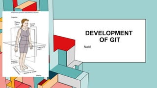
Sagittal Plane
Transverse Plane
Before After
Longitudinal & lateral folding of
embryo
Primitive gut formation which
then differentiates to FG, MG
and HG
Before After
End of 4th Week
• Rupture of
oropharyngeal
membrane at FG
Mouth
End of 5th Week
• Connection btw Yolk
Sac & MG narrows
as embryo folds
Vitelline duct
7th Week
• Rupture of cloacal
membrane at HG
Urogenital tract & Anal
openings
7/1/20XX 3
DEVELOPMENT OF GIT
Derivatives
Dorsal mesentery Dorsal mesogastrium 
greater omentum
Dorsal mesoduodenum
Mesentry proper
Dorsal mesocolon
Ventral mesentery Lesser omentum
Falciform ligament
Derivatives of FG in Digestive System
Mnemonics :
DOn’t (Duodenum) MES Personal Live with your BP
• Duodenum
• Mouth
• Esophagus
• Stomach
• Pancreas
• Liver
• Biliary apparatus
• Pharynx (Primordial)
4
FOREGUT DERIVATIVES
Before After
Part of Stomodeum
(Ectoderm) & FG
(endoderm) when
Buccopharyngeal
membrane ruptures
Mouth
• Epithelium of lips,
cheeks & palate
(Ecto)
• Epithelium of tongue
(Endo)
Cranial most part of FG
(Pharyngeal gut)
Pharynx
Before After
Dorsal portion of FG Esophagus
• Reaches its final
relative length by 7th
week
• Epithelium & glands
(endoderm)
• Muscular coat
(splanchnic
mesoderm)
7/1/20XX 5
Before After
Space behind the stomach due to the
pull of dorsal mesogastrium to the left
Omental busa
Developed btw 2 layers of dorsal
mesogastrium
Spleen
Dorsal Mesogastrium Liorenal ligament
• Btw posterior body wall (kidney) &
spleen
Gastrolienal ligament
• Btw spleen & stomach
Developed btw ventral mesogastrium Liver
• As liver cords grow into it, it thins to
form peritoneum of liver, falciform
ligament and lesser omentum
1st Rotation
(90’ clockwise around longitudinal
axis)
Before After
Bulging down of
dorsal mesogastrium
Greater omentum
2nd Rotation
(clockwise around anteroposterior
axis)
 Original dorsal border grows faster than
ventral border  greater curvature and
lesser curvature respectively
DEVELOPMENT OF STOMACH
6
DEVELOPMENT OF LIVER, BILIARY APPARATUS & GALLBLADDER
Liver & Biliary Apparatus
Before After
Enlargement of liver buds or
hepatic diverticulum
• Pars hepatica  liver cords
continue to penetrate septum
transversum
• Pars cystica
Vitelline & Umbilical Vein Liver sinusoids
Mesoderm of septum
transversum
Hematopoietic cells, Kupffer
cells & connective tissue cells
Septum transversum • Peritoneum of liver except
bare area
• Falciform ligament
• Lesser omentum
Cranial surface of liver
uncovered by peritoneum
Bare area of liver
Obliteration of umbilical vein in a
free margin of falciform ligament
Ligamentum teres hepatis
Pars cystica Gallbladder & cystic duct
Stalk connecting hepatic &
cystic duct to FG
Bile duct
Growth & rotation of duodenum Opening of bile duct carried to
posteromedial position from
ventral position
Gallbladder
7
DEVELOPMENT OF PANCREAS & DUODENUM
Duodenum Before After
Terminal part of FG & Proximal
part of MG
Duodenum
• Receive blood from celiac trunk (FG
artery) & superior mesenteric artery
(MG artery)
Rotation of stomach & rapid
growth of pancreatic head
Duodenum swinged from mid to right
• Mesoduodenum disappears
• Small portion near pylorus
• Duodenum become fixed at
retroperitoneal position
• Remains intraperitoneally
2 endodermal buds (dorsal &
ventral pancreatic buds ) arise
from caudal part of FG
Pancreas
Rotation of duodenum VPB moves dorsally and lie below & behind
DPB before fusing forming a single mass
Ventral Pancreatic Bud Uncinate process and inferior part of
pancreas head
Remaining part is from DPB
Distal part of Dorsal pancreatic
duct and entire ventral pancreatic
duct
Main pancreatic duct
Proximal part of dorsal pancreatic
duct
Accessory pancreatic duct
Pancreas
8
DEVELOPMENTAL ERRORS
Congenital anomalies
• Accessory spleens
(Polysplenia) –may exists in
one of the peritoneal folds.
• Congenital hypertrophic
pyloric stenosis
• Duodenal stenosis
• Duodenal atresia
Spleen
Accessory spleen
Duodenal stenosis
• Oesophageal atresia and/or
tracheoesophageal fistula.
• Polyhydramnios
• Oesophageal stenosis.
• Congenital hiatal hernia -
Short oesophagus
VACTERL association
9
MIDGUT DERIVATIVES
Before After
Elongation of
MG
Primary Intestinal Loop (cephalic limb &
caudal limb)
Superior
mesenteric
artery
Axis of Primary Intestinal Loop
Cephalic limb
(pre-arterial)
Distal part of Duodenum, Jejunum & Ileum
Caudal limb
(post-arterial)
Lower part of ileum, appendix, cecum,
ascending colon & right proximal 2/3 of
transverse colon
Before After
At 6th week
• Rapid elongation of Primary IL
especially cephalic limb
• Rapid growth & expansion of liver
• Lack of room in abdominal cavity to
accommodate all the intestinal loops
Physiological umbilical herniation
7/1/20XX Pitch deck title 10
ROTATION OF MG Process Description
1
(8th W)
Brings the :
• Cephalic limb to the right
• Caudal limb to the left
The loop lies outside the body cavity
2
(10th–11th W)
Elongation of Cephalic limb > Caudal limb
3
(10th W)
Return of the herniated intestinal loop back into abdominal cavity due
to
• Expansion of abdominal cavity with the embryo growth
• Reduced liver growth
• Regression of mesonephric kidney
Proximal portion of jejunum (cephalic limb) returns first and lie on left
side on abdominal cavity
Remaining coils of cephalic limb (distal part of jejunum & ileum) returns
gradually and occupies more towards right side
Cecal bud (caudal limb) return to abdominal cavity and temporarily
occupies the right upper quadrant below right lobe of liver  descent
to its adult position of R iliac fossa  elongation of caudal limb
downwards  formation of ascending colon and hepatic flexure of
colon
Remaining part of caudal limb  2/3 transverse colon
7/1/20XX 11
Before After
Cecal bud Cecum and Appendix
Distal end (apex) Appendix
After birth, growth of
lateral wall > medial wall
of cecum
Appendix comes to open on its
medial side
Posterior to cecum (retrocecal0 or
colon (retrocolic)
1. Large intestine enlarges &
lengthen  their mesenteries &
duodenal mesentery (duodenum &
pancreas) are pressed against the
peritoneum of posterior abdominal
wall and get fused except the first
part of duodenum
2. Ascending & descending colon are
permanently anchored
retroperitoneally
3. Transverse colon fuses with post.
Wall of greater omenta  mobile
4. Jejunoileal loop, appendix, lower
end of cecum and sigmoid colon
retain their mesenteries
MIDGUT
DERIVATIVES
FIXATION OF INTESTINE
12
HINDGUT DERIVATIVES
Before After
Dorsal part of cloaca Rectum and upper part of anal canal
Ventral part of cloaca Urogenital sinus
At 7th week
• Growth of urorectal septum
towards cloacal membrane and
fuses
Dorsal anal membrane and ventral
urogenital membrane
Perineal body between two
membranes
proctodeum
13
DEVELOPMENTAL ERRORS
Pathological Disorder Characteristic Diagram
Congenital Omphalocele • Abdominal viscera herniates via an
enlarged umbilical ring
• May included Liver, Intestines, Stomach,
Spleen & Gallbladder
• Covered by amnion
Umbilical Hernia • Herniation via an imperfectly closed
umbilicus
• Greater omentum & part of small intestine
• Covered by SC tissue & Skin
Gastroschisis • Abdnominal contents herniate via body
wall directly into amniotic cavity
• Lateral to umbilicus & on right side
• Viscera not covered by peritoneum or
amnion
14
VITELLINE DUCT ABNORMALITY
Pathological
Disorder
Characteristic
Meckel’s
diverticulum
• 2ft from ileo-cecal valve
• Gastric or pancreatic type
of ectopic mucosa
• 2 inch long
Vitelline cyst
Vitelline fistula
GUT ROTATION DEFECTS
Pathologica
l Disorder
Characteristic
Malrotation • Incomplete 270’ counterclockwise
rotation or may rotate only 90’
• Colon & cecum firstly return to abdomen
& lie on left side
Reverse Rotation • Primary intestinal loop rotates 90’
clockwise
• Transverse colon lies behind the
duodenum & superior mesenteric artery
• Result in volvulus and gangrene
Subhepatic
cecum &
appendix
• Anomalies of midgut rotation
Mobile cecum • Persistence of mesocolon portion 
Incomplete fixation of ascending colon
Internal hernia • Portion of small intestine passes into
mesentery & entrapped in it
Stenosis and
atresia of intestine
• Failure of formation of enough number of
vacuoles during recanalization
15
HINDGUT ABNORMALITIES
Pathological Disorder Characteristic Diagram
Congenital megacolon /
Hirschsprung’s disease
• Absent parasympathetic ganglia in the
bowel wall
Imperforate anus • Failure of rupture of anal membrane at end
of 8th week  Thin layer of anal membrane
separates the anal canal from exterior 
no anal opening
Rectovaginal fistula • Incomplete separation of cloaca by
urorectal septum
Anorectal atresia
Rectourethral/Urorectal
fistula

More Related Content

Similar to GIT Development Summary (Embroyology).pptx

GIT embryology By Dr Parashuram Waddar Pediatrician
GIT embryology By Dr Parashuram Waddar Pediatrician GIT embryology By Dr Parashuram Waddar Pediatrician
GIT embryology By Dr Parashuram Waddar Pediatrician ParasuramWaddar2
 
DEVELOPMENT stomach.pptx
DEVELOPMENT stomach.pptxDEVELOPMENT stomach.pptx
DEVELOPMENT stomach.pptxHabibAhmad80
 
Development of stomach
Development of stomachDevelopment of stomach
Development of stomachdrasarma1947
 
GI development embryology & Concept of Peritoneum
GI development embryology & Concept of PeritoneumGI development embryology & Concept of Peritoneum
GI development embryology & Concept of PeritoneumProf. Dr. Hironmoy Roy
 
SURGICAL ANATOMY OF STOMACH AND DUODENUM dr. bruhath^final. .pdf
SURGICAL ANATOMY OF STOMACH AND DUODENUM dr. bruhath^final. .pdfSURGICAL ANATOMY OF STOMACH AND DUODENUM dr. bruhath^final. .pdf
SURGICAL ANATOMY OF STOMACH AND DUODENUM dr. bruhath^final. .pdfmadhurikakarnati
 
Alimentary Canal - Foregut - Dr Joshua Tadayo.pdf
Alimentary Canal - Foregut - Dr Joshua Tadayo.pdfAlimentary Canal - Foregut - Dr Joshua Tadayo.pdf
Alimentary Canal - Foregut - Dr Joshua Tadayo.pdfJoshuaTadayo
 
Development of Hepatobiliary System
Development of Hepatobiliary System Development of Hepatobiliary System
Development of Hepatobiliary System Prabhakar Yadav
 
PMDC NEB Step-1 Day-6 (Review of Abdomen)
PMDC NEB Step-1 Day-6 (Review of Abdomen)PMDC NEB Step-1 Day-6 (Review of Abdomen)
PMDC NEB Step-1 Day-6 (Review of Abdomen)DrSaeed Shafi
 
Chap 16.pptx english chapter for students
Chap 16.pptx english chapter for studentsChap 16.pptx english chapter for students
Chap 16.pptx english chapter for studentsAneerSha
 
GIT- Embryology - 1 and 2.pdf
GIT- Embryology - 1 and 2.pdfGIT- Embryology - 1 and 2.pdf
GIT- Embryology - 1 and 2.pdfSundip Charmode
 
Pathology of the large intestines
Pathology of the large intestinesPathology of the large intestines
Pathology of the large intestinesOrato Ogoti
 
Development of the foregut (esophagus and stomach
Development of the foregut (esophagus and stomachDevelopment of the foregut (esophagus and stomach
Development of the foregut (esophagus and stomachSahar Hafeez
 
Development & histology of GIT & clinical considerations by Shapi. MD.pdf
Development & histology of GIT & clinical considerations by Shapi. MD.pdfDevelopment & histology of GIT & clinical considerations by Shapi. MD.pdf
Development & histology of GIT & clinical considerations by Shapi. MD.pdfShapi. MD
 
Embryology of GIT malformations by Dr. Yeneneh
Embryology of GIT malformations by Dr. Yeneneh Embryology of GIT malformations by Dr. Yeneneh
Embryology of GIT malformations by Dr. Yeneneh Jimma University
 
DEVELOPMENT OF DIGESTIVE SYSTEM.pptx
DEVELOPMENT OF DIGESTIVE SYSTEM.pptxDEVELOPMENT OF DIGESTIVE SYSTEM.pptx
DEVELOPMENT OF DIGESTIVE SYSTEM.pptxwmdpr4x64f
 
Digestive system by dr tayyaba......pptx
Digestive system by dr tayyaba......pptxDigestive system by dr tayyaba......pptx
Digestive system by dr tayyaba......pptxBIANOOR123
 

Similar to GIT Development Summary (Embroyology).pptx (20)

GIT embryology By Dr Parashuram Waddar Pediatrician
GIT embryology By Dr Parashuram Waddar Pediatrician GIT embryology By Dr Parashuram Waddar Pediatrician
GIT embryology By Dr Parashuram Waddar Pediatrician
 
DEVELOPMENT stomach.pptx
DEVELOPMENT stomach.pptxDEVELOPMENT stomach.pptx
DEVELOPMENT stomach.pptx
 
Foregut
ForegutForegut
Foregut
 
Foregut
ForegutForegut
Foregut
 
Development of stomach
Development of stomachDevelopment of stomach
Development of stomach
 
GI development embryology & Concept of Peritoneum
GI development embryology & Concept of PeritoneumGI development embryology & Concept of Peritoneum
GI development embryology & Concept of Peritoneum
 
Duodenum
DuodenumDuodenum
Duodenum
 
SURGICAL ANATOMY
SURGICAL ANATOMYSURGICAL ANATOMY
SURGICAL ANATOMY
 
SURGICAL ANATOMY OF STOMACH AND DUODENUM dr. bruhath^final. .pdf
SURGICAL ANATOMY OF STOMACH AND DUODENUM dr. bruhath^final. .pdfSURGICAL ANATOMY OF STOMACH AND DUODENUM dr. bruhath^final. .pdf
SURGICAL ANATOMY OF STOMACH AND DUODENUM dr. bruhath^final. .pdf
 
Alimentary Canal - Foregut - Dr Joshua Tadayo.pdf
Alimentary Canal - Foregut - Dr Joshua Tadayo.pdfAlimentary Canal - Foregut - Dr Joshua Tadayo.pdf
Alimentary Canal - Foregut - Dr Joshua Tadayo.pdf
 
Development of Hepatobiliary System
Development of Hepatobiliary System Development of Hepatobiliary System
Development of Hepatobiliary System
 
PMDC NEB Step-1 Day-6 (Review of Abdomen)
PMDC NEB Step-1 Day-6 (Review of Abdomen)PMDC NEB Step-1 Day-6 (Review of Abdomen)
PMDC NEB Step-1 Day-6 (Review of Abdomen)
 
Chap 16.pptx english chapter for students
Chap 16.pptx english chapter for studentsChap 16.pptx english chapter for students
Chap 16.pptx english chapter for students
 
GIT- Embryology - 1 and 2.pdf
GIT- Embryology - 1 and 2.pdfGIT- Embryology - 1 and 2.pdf
GIT- Embryology - 1 and 2.pdf
 
Pathology of the large intestines
Pathology of the large intestinesPathology of the large intestines
Pathology of the large intestines
 
Development of the foregut (esophagus and stomach
Development of the foregut (esophagus and stomachDevelopment of the foregut (esophagus and stomach
Development of the foregut (esophagus and stomach
 
Development & histology of GIT & clinical considerations by Shapi. MD.pdf
Development & histology of GIT & clinical considerations by Shapi. MD.pdfDevelopment & histology of GIT & clinical considerations by Shapi. MD.pdf
Development & histology of GIT & clinical considerations by Shapi. MD.pdf
 
Embryology of GIT malformations by Dr. Yeneneh
Embryology of GIT malformations by Dr. Yeneneh Embryology of GIT malformations by Dr. Yeneneh
Embryology of GIT malformations by Dr. Yeneneh
 
DEVELOPMENT OF DIGESTIVE SYSTEM.pptx
DEVELOPMENT OF DIGESTIVE SYSTEM.pptxDEVELOPMENT OF DIGESTIVE SYSTEM.pptx
DEVELOPMENT OF DIGESTIVE SYSTEM.pptx
 
Digestive system by dr tayyaba......pptx
Digestive system by dr tayyaba......pptxDigestive system by dr tayyaba......pptx
Digestive system by dr tayyaba......pptx
 

Recently uploaded

Gallbladder Double-Diverticular: A Case Report المرارة مزدوجة التج: تقرير حالة
Gallbladder Double-Diverticular: A Case Report  المرارة مزدوجة التج: تقرير حالةGallbladder Double-Diverticular: A Case Report  المرارة مزدوجة التج: تقرير حالة
Gallbladder Double-Diverticular: A Case Report المرارة مزدوجة التج: تقرير حالةMohamad محمد Al-Gailani الكيلاني
 
A thorough review of supernormal conduction.pptx
A thorough review of supernormal conduction.pptxA thorough review of supernormal conduction.pptx
A thorough review of supernormal conduction.pptxSergio Pinski
 
SURGICAL ANATOMY OF ORAL IMPLANTOLOGY.pptx
SURGICAL ANATOMY OF ORAL IMPLANTOLOGY.pptxSURGICAL ANATOMY OF ORAL IMPLANTOLOGY.pptx
SURGICAL ANATOMY OF ORAL IMPLANTOLOGY.pptxSuresh Kumar K
 
Cervical screening – taking care of your health flipchart (Vietnamese)
Cervical screening – taking care of your health flipchart (Vietnamese)Cervical screening – taking care of your health flipchart (Vietnamese)
Cervical screening – taking care of your health flipchart (Vietnamese)Cancer Institute NSW
 
hypo and hyper thyroidism final lecture.pptx
hypo and hyper thyroidism  final lecture.pptxhypo and hyper thyroidism  final lecture.pptx
hypo and hyper thyroidism final lecture.pptxdr shahida
 
PREPARATION FOR EXAMINATION FON II .pptx
PREPARATION FOR EXAMINATION FON II .pptxPREPARATION FOR EXAMINATION FON II .pptx
PREPARATION FOR EXAMINATION FON II .pptxPupayumnam1
 
CAS 110-63-4 BDO Liquid 1,4-Butanediol 1 4 BDO Warehouse Supply For Excellent...
CAS 110-63-4 BDO Liquid 1,4-Butanediol 1 4 BDO Warehouse Supply For Excellent...CAS 110-63-4 BDO Liquid 1,4-Butanediol 1 4 BDO Warehouse Supply For Excellent...
CAS 110-63-4 BDO Liquid 1,4-Butanediol 1 4 BDO Warehouse Supply For Excellent...ocean4396
 
Hemodialysis: Chapter 2, Extracorporeal Blood Circuit - Dr.Gawad
Hemodialysis: Chapter 2, Extracorporeal Blood Circuit - Dr.GawadHemodialysis: Chapter 2, Extracorporeal Blood Circuit - Dr.Gawad
Hemodialysis: Chapter 2, Extracorporeal Blood Circuit - Dr.GawadNephroTube - Dr.Gawad
 
Tips and tricks to pass the cardiovascular station for PACES exam
Tips and tricks to pass the cardiovascular station for PACES examTips and tricks to pass the cardiovascular station for PACES exam
Tips and tricks to pass the cardiovascular station for PACES examJunhao Koh
 
Factors Affecting child behavior in Pediatric Dentistry
Factors Affecting child behavior in Pediatric DentistryFactors Affecting child behavior in Pediatric Dentistry
Factors Affecting child behavior in Pediatric DentistryDr Simran Deepak Vangani
 
Case presentation on Antibody screening- how to solve 3 cell and 11 cell panel?
Case presentation on Antibody screening- how to solve 3 cell and 11 cell panel?Case presentation on Antibody screening- how to solve 3 cell and 11 cell panel?
Case presentation on Antibody screening- how to solve 3 cell and 11 cell panel?DrShinyKajal
 
Denture base resins materials and its mechanism of action
Denture base resins materials and its mechanism of actionDenture base resins materials and its mechanism of action
Denture base resins materials and its mechanism of actionDr.shiva sai vemula
 
Renal Replacement Therapy in Acute Kidney Injury -time modality -Dr Ayman Se...
Renal Replacement Therapy in Acute Kidney Injury -time  modality -Dr Ayman Se...Renal Replacement Therapy in Acute Kidney Injury -time  modality -Dr Ayman Se...
Renal Replacement Therapy in Acute Kidney Injury -time modality -Dr Ayman Se...Ayman Seddik
 
CT scan of penetrating abdominopelvic trauma
CT scan of penetrating abdominopelvic traumaCT scan of penetrating abdominopelvic trauma
CT scan of penetrating abdominopelvic traumassuser144901
 
5cladba raw material 5CL-ADB-A precursor raw
5cladba raw material 5CL-ADB-A precursor raw5cladba raw material 5CL-ADB-A precursor raw
5cladba raw material 5CL-ADB-A precursor rawSherrylee83
 
TEST BANK for The Nursing Assistant Acute, Subacute, and Long-Term Care, 6th ...
TEST BANK for The Nursing Assistant Acute, Subacute, and Long-Term Care, 6th ...TEST BANK for The Nursing Assistant Acute, Subacute, and Long-Term Care, 6th ...
TEST BANK for The Nursing Assistant Acute, Subacute, and Long-Term Care, 6th ...marcuskenyatta275
 
Vaccines: A Powerful and Cost-Effective Tool Protecting Americans Against Dis...
Vaccines: A Powerful and Cost-Effective Tool Protecting Americans Against Dis...Vaccines: A Powerful and Cost-Effective Tool Protecting Americans Against Dis...
Vaccines: A Powerful and Cost-Effective Tool Protecting Americans Against Dis...PhRMA
 
TEST BANK For Timby's Introductory Medical-Surgical Nursing, 13th Edition by ...
TEST BANK For Timby's Introductory Medical-Surgical Nursing, 13th Edition by ...TEST BANK For Timby's Introductory Medical-Surgical Nursing, 13th Edition by ...
TEST BANK For Timby's Introductory Medical-Surgical Nursing, 13th Edition by ...marcuskenyatta275
 
Video capsule endoscopy (VCE ) in children
Video capsule endoscopy (VCE ) in childrenVideo capsule endoscopy (VCE ) in children
Video capsule endoscopy (VCE ) in childrenRaju678948
 
Integrated Neuromuscular Inhibition Technique (INIT)
Integrated Neuromuscular Inhibition Technique (INIT)Integrated Neuromuscular Inhibition Technique (INIT)
Integrated Neuromuscular Inhibition Technique (INIT)Anjali Parmar
 

Recently uploaded (20)

Gallbladder Double-Diverticular: A Case Report المرارة مزدوجة التج: تقرير حالة
Gallbladder Double-Diverticular: A Case Report  المرارة مزدوجة التج: تقرير حالةGallbladder Double-Diverticular: A Case Report  المرارة مزدوجة التج: تقرير حالة
Gallbladder Double-Diverticular: A Case Report المرارة مزدوجة التج: تقرير حالة
 
A thorough review of supernormal conduction.pptx
A thorough review of supernormal conduction.pptxA thorough review of supernormal conduction.pptx
A thorough review of supernormal conduction.pptx
 
SURGICAL ANATOMY OF ORAL IMPLANTOLOGY.pptx
SURGICAL ANATOMY OF ORAL IMPLANTOLOGY.pptxSURGICAL ANATOMY OF ORAL IMPLANTOLOGY.pptx
SURGICAL ANATOMY OF ORAL IMPLANTOLOGY.pptx
 
Cervical screening – taking care of your health flipchart (Vietnamese)
Cervical screening – taking care of your health flipchart (Vietnamese)Cervical screening – taking care of your health flipchart (Vietnamese)
Cervical screening – taking care of your health flipchart (Vietnamese)
 
hypo and hyper thyroidism final lecture.pptx
hypo and hyper thyroidism  final lecture.pptxhypo and hyper thyroidism  final lecture.pptx
hypo and hyper thyroidism final lecture.pptx
 
PREPARATION FOR EXAMINATION FON II .pptx
PREPARATION FOR EXAMINATION FON II .pptxPREPARATION FOR EXAMINATION FON II .pptx
PREPARATION FOR EXAMINATION FON II .pptx
 
CAS 110-63-4 BDO Liquid 1,4-Butanediol 1 4 BDO Warehouse Supply For Excellent...
CAS 110-63-4 BDO Liquid 1,4-Butanediol 1 4 BDO Warehouse Supply For Excellent...CAS 110-63-4 BDO Liquid 1,4-Butanediol 1 4 BDO Warehouse Supply For Excellent...
CAS 110-63-4 BDO Liquid 1,4-Butanediol 1 4 BDO Warehouse Supply For Excellent...
 
Hemodialysis: Chapter 2, Extracorporeal Blood Circuit - Dr.Gawad
Hemodialysis: Chapter 2, Extracorporeal Blood Circuit - Dr.GawadHemodialysis: Chapter 2, Extracorporeal Blood Circuit - Dr.Gawad
Hemodialysis: Chapter 2, Extracorporeal Blood Circuit - Dr.Gawad
 
Tips and tricks to pass the cardiovascular station for PACES exam
Tips and tricks to pass the cardiovascular station for PACES examTips and tricks to pass the cardiovascular station for PACES exam
Tips and tricks to pass the cardiovascular station for PACES exam
 
Factors Affecting child behavior in Pediatric Dentistry
Factors Affecting child behavior in Pediatric DentistryFactors Affecting child behavior in Pediatric Dentistry
Factors Affecting child behavior in Pediatric Dentistry
 
Case presentation on Antibody screening- how to solve 3 cell and 11 cell panel?
Case presentation on Antibody screening- how to solve 3 cell and 11 cell panel?Case presentation on Antibody screening- how to solve 3 cell and 11 cell panel?
Case presentation on Antibody screening- how to solve 3 cell and 11 cell panel?
 
Denture base resins materials and its mechanism of action
Denture base resins materials and its mechanism of actionDenture base resins materials and its mechanism of action
Denture base resins materials and its mechanism of action
 
Renal Replacement Therapy in Acute Kidney Injury -time modality -Dr Ayman Se...
Renal Replacement Therapy in Acute Kidney Injury -time  modality -Dr Ayman Se...Renal Replacement Therapy in Acute Kidney Injury -time  modality -Dr Ayman Se...
Renal Replacement Therapy in Acute Kidney Injury -time modality -Dr Ayman Se...
 
CT scan of penetrating abdominopelvic trauma
CT scan of penetrating abdominopelvic traumaCT scan of penetrating abdominopelvic trauma
CT scan of penetrating abdominopelvic trauma
 
5cladba raw material 5CL-ADB-A precursor raw
5cladba raw material 5CL-ADB-A precursor raw5cladba raw material 5CL-ADB-A precursor raw
5cladba raw material 5CL-ADB-A precursor raw
 
TEST BANK for The Nursing Assistant Acute, Subacute, and Long-Term Care, 6th ...
TEST BANK for The Nursing Assistant Acute, Subacute, and Long-Term Care, 6th ...TEST BANK for The Nursing Assistant Acute, Subacute, and Long-Term Care, 6th ...
TEST BANK for The Nursing Assistant Acute, Subacute, and Long-Term Care, 6th ...
 
Vaccines: A Powerful and Cost-Effective Tool Protecting Americans Against Dis...
Vaccines: A Powerful and Cost-Effective Tool Protecting Americans Against Dis...Vaccines: A Powerful and Cost-Effective Tool Protecting Americans Against Dis...
Vaccines: A Powerful and Cost-Effective Tool Protecting Americans Against Dis...
 
TEST BANK For Timby's Introductory Medical-Surgical Nursing, 13th Edition by ...
TEST BANK For Timby's Introductory Medical-Surgical Nursing, 13th Edition by ...TEST BANK For Timby's Introductory Medical-Surgical Nursing, 13th Edition by ...
TEST BANK For Timby's Introductory Medical-Surgical Nursing, 13th Edition by ...
 
Video capsule endoscopy (VCE ) in children
Video capsule endoscopy (VCE ) in childrenVideo capsule endoscopy (VCE ) in children
Video capsule endoscopy (VCE ) in children
 
Integrated Neuromuscular Inhibition Technique (INIT)
Integrated Neuromuscular Inhibition Technique (INIT)Integrated Neuromuscular Inhibition Technique (INIT)
Integrated Neuromuscular Inhibition Technique (INIT)
 

GIT Development Summary (Embroyology).pptx

  • 2. DEVELOPMENT OF GIT Sagittal Plane Transverse Plane Before After Longitudinal & lateral folding of embryo Primitive gut formation which then differentiates to FG, MG and HG Before After End of 4th Week • Rupture of oropharyngeal membrane at FG Mouth End of 5th Week • Connection btw Yolk Sac & MG narrows as embryo folds Vitelline duct 7th Week • Rupture of cloacal membrane at HG Urogenital tract & Anal openings
  • 3. 7/1/20XX 3 DEVELOPMENT OF GIT Derivatives Dorsal mesentery Dorsal mesogastrium  greater omentum Dorsal mesoduodenum Mesentry proper Dorsal mesocolon Ventral mesentery Lesser omentum Falciform ligament Derivatives of FG in Digestive System Mnemonics : DOn’t (Duodenum) MES Personal Live with your BP • Duodenum • Mouth • Esophagus • Stomach • Pancreas • Liver • Biliary apparatus • Pharynx (Primordial)
  • 4. 4 FOREGUT DERIVATIVES Before After Part of Stomodeum (Ectoderm) & FG (endoderm) when Buccopharyngeal membrane ruptures Mouth • Epithelium of lips, cheeks & palate (Ecto) • Epithelium of tongue (Endo) Cranial most part of FG (Pharyngeal gut) Pharynx Before After Dorsal portion of FG Esophagus • Reaches its final relative length by 7th week • Epithelium & glands (endoderm) • Muscular coat (splanchnic mesoderm)
  • 5. 7/1/20XX 5 Before After Space behind the stomach due to the pull of dorsal mesogastrium to the left Omental busa Developed btw 2 layers of dorsal mesogastrium Spleen Dorsal Mesogastrium Liorenal ligament • Btw posterior body wall (kidney) & spleen Gastrolienal ligament • Btw spleen & stomach Developed btw ventral mesogastrium Liver • As liver cords grow into it, it thins to form peritoneum of liver, falciform ligament and lesser omentum 1st Rotation (90’ clockwise around longitudinal axis) Before After Bulging down of dorsal mesogastrium Greater omentum 2nd Rotation (clockwise around anteroposterior axis)  Original dorsal border grows faster than ventral border  greater curvature and lesser curvature respectively DEVELOPMENT OF STOMACH
  • 6. 6 DEVELOPMENT OF LIVER, BILIARY APPARATUS & GALLBLADDER Liver & Biliary Apparatus Before After Enlargement of liver buds or hepatic diverticulum • Pars hepatica  liver cords continue to penetrate septum transversum • Pars cystica Vitelline & Umbilical Vein Liver sinusoids Mesoderm of septum transversum Hematopoietic cells, Kupffer cells & connective tissue cells Septum transversum • Peritoneum of liver except bare area • Falciform ligament • Lesser omentum Cranial surface of liver uncovered by peritoneum Bare area of liver Obliteration of umbilical vein in a free margin of falciform ligament Ligamentum teres hepatis Pars cystica Gallbladder & cystic duct Stalk connecting hepatic & cystic duct to FG Bile duct Growth & rotation of duodenum Opening of bile duct carried to posteromedial position from ventral position Gallbladder
  • 7. 7 DEVELOPMENT OF PANCREAS & DUODENUM Duodenum Before After Terminal part of FG & Proximal part of MG Duodenum • Receive blood from celiac trunk (FG artery) & superior mesenteric artery (MG artery) Rotation of stomach & rapid growth of pancreatic head Duodenum swinged from mid to right • Mesoduodenum disappears • Small portion near pylorus • Duodenum become fixed at retroperitoneal position • Remains intraperitoneally 2 endodermal buds (dorsal & ventral pancreatic buds ) arise from caudal part of FG Pancreas Rotation of duodenum VPB moves dorsally and lie below & behind DPB before fusing forming a single mass Ventral Pancreatic Bud Uncinate process and inferior part of pancreas head Remaining part is from DPB Distal part of Dorsal pancreatic duct and entire ventral pancreatic duct Main pancreatic duct Proximal part of dorsal pancreatic duct Accessory pancreatic duct Pancreas
  • 8. 8 DEVELOPMENTAL ERRORS Congenital anomalies • Accessory spleens (Polysplenia) –may exists in one of the peritoneal folds. • Congenital hypertrophic pyloric stenosis • Duodenal stenosis • Duodenal atresia Spleen Accessory spleen Duodenal stenosis • Oesophageal atresia and/or tracheoesophageal fistula. • Polyhydramnios • Oesophageal stenosis. • Congenital hiatal hernia - Short oesophagus VACTERL association
  • 9. 9 MIDGUT DERIVATIVES Before After Elongation of MG Primary Intestinal Loop (cephalic limb & caudal limb) Superior mesenteric artery Axis of Primary Intestinal Loop Cephalic limb (pre-arterial) Distal part of Duodenum, Jejunum & Ileum Caudal limb (post-arterial) Lower part of ileum, appendix, cecum, ascending colon & right proximal 2/3 of transverse colon Before After At 6th week • Rapid elongation of Primary IL especially cephalic limb • Rapid growth & expansion of liver • Lack of room in abdominal cavity to accommodate all the intestinal loops Physiological umbilical herniation
  • 10. 7/1/20XX Pitch deck title 10 ROTATION OF MG Process Description 1 (8th W) Brings the : • Cephalic limb to the right • Caudal limb to the left The loop lies outside the body cavity 2 (10th–11th W) Elongation of Cephalic limb > Caudal limb 3 (10th W) Return of the herniated intestinal loop back into abdominal cavity due to • Expansion of abdominal cavity with the embryo growth • Reduced liver growth • Regression of mesonephric kidney Proximal portion of jejunum (cephalic limb) returns first and lie on left side on abdominal cavity Remaining coils of cephalic limb (distal part of jejunum & ileum) returns gradually and occupies more towards right side Cecal bud (caudal limb) return to abdominal cavity and temporarily occupies the right upper quadrant below right lobe of liver  descent to its adult position of R iliac fossa  elongation of caudal limb downwards  formation of ascending colon and hepatic flexure of colon Remaining part of caudal limb  2/3 transverse colon
  • 11. 7/1/20XX 11 Before After Cecal bud Cecum and Appendix Distal end (apex) Appendix After birth, growth of lateral wall > medial wall of cecum Appendix comes to open on its medial side Posterior to cecum (retrocecal0 or colon (retrocolic) 1. Large intestine enlarges & lengthen  their mesenteries & duodenal mesentery (duodenum & pancreas) are pressed against the peritoneum of posterior abdominal wall and get fused except the first part of duodenum 2. Ascending & descending colon are permanently anchored retroperitoneally 3. Transverse colon fuses with post. Wall of greater omenta  mobile 4. Jejunoileal loop, appendix, lower end of cecum and sigmoid colon retain their mesenteries MIDGUT DERIVATIVES FIXATION OF INTESTINE
  • 12. 12 HINDGUT DERIVATIVES Before After Dorsal part of cloaca Rectum and upper part of anal canal Ventral part of cloaca Urogenital sinus At 7th week • Growth of urorectal septum towards cloacal membrane and fuses Dorsal anal membrane and ventral urogenital membrane Perineal body between two membranes proctodeum
  • 13. 13 DEVELOPMENTAL ERRORS Pathological Disorder Characteristic Diagram Congenital Omphalocele • Abdominal viscera herniates via an enlarged umbilical ring • May included Liver, Intestines, Stomach, Spleen & Gallbladder • Covered by amnion Umbilical Hernia • Herniation via an imperfectly closed umbilicus • Greater omentum & part of small intestine • Covered by SC tissue & Skin Gastroschisis • Abdnominal contents herniate via body wall directly into amniotic cavity • Lateral to umbilicus & on right side • Viscera not covered by peritoneum or amnion
  • 14. 14 VITELLINE DUCT ABNORMALITY Pathological Disorder Characteristic Meckel’s diverticulum • 2ft from ileo-cecal valve • Gastric or pancreatic type of ectopic mucosa • 2 inch long Vitelline cyst Vitelline fistula GUT ROTATION DEFECTS Pathologica l Disorder Characteristic Malrotation • Incomplete 270’ counterclockwise rotation or may rotate only 90’ • Colon & cecum firstly return to abdomen & lie on left side Reverse Rotation • Primary intestinal loop rotates 90’ clockwise • Transverse colon lies behind the duodenum & superior mesenteric artery • Result in volvulus and gangrene Subhepatic cecum & appendix • Anomalies of midgut rotation Mobile cecum • Persistence of mesocolon portion  Incomplete fixation of ascending colon Internal hernia • Portion of small intestine passes into mesentery & entrapped in it Stenosis and atresia of intestine • Failure of formation of enough number of vacuoles during recanalization
  • 15. 15 HINDGUT ABNORMALITIES Pathological Disorder Characteristic Diagram Congenital megacolon / Hirschsprung’s disease • Absent parasympathetic ganglia in the bowel wall Imperforate anus • Failure of rupture of anal membrane at end of 8th week  Thin layer of anal membrane separates the anal canal from exterior  no anal opening Rectovaginal fistula • Incomplete separation of cloaca by urorectal septum Anorectal atresia Rectourethral/Urorectal fistula