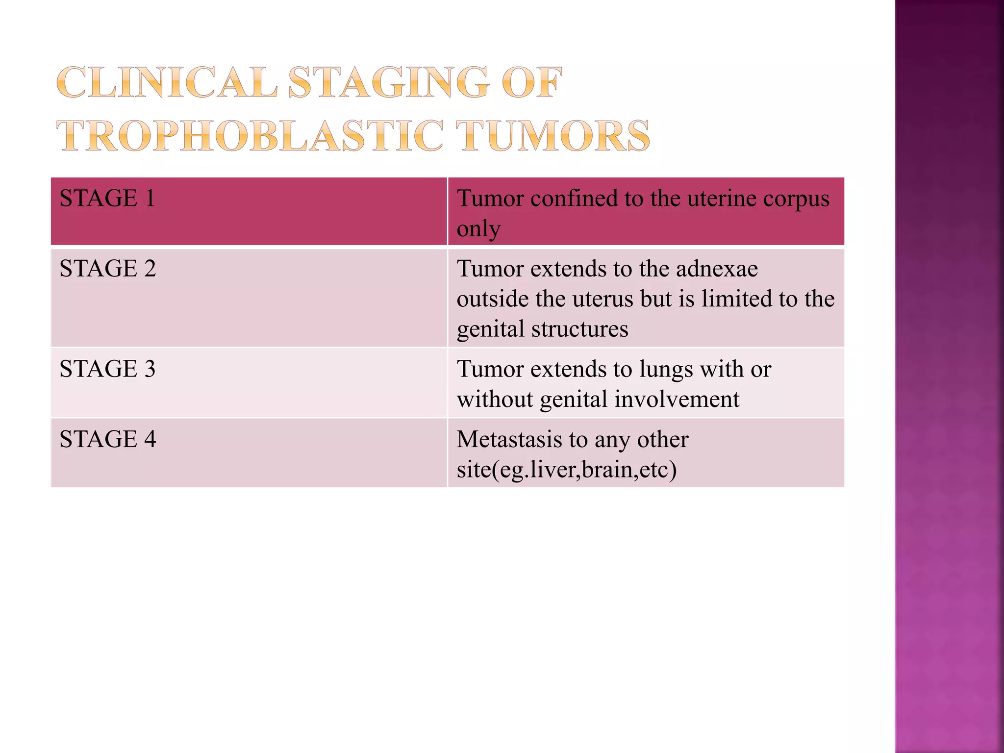
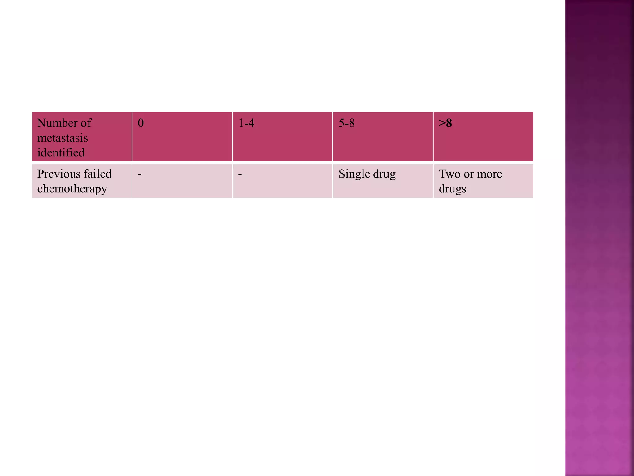
DR.PRIYA SAXENA
 Gestational trophoblastic tumors include:
o Invasive mole
o Choriocarcinoma
o Placental site trophoblastic tumor
 H.mole which has invaded the myometrium
 It can progress to choriocarcinoma but may regress spontaneously
 Locally invasive but do not usually metastasize
 If serosa is involved-it may cause intraperitoneal hemorrhage
 It may need hysterectomy as treatment for intractable bleeding.
 Special tumor arising from the placental implantation site
 It can be of:
o Low or
o High grade malignancy
 Characteristic histology-composed mainly of cytotrophoblastic elements
arising from intermediate trophoblast of placental bed.
 Bleeding-presenting symptom
 Serum beta HCG-may be only marginally increased
 Immunohistochemistry stains of the tumor tissue-positive for HPL
 Chemotherapy-usually not effective
 It requires hysterectomy for treatment.
 Carcinoma of the chorionic epithelium.
 Half the cases follow a hydatiform mole,25% follow an abortion and 25%
follow a normal pregnancy.
 There is absence of villus pattern on histology which differentiates it
from hydatiform mole.
 There is marked elevation of beta-HCG levels.
 Abnormal uterine bleeding
 Symptoms due to metastasis like hemoptysis or features suggestive of an
intracranial space occupying lesion. The common sites of metastasis are
the lungs in 75% cases and suburethral vaginal metastasis in 50% cases.
 Theca-lutein cysts of the ovaries are usually seen.
 Rapidly increasing levels of beta-HCG.
 Histopathological diagnosis of choriocarcinoma on curettage for irregular
bleeding.
STAGE 1 Tumor confined to the uterine corpus
only
STAGE 2 Tumor extends to the adnexae
outside the uterus but is limited to the
genital structures
STAGE 3 Tumor extends to lungs with or
without genital involvement
STAGE 4 Metastasis to any other
site(eg.liver,brain,etc)
Prognostic
factors
Score
0 1 2 4
Age(years) ≤39 >39
Antecedent
pregnancy
Hydatiform mole Abortion Term pregnancy
Interval from
antecedent
pregnancy(month
s)
<4 4-6 7-12 >12
Pre-treatment
HCG(IU/L)
<103 103-104 104-105 >105
Largest tumor
size including
uterus
3-4cm 5cm
Site of metastasis lungs Spleen,kidneys Gastrointestinal
tract
Brain,liver
Number of
metastasis
identified
0 1-4 5-8 >8
Previous failed
chemotherapy
- - Single drug Two or more
drugs
 A score of 0-6 indicates low risk
 A score ≥7 indicates high risk
 Low risk disease:
 Methotrexate is given for low risk disease
 The recommended regimen is Methotrexate alternating with folinic acid
 Methotrexate 1mg/kg body weight is given i.m. on days 1,3,5 and 7 and
folinic acid 0.1 mg/kg body weight on days 2,4,6 and 8
 The courses are repeated at intervals of every 2 weeks.
 Chemotherapy is continued for three courses after the beta-HCG levels
become normal
 Thereafter, levels are checked monthly
 Actinomycin-D in dose of 1.25 mg/m2 every 2 weeks is used for
methotrexate resistance or where methotrexate is contraindicated.
 High risk disease:
 The most commonly used regime is the EMACO regime which consists
of Etoposide,Methotrexate,Actinomycin-D, Cyclophosphamide and
Vincristine
 Cycle is repeated every 2 weeks until the beta-HCG is negative,
following which 3 more cycles are given
 Invasive mole perforating the serosa causing an intraperitoneal bleeding
 Uncontrollable vaginal bleeding
 Drug resistance
 Placental site trophoblastic tumor
 Follow-up and avoidance of pregnancy for atleast 1 year is essential after
chemotherapy
 Women with any form of trophoblastic disease are at increased risk of
developing trophoblastic disease in a subsequent pregnancy.
 It is important to send the placenta for histopathological examination in a
subsequent pregnancy and to do beta-HCG estimations 6 weeks after
delivery to rule out the possibility of developing any gestational
trophoblastic neoplasia.
THANK YOU

Gestational trophoblastic neoplasm

  • 1.
  • 2.
     Gestational trophoblastictumors include: o Invasive mole o Choriocarcinoma o Placental site trophoblastic tumor
  • 3.
     H.mole whichhas invaded the myometrium  It can progress to choriocarcinoma but may regress spontaneously  Locally invasive but do not usually metastasize  If serosa is involved-it may cause intraperitoneal hemorrhage  It may need hysterectomy as treatment for intractable bleeding.
  • 4.
     Special tumorarising from the placental implantation site  It can be of: o Low or o High grade malignancy  Characteristic histology-composed mainly of cytotrophoblastic elements arising from intermediate trophoblast of placental bed.  Bleeding-presenting symptom  Serum beta HCG-may be only marginally increased  Immunohistochemistry stains of the tumor tissue-positive for HPL  Chemotherapy-usually not effective  It requires hysterectomy for treatment.
  • 5.
     Carcinoma ofthe chorionic epithelium.  Half the cases follow a hydatiform mole,25% follow an abortion and 25% follow a normal pregnancy.  There is absence of villus pattern on histology which differentiates it from hydatiform mole.  There is marked elevation of beta-HCG levels.
  • 6.
     Abnormal uterinebleeding  Symptoms due to metastasis like hemoptysis or features suggestive of an intracranial space occupying lesion. The common sites of metastasis are the lungs in 75% cases and suburethral vaginal metastasis in 50% cases.  Theca-lutein cysts of the ovaries are usually seen.  Rapidly increasing levels of beta-HCG.  Histopathological diagnosis of choriocarcinoma on curettage for irregular bleeding.
  • 7.
    STAGE 1 Tumorconfined to the uterine corpus only STAGE 2 Tumor extends to the adnexae outside the uterus but is limited to the genital structures STAGE 3 Tumor extends to lungs with or without genital involvement STAGE 4 Metastasis to any other site(eg.liver,brain,etc)
  • 8.
    Prognostic factors Score 0 1 24 Age(years) ≤39 >39 Antecedent pregnancy Hydatiform mole Abortion Term pregnancy Interval from antecedent pregnancy(month s) <4 4-6 7-12 >12 Pre-treatment HCG(IU/L) <103 103-104 104-105 >105 Largest tumor size including uterus 3-4cm 5cm Site of metastasis lungs Spleen,kidneys Gastrointestinal tract Brain,liver
  • 9.
    Number of metastasis identified 0 1-45-8 >8 Previous failed chemotherapy - - Single drug Two or more drugs
  • 10.
     A scoreof 0-6 indicates low risk  A score ≥7 indicates high risk
  • 11.
     Low riskdisease:  Methotrexate is given for low risk disease  The recommended regimen is Methotrexate alternating with folinic acid  Methotrexate 1mg/kg body weight is given i.m. on days 1,3,5 and 7 and folinic acid 0.1 mg/kg body weight on days 2,4,6 and 8  The courses are repeated at intervals of every 2 weeks.  Chemotherapy is continued for three courses after the beta-HCG levels become normal  Thereafter, levels are checked monthly  Actinomycin-D in dose of 1.25 mg/m2 every 2 weeks is used for methotrexate resistance or where methotrexate is contraindicated.
  • 12.
     High riskdisease:  The most commonly used regime is the EMACO regime which consists of Etoposide,Methotrexate,Actinomycin-D, Cyclophosphamide and Vincristine  Cycle is repeated every 2 weeks until the beta-HCG is negative, following which 3 more cycles are given
  • 13.
     Invasive moleperforating the serosa causing an intraperitoneal bleeding  Uncontrollable vaginal bleeding  Drug resistance  Placental site trophoblastic tumor
  • 14.
     Follow-up andavoidance of pregnancy for atleast 1 year is essential after chemotherapy  Women with any form of trophoblastic disease are at increased risk of developing trophoblastic disease in a subsequent pregnancy.  It is important to send the placenta for histopathological examination in a subsequent pregnancy and to do beta-HCG estimations 6 weeks after delivery to rule out the possibility of developing any gestational trophoblastic neoplasia.
  • 15.