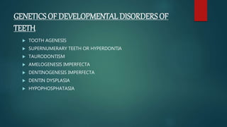
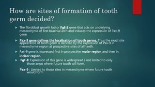
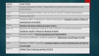
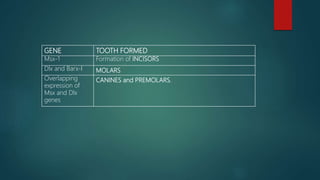
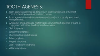
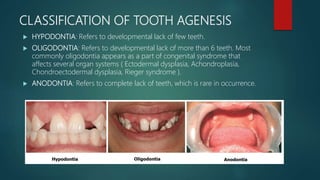
GENETICS
PRESENTED BY
DR.P.B.N.MOUNIKA
1st YEAR POST GRADUATE IN
DEPARTMENT OF
PEDODONTICS AND
PREVENTIVE DENTISTRY
GENETICS
 GENETICS can be defined as the study of genes and of statistical laws that
govern the passage of genes from one generation to next.
HEREDITARY:
The process of transmission of characters from one generation to next
(parents to children) is called hereditary or inheritance.
 GENE:
The functional unit of DNA is called gene.
 GENOME:
The total genetic information present in a cell is called genome.
 GENOTYPE:
The genetic constitution or makeup of an individual.
 PHENOTYPE:
It is the physical, mental or biochemical manifestation of an individual in
relation to a particular character resulting from expression of associated
genes.
 LOCUS:
The position of a gene on chromosome is called locus.
 ALLELE:
Alternate form of gene present at any particular locus.
 DIPLOID CELL:
A cell with 23 pairs of chromosomes.
 HAPLOID CELL:
A cell with 23 chromosomes.
 CHROMOSOME:
The chromosomes are deeply stained minute rod like structures in nucleus of
cell formed by condensation of chromatin during cell division.
• They contain DNA encoding genetic information inherited from parents.
• The individual chromosomes are best visible under microscope only
metaphase stage of cell division.
 The complement of 46 chromosomes in each human cell is classified into 44 autosomes
(22 pairs) and 2 sex chromosomes (1 pair).
 One member of each pair of autosomes and sex chromosome is contributed by either the
father (paternal) or the mother (maternal).
 The sex chromosomes are of two different types : X and Y chromosomes.
FEMALE: 44 autosomes+ two X chromosomes (44+ XX) forming
homomorphic set of sex chromosomes.
MALE: 44 autosomes + two dissimilar sex chromosome (44+ XY)
forming heteromorphic set of sex chromosome.
• Chromosome look like an entangled mesh of chromatin thread in
interphase.
• Prior to onset of cell division and progressive thickening of individual
chromosome, each chromosome undergoes duplication of its DNA
content and appears as two closely placed free strands attached together
near their waists. This event is called DNA REPLICATION.
• Subsequently during the later stages of cell division , as chromosome gets
condensed further, each of them looks like thick rod in Metaphase.
• Or letters like J or V in Anaphase.
STRUCTURE:
 CHROMATIDS:
Each metaphase chromosome comprises
Of two identical components after DNA
replication. These two symmetrical halves
are called SISTER CHROMATIDS.
 CENTROMERE OR KINETOCHORE:
The chromatids are attached together at a constricted region that stains
called CENTROMERE. The centromere defines the PRIMARY CONSTRICTION
of the chromosome and divides the chromosome into a short arm (p) and a
long arm (q).
ROLE:
It plays a major role during movement of chromosome in cell division.
 TELOMERES:
The free ends of chromatids are known as telomeres.
 SECONDARY CONSTRICTION:
Chromosomes usually carry an additional secondary constriction in one or
both ends of chromatids. These constrictions are linked to formation of
nucleolus and hence referred to as NUCLEOLAR ORGANISING REGION.
The secondary constriction may lie at distal end of chromatid giving rise to a
small fragment of chromosome at extreme end of the chromosome called
SATELLITE. Such chromosomes are called SAT- chromosomes.
 In active state it is lightly stained and most extended and is called
EUCHROMATIN.
 In inactive state it is darkly stained and remains highly coiled and is called
HETEROCHROMATIN.
HETEROCHROMATIN
EUCHROMATIN
CLASSIFICATION OF
CHROMOSOMES:
 STANDARD ( DENVER) CLASSIFICATION:
 Chromosome are classified into seven groups in an arrangement in
descending order of their lengths.
 Groups are designated alphabetically from groups A TO G.
 The longer female sex chromosome X is included in GROUP C.
 Thee smaller male sex chromosome Y is included in GROUP G.
GROUP PAIRS OF
CHROMOSOME
FEATURES
A 1,2,3 Long and metacentric
B 4,5 Fairly long and submetacentric
C 6 to 12 + X chromosome Medium sized and
submetacentric
D 13,14,15 Medium sized and acrocentric
A SATELLITE BODY is attached
to free end of short arm of
chromosome.
E 16,17,18 Fairly short and submetacentric
F 19,20 Short and metacentric
G 21,22 + Y chromosome Very short and acrocentric with
SATELLITE BODIES on their short
arms.
 CLASSIFICATION BASED ON POSITION OF CENTROMERE:
 METACENTRIC: Centromere located near the middle of the chromosome;
length of p=q.
 SUBMETACENTRIC: Centromere located slightly away from the middle;
length of p<q.
 ACROCENTRIC: Centromere located very near to the end ; the length of
p<<q.
 TELOCENTRIC: Centromere located at one end of chromosome;
effectively having only a single arm.
KARYOTYPE:
 The chromosomal make-up of an individual is called his or her karyotype.
 Karyotype is essentially a photograph of an individual’s chromosomes
arranged to the standard classification.
 Diagrammatic representation of karyotype is called as ideogram.
 A karyotype is done to:
a. Identify and number the chromosomes.
b. Detect numerical and structural anomalies of chromosomes.
SEX CHROMATIN ( BARR BODY)
 During interphase , in females, out of two X chromosome one becomes
highly coiled ( genetically inactive) and other remains uncoiled (
active).
 The coiled, genetically inactive X chromosome is seen as heterochromatic
body called SEX CHROMATIN OR BARR BODY.
 It was first noticed by Barr and Betram (1949) in nucleus of nerve cell of
female cat.
 Barr body is located on inner surface of nuclear membrane as dark
basophilic body of chromatin in most of cells of body.
 It is generally ovoid or planoconvex.
 Appearance:
Neurons: small dark body opposite to nucleolus
Neutrophils : knob of 1.5 micrometers in diameter known as
drumstick.
IMPORTANCE OF X CHROMOSOME
INACTIVATION:
 At its onset, embryogenesis in the females requires active participation of
both the X chromosomes.
 Thereafter one of the chromosome is inactivated in subsequent courses
of development.
 The presence of only a single active X chromosome is sufficient to
maintain the protein levels expressed by genes on X chromosome.
 The presence of extra active X chromosome causes the dose of gene
products to be double and are eventually deleterious or fatal.
 So nature has evolved a mechanism of inactivation of X- chromosome .
This mechanism is called DOSAGE COMPENSATION.
LYONS HYPOTHESIS:
 According to hypothesis formulated by Mary F Lyon in 1962, the
of Barr bodies in a cell is equal to total number of X chromosomes minus
1.
 It is also known as n-1 rule, where n represents the number of X
chromosome in the cell.
INDIVIDUAL SEX CHROMOSOME
CONSTITUTION
NUMBER OF BARR
BODIES
Normal male XY NIL
Turners syndrome XO NIL
Normal female XX ONE
Klinefelter’s syndrome XXY ONE
Triple X chromosome
super female)
XXX TWO
DEOXYRIBONUCLEIC ACID(DNA):
 The DNA is a double stranded molecule,
made up of chain of nucleotides , coiled
around each other , forming what is
commonly called DOUBLE HELIX.
 The helix model of DNA was first introduced
by JAMES WATSON AND FRANCIS CRICK
in 1953.
 The NUCLEOTIDES are the basic structural
units of DNA
 Each nucleotide consists of three subunits:
a. A sugar
b. A phosphate group
c. A nitrogenous base
 Each strand of double helix consists of alternate units of sugar and
phosphate.
 SUGAR- DEOXYRIBOSE- 5 CARBON PENTOSE SUGAR
 The two strands are held together by hydrogen bonds.
 NITROGENOUS BASES:
 ADENINE binds with THYMINE by two hydrogen bonds.
 GUANINE binds with CYTOSINE by three hydrogen bonds.
 The two strands of DNA are complimentary to each other i.e. if the base
sequence of one strand is known , the base sequence of other strand can
be formulated.
FUNCTIONS OF DNA:
 SELF REPLICATION:
During nuclear division the two strands of DNA separate and each
acts as a template to form a new complimentary strand.
 SYNTHESIS OF RNA AND PROTEINS:
The certain regions of DNA serves as a template for synthesis of RNA
in turn into proteins.
 RECOMBINATION:
During crossing over in meiosis, there is exchange of genetic material
between homologous chromosomes, which leads to shuffling of genes
and process is called recombination.
 MUTATION:
Major source of genetic variation. The change of base sequence in DNA
molecule is called GENE MUTATION. It may be spontaneous or induced
by chemicals, radiation etc.
 DNA MOLECULES ARE ASSOCIATED WITH GLOBULAR HISTONE
PROTEINS TO FORM CHROMATIN. HISTONE PROTEINS ARE INVOLVED
IN REGULATING FUNCTION OF DNA. NORMALLY CHROMATIN IS
ORGANISED AS STRING WITH BEADS WITHIN NUCLEUS BUT DURING
DIVISION THE CHROMATIN CONDENSES INTO STRUCTURES CALLED
CHROMOSOMES.
RIBONUCLEIC ACID(RNA)
 RNA is synthesized in nucleus by transcription of one strand of DNA.
 Its structure is similar to single stranded DNA with difference that sugar is
RIBOSE and URACIL substitutes for thymine. Uracil binds only to Adenine.
 RNA PLAYS A MAJOR ROLE IN GENE EXPRESSION.
 TYPES OF RNA:
1. MESSENGER RNA:
Synthesized in nucleus from DNA and copies nucleotide sequence
from DNA. It moves into cytoplasm through nuclear pores and dictates
sequence of AMINOACIDS IN POLYPEPTIDE CHAIN. This information
about sequence of amino acid is called GENETIC CODE- carried in groups
of three nucleotides known as CODONS.
2. TRANSFER RNA:
Function is to match a specific amino acid to a specific codon of m
RNA. Each t RNA can pickup particular amino acid. In other words, before
formation of peptide linkage the amino acid are activated and
one end of specific transfer RNA.
3. RIBOSOMAL RNA:
It is rich in Guanine and Cytosine in comparison to other RNA’s. It
plays a major role in formation of protein at the ribosome.
GENETICS OF DEVELOPMENTAL DISORDERS OF
TEETH
 TOOTH AGENESIS
 SUPERNUMERARY TEETH OR HYPERDONTIA
 TAURODONTISM
 AMELOGENESIS IMPERFECTA
 DENTINOGENESIS IMPERFECTA
 DENTIN DYSPLASIA
 HYPOPHOSPHATASIA
How are sites of formation of tooth
germ decided?
 The fibroblast growth factor Fgf-8 gene that acts on underlying
mesenchyme of first brachial arch and induces the expression of Pax-9
gene.
 Pax-9 gene defines the localisation of tooth germs. Thus the exact site
appearance of tooth germ is decided by the expression of Pax-9 in
mesenchyme region at prospective sites of all teeth.
 Pax-9 gene is expressed first in prospective molar region and then in
incisor region.
 Fgf-8: Expression of this gene is widespread ( not limited to only
those areas where future tooth will form.
Pax-9 : Limited to those sites in mesenchyme where future tooth
would form
GENES FUNCTIONS
Fgf-4 Induces expression of Msx-1 responsible for growth of dental epithelium and
mesenchyme.
Fgf-8 Induces expression of Msx-1, Dlx-1/2, Pax-9, Lhx-6/7 in dental mesenchyme.
Fgf-9 Same as Fgf-4/8
Pax-9 Maintains BMP-4 expression in dental mesenchyme. Mutation results in failure of
development of all teeth.
Pax-6 Mutation develops additional upper incisors.
Msx-1/2 Required for expression of Bmp-4, Fgf-3 and Dlx-2 in dental mesenchyme.
Mutations results in failure to develop all teeth.
Dlx-3 Mutation result in Enamel hypoplasia and Taurodontism.
Shh Stimulates proliferation of dental epithelium. Determines morphology of tooth.
Lef-1 Mediator of BMP-4 signalling. Mutation leads to arrest of development of tooth at
at bud stage
BMP-4 Inhibits ‘Pax-9 inducing activity of Fgf-8.
GENE TOOTH FORMED
Msx-1 Formation of INCISORS
Dlx and Barx-I MOLARS
Overlapping
expression of
Msx and Dlx
genes
CANINES and PREMOLARS.
TOOTH AGENESIS:
 Tooth agenesis is defined as deficiency in tooth number and is the most
common developmental anomalies in human.
 Tooth agenesis is usually isolated(non-syndromic) or it is usually associated
with syndromes.
 Syndromes and Congenital malformation in which tooth agenesis is found in
conjugation with other developmental anomalies:
• Cleft lip/ palate
• Ectodermal dysplasia
• Chondroectodermal dysplasia
• Achondroplasia
• Rieger’s syndrome
• Wolf- Hirschhorn syndrome
• Williams syndrome
CLASSIFICATION OF TOOTH AGENESIS
 HYPODONTIA: Refers to developmental lack of few teeth.
 OLIGODONTIA: Refers to developmental lack of more than 6 teeth. Most
commonly oligodontia appears as a part of congenital syndrome that
affects several organ systems ( Ectodermal dysplasia, Achondroplasia,
Chondroectodermal dysplasia, Rieger syndrome ).
 ANODONTIA: Refers to complete lack of teeth, which is rare in occurrence.
 Shapes and positions of existing teeth may also be abnormal in association
with missing teeth. The features include ‘ PEG- SHAPED’ maxillary lateral
incisors, ‘TAURODONTISM’ of molars and ‘MALPOSITIONS’.
 Pax-9 and Msx-1 are two key genes involved in embryological
development of teeth and their mutation leads to tooth agenesis.
 The responsible genetic factors may be of dominant, recessive or
multifactorial ( genetic and environment) patterns in terms of inheritance.
 Both Hypodontia and Oligodontia due to mutation Pax9 and Msx1 genes
have AUTOSOMAL DOMINANT mode of inheritance.
 Many studies have suggested that most cases of hypodontia have a
polygenic inheritance pattern.
 In some cases of hypodontia autosomal recessive and X-linked inheritance
have also been reported.
SUPERNUMERARY TEETH OR HYPERDONTIA:
 HYPERDONTIA is the condition of having SUPERNUMERARY TEETH or teeth
which appear in addition to the regular number of teeth.
 The most common supernumerary tooth is a MESIODENS which is a
malformed, peg like tooth that occurs between the maxillary central incisors.
 HYPERDONTIA can be syndromic associated with:
 Gardner’s syndrome
 Cleft lip and Palate – presence of extra teeth often create problem in
surgical correction of cleft palate itself.
 Cleidocranial dysostosis
 TYPES OF SUPRNUMERARY TEETH:
A. ACCORDING TO SITE:
 Mesiodens
 Distomolars
 Paramolars
 Extralateral incisors
B. ACCORDING TO MORPHOLOGY:
 Conical
 Tuberculate
 Supplemental
 Odontome associated
 Supernumerary teeth in deciduous dentition are less common than seen
in permanent dentition.
 It is thought that supernumerary tooth is created as a result of branching
of tooth bud or from fragmentation of the dental lamina.
 These are most commonly seen among the family members of affected
individuals as compared to general population.
 Many studies have indicated that anomaly follows autosomal dominant
pattern of inheritance while some other studies indicate that no such
definite pattern of inheritance exists.
 Supernumerary tooth may be impacted or erupted.
 Presence of these extra teeth may create problems in orthodontic
correction of teeth and it may cause resorption of normal teeth in jaw.
TREATMENT:
 Supernumerary teeth are mostly non-functional and they should be
extracted especially if they are causing displacement or delayed eruption
of normal teeth.
 Impacted supernumerary teeth should also be removed surgically since
they can interfere with normal tooth alignment or may develop some
pathology.
TAURODONTISM:
 Taurodontism or ‘bull-like’ tooth is a peculiar developmental condition in which
, the crown portion of the tooth is enlarged at the expanse of its roots.
 Studies suggest that it may be dominant, recessive and some other as X linked
trait.
 It occurs due to failure of the Hertwig’s epithelial root sheath to invaginate at
the proper horizontal level during tooth development.
CLINICAL FEATURES:
 The affected tooth exhibits large crown with
elongated pulp chamber and short rudimentary
root.
 The affected tooth is usually rectangular in shape with minimum
constriction at the cervical area; moreover the furcation area of tooth is more
apically placed than normal.
 Taurdontism commonly affects the multirooted permanent molars and
sometimes premolars, it is rarely seen in primary dentition.
 This anomaly may be sometimes associated with some craniofacial
deformities e.g. Down’s syndrome, Klinefelter syndrome, Amelogenesis
imperfecta etc.
TREATMENT:
No treatment is required for taurodontism; however this anomaly can pose
some difficulty during root canal treatment.
AMELOGENESIS IMPERFECTA:
 Amelogenesis imperfecta is a heterogenous group
of hereditary disorders of enamel formation affecting
both deciduous and permanent dentition.
 This condition is caused by mutation in genes which
encode for enamel matrix proteins (Santos et al, 2005).
These enamel proteins (Enamelin, Ameloblastin, Tuftelin)
are needed for formation of normal enamel.
 Genes like AMELX, ENAM, KLK4, MMP20 and DLX3 code for major protein
components involved in formation of enamel.
GENE PROTEIN TYPE OF AI DUE TO
MUTATION
ENAM Enamelin Hypoplastic type
AMELX Amelogenin Hypoplastic type
Hypomaturation type
DLX3 Transcription factor Hypomaturation/
Hypoplastic
MMP20 Enamelysins Hypomaturation type
KLK4 Kallikrein-4 Hypomaturation
TYPES OF AMELOGENESIS IMPERFECTA:
1.HYPOPLASTIC TYPE:
 The enamel thickness is usually far below normal since the disease affects
the stage of matrix formation.
 The teeth exhibit either complete absence of enamel from the crown
surface or there may be a very thin layer of enamel on small focal areas
crown.
 When the enamel is thin the teeth are small size
and they may not contact each other mesiodistally.
2. HYPOMATURATION TYPE:
 This occurs due to interruption in process of maturation.
 Here enamel is of normal thickness but it doesn’t have normal hardness and
translucency ( snow capped tooth).
 Enamel can be pierced with an explorer tip with firm pressure.
3. HYPOCALCIFICATION TYPE:
 It represents the disturbance in process of
early mineralisation of enamel.
 In this type enamel is of normal thickness
but it is soft and can be easily removed
with blunt instrument.
4. HYPOMATURATION- HYPOPLASTIC TYPE WITH TAURODONTISM:
 Enamel appears mottled
 Teeth may be pitted on facial surface with yellowish brown colour.
 Molar teeth may show Taurodontism.
TREATMENT:
There is no definitive treatment, composite veneering can be done to improve
overall aesthetics of teeth.
DENTINOGENESIS IMPERFECTA
 Dentinogenesis imperfecta is an inherited disorder
of dentin formation, characterised by excessive
formation of defective dentin, which results in
obliteration of pulp chamber and root canals of
tooth.
 It is inherited as an autosomal dominant trait and
an affected person has one affected parent with
DGI.
 This is caused due to mutation in gene DSPP ( Zhang et al, 2001) that
codes for dental sialophosphoprotein.
TYPE I: DENTINOGENESIS IMPERFECTA ASSOCIATED WITH OSTEOGENESIS
IMPERFECTA:
 Involves deciduous teeth more often than permanent teeth.
 Teeth will usually have opalescent colour.
 Patient exhibits features of osteogenesis imperfecta with blue sclera of eyes
and several bony defects.
TYPE II: DENTINOENESIS IMPERFECTA NOT ASSOCIATED WITH
OSTEOGENESIS IMPERFECTA:
 This type is often known as HEREDITARY OPALESCENT DENTIN.
 Most common among the three types.
 Involves deciduous and permanent teeth with equal frequency.
TYPE III: DENTINOGENESIS IMPERFECTA TYPE III OR BRADYWINE TYPE:
 It is characterised by too little dentin formation in tooth with presence of
abnormally large pulp chambers.
 Clinically the disease is same as type I and II , however it often exhibits multiple
pulp exposures and periapical lesions in deciduous teeth.
 Presence of little or no dentin in the tooth with large pulp chamber results in a
classic ’SHELL TEETH’ appearance of affected tooth.
TREATMENT:
Treatment is at aimed at preventing excessive tooth attrition and improving esthetics
of patients.
 Metal or ceramic crowns may be given.
 These teeth are not suitable candidates for bridge since the roots are small and
tend to fracture under tensional stress.
 In case of generalised attrition, complete denture prosthesis may be necessary.
DENTIN DYSPLASIA
 Dentin dysplasia is an autosomal dominant inherited disorder characterised
by defective dentin formation and abnormal pulpal morphology, however
enamel is absolutely normal.
 This condition is due to mutation in gene DSPP.
TYPE I : RADICULAR
 Common than type II
 Affects both deciduous and permanent dentition.
 Although roots are defective, crowns are normal structurally and
morphologically.
 Unlike Dentinogenesis imperfecta, enamel doesn’t chip off from the
surface.
 Because of presence of functionally unstable short roots, affected tooth
exhibits mobility and they may exfoliate prematurely due to minor
TYPE II: CORONAL
 Both dentitions are affected.
 Permanent teeth are of normal colour whereas deciduous teeth exhibit
‘amber grey’ colour with translucent or opalescent appearance.
HYPOPHOSPHATASIA:
 Severe form : Autosomal recessive inheritance
Mild form: Autosomal dominant inheritance
 Affects development of bones and teeth due to faulty mineralisation.
 This condition occurs due to deficiency of enzyme alkaline phosphatase
encoded by ALPL gene.
 Persons with severe form of disease may show early loss of primary teeth.
 Affected child has short stature with bowed legs, abnormal shape of skull,
enlarged ankle and wrist joints.
 Mildest form of disease is called Odontohypophosphatasia – people have
abnormal tooth development and premature tooth loss.
GENETICS OF CRANIOFACIAL DISORDERS AND
SYNDROMES
 Ectodermal dysplasia
 Mandibulofacial dysostosis (Treacher Collins- Franceschetti syndrome)
 Cleidocranial dysplasia
 Apert syndrome
 Crouzon syndrome ( Craniofacial dysostosis)
 Cherubism
 Van Der Woude syndrome
 Gorlin- Goltz syndrome
 Osteogenesis imperfecta
 Downs syndrome
 Achondroplasia
ECTODERMAL DYSPLASIA
 Characterised by defective formation of ectodermal structures of body ( skin,
teeth, nails, sweat glands, sebaceous glands and hair follicles).
 The 3 most outstanding features are:
HYPOHYDROSIS: LACK OF SWEATING
HYPOTRICHOSIS: ABSENCE OF HAIR
HYPODONTIA: ABSENCE OF TEETH
 Patients have soft, dry and smooth skin with little or
no tendency of sweating; due to decreased number of sweat glands.
 Patient may have an unexplained fever as release of heat from body through
sweating is not possible.
 Hair over scalp, eyelashes and eyebrows are fine, scanty and blond.
 They have malformed nails.
 Complete or partial anodontia – teeth are often small and abnormal shape.
 XEROSTOMIA may be present due to decreased salivary glands.
 Incisors are tapered, conical or pointed while molars look narrow and much
smaller in diameter.
TREATMENT:
Artificial dentures (with soft liners) are constructed and are changed from time to time
to cope up with growth of jaws and artificial saliva is given to keep mouth moist.
TYPE OF ED INHERITANCE GENE PROTEIN
Hypohidrotic ED XR EDA Ectodysplasmin -A
Hypohidrotic ED AR/AD EDAR Ectodysplasmin -A receptor
Hypohidrotic ED AR/AD EDARADD EDAR associated death domain
protein
Hidrotic ED AD GJB6 Gap junction protein
AEC ( Hay- wells
syndrome)
AD p63 p63 protein
ECC syndrome AD p63 p63 protein
MANDIBULOFACIAL DYSOSTOSIS:
 Autosomal dominant inheritance
 Syndrome is due to mutation in gene Treacher Collins- Franceshetti
syndrome1 (TCOF1)
 BIRD FACE or FISH FACE like appearance.
 Oral manifestations: crowding , malocclusion of teeth, high arched palate
and occasional clefts.
 Parotid hypoplasia.
MICROTIA: CONGENITAL
DEFORMITY IN WHICH THE
PINNA( EXTERNAL EAR) IS
UNDERDEVELOPED.
COLOBOMA: CONGENITAL
MALFORMATION OF EYE
CAUSING DEFECTS IN LENS,
RETINA AND IRIS.
CLEIDOCRANIAL DYSPLASIA:
 Autosomal dominant inheritance.
 Due to mutation of gene Core binding factor alpha-1 (CBFA1) located on
chromosome 6 ( Mundlos, et al 1995). It controls differentiation of precursor
cells into osteoblasts.
 Nose is flat, broad based and lacks the bridge.
 Although, mandible is of normal size, it appears elongated because of
hypoplastic maxilla.
 High and narrow arched palate with increased incidence of clefts.
 Multiple impacted and embedded permanent teeth are found in jaws
 Large number of supernumerary teeth with defective crowns and
abnormal root patterns are seen.
 Roots are thin, short with absence of cellular cementum.
 Partial or complete anodontia.
 Cystic lesions may develop in jaws, mostly in association with impacted or
embedded teeth.
APERT’S SYNDROME
 Autosomal dominant due to mutation in FIBROBLAST GROWTH FACTOR
RECEPTOR 2 (FGFR2).
 Syndrome is characterised by:
1.Craniosyntosis
( premature fused cranial sutures)
2.Craniofacial anomalies
like midface hypoplasia
3.Syndactyly (the condition of having
some or all of the fingers or toes
wholly or partly united)
 Early synostosis of cranial sutures.
 High arched palate
 Mandibular prognathism
 Shovel shaped incisors
 Malocclusion
 Parrot beak type nose.
CROUZON SYNDROME
(CRANIOFACIAL DYSOSTOSIS)
 Similar to Apert syndrome except that it is not associated with Syndactyly.
 It is also known as branchial arch syndrome, specifically affects the first
branchial arch; structures that are precursors of maxilla and mandible.
 Coronal and sagittal sutures are obliterated
 Short and broad head( brachycephaly)
 Frontal bossing (Unusually pronounced forehead)
 Bulging eyes ( Exophthalmos)
 Hypertelorism ( Increased distance between orbits)
 Underdeveloped maxilla, high arched palate, posterior
bilateral crossbite
 Mandibular hypergnathism, hypodontia, malocclusion
CHERUBISM
 Autosomal dominant inheritance which affects the jaw bones due to mutation
in gene SH3BP2 that triggers production of osteoclasts.
 Characterised by “ bilaterally symmetrical enlargement” of mandible or
sometimes maxilla.
 Bilateral, painless, symmetrical swelling.
 Heavenward look, which often gives Angelic
appearance
 Increased cheek fullness, expansion and widening
of alveolar ridge, flattening of palatal vault.
 Premature exfoliation of deciduous teeth and delayed
eruption of permanent teeth.
 Malalignment of teeth
RADIOGRAPHIC FEATURES:
 Well defined, multilocular, “cyst like” radiolucent areas on both sides of
mandible.
 Floating tooth syndrome.
TREATMENT:
No treatment is required as Cherubism is a self- limiting disease and with
skeletal maturation the disease regresses spontaneously after puberty.
VAN DER WOUDE SYNDROME
 Autosomal dominant inheritance due to mutation of interferon regulatory
factor6 ( IRF6).
 FEATURES: Cleft lip, Cleft palate and pits in lower lip.
TREATMENT:
 Surgical repair of cleft lip and palate.
 Surgical excision of lip pits is often performed for
cosmetic reasons.
GORLIN- GOLTZ SYNDROME
 Autosomal dominant inheritance, mutation in gene PTCH1.
MAJOR CRITERIA:
 Two or more basal cell carcinomas in persons younger than 20 years.
 Odontogenic keratocysts of jaws
 Bifid, fused ribs
 Calcification of falx cerebri
 Palmar or plantar keratosis
MINOR CRITERIA:
 Syndactyly of digits
 Macrocephaly
 Hypertelorism, frontal bossing, cleft lip and palate, scoliosis
OSTEOGENESIS IMPERFECTA
 It is genetically transmitted disease of bone characterised by defective matrix
formation and lack of mineralisation, which results in increased bone fragility.
 Occurs due to mutation in COL1A1, COL1A2, CRTAP, LEPRE 1 genes.
 Mutation of COL1A1 (Collagen type1 alpha1): Reduces the amount of
collagen formed and may interfere with assembly of collagen
molecules.
 Mutation of COL1A: It produces abnormal collagen
 Mutation of CRTAP( Cartilage associated protein): produces abnormal
collagen
 Mutation of LEPRE1: Leads to incorrect assembly and folding of collagen
molecules.
 Types: Neonatal lethal type
Severe nonlethal type
Moderate and deforming type
Mild non- deforming type
MANIFESTATIONS:
 Bowing deformity of bone with multiple fractures due to increased fragility.
 Blue sclera with defective teeth in the form of bulbous crowns,
Dentinogenesis imperfecta and blue or brown translucency
( opalescent teeth).
 Loss of hearing due to obliteration of the cranial foramen with
compression of nerves.
 Hypermobility of joints.
 Increased incidence of Class-III malocclusion due to maxillary hypoplasia.
 Severe attrition of deciduous teeth.
 Multiple impacted permanent teeth.
TREATMENT:
No treatment is possible to alter the course of disease. Care should be taken
during extraction of tooth, so that alveolar bone isn’t fractured.
DOWN SYNDROME
 In this syndrome, children are mentally retarded with low I.Q. scores
( range from 25-75).
Down syndrome is a genetic disorder and the most common autosomal
chromosome abnormality in humans, where extra genetic material from
chromosome 21 is transferred to a newly formed embryo. These extra genes
and DNA cause changes in development of the embryo and fetus resulting in
physical and mental abnormalities.
 Short head, small and open mouth with protrusion of tongue due to
macroglossia.
 High arched palate, hypoplastic maxilla.
 Mandibular prognathism with class III malocclusion, delayed eruption of teeth.
 There may be presence of cleft lip and palate with difficulty in speech.
 Scrotal tongue ( Deep grooves on tongue).
 Malocclusion, partial anodontia and microdontia with short roots of teeth.
 Malformed teeth, supernumerary teeth, enamel hypoplasia are frequently seen.
 Low caries activity.
 They have increased tendency to develop ANUG and Juvenile periodontitis.
ACHONDROPLASIA
 It is the hereditary defect of endochondral ossification.
ORAL MANIFESTATIONS:
 Short maxilla
 Depressed nasal bridge
 Mandibular prognathism
 Malocclusion
TREATMENT:
No treatment is possible. Malocclusion can be corrected with orthodontic
treatment.
GENETICS OF CLEFT LIP AND PALATE
 CLEFT LIP:
It is a developmental anomaly characterised by wedge shaped defect in the lip,
which results from failure of two parts of lip to fuse together at the time of
development.
 CLEFT PALATE:
It is a developmental defect of palate characterised by lack of complete fusion of
two lateral halves of the palate resulting in cleft. Cleft in palate leads to
communication between oral and nasal cavities.
KERNAHAN’S STRIPED ‘Y’
CLASSIFICATION
MILLIARD’D
MODIFICATION OF
STRIPED ‘Y’
The following genes play a major role in the development of palate:
 SHH- Sonic hedgehog
 BMP- Bone morphogenic proteins
 FGF- Fibroblast growth factors
 Members of TGFβ – Transforming growth factorβ.
MECHANISM:
 The mesenchyme of the palatal shelf expresses MSX-1 that stimulates BMP-
4 signalling in mesenchyme.
 This leads to expression of SHH signalling in apical ectoderm.
 SHH further induces BMP-2 signalling in underlying mesenchyme.
 Both BMP-2 and BMP-4 stimulate mesenchyme proliferation leading to
growth of shelf like palate.
 Epidermal growth factor (EGF) stimulates glycosaminoglycan production within
palatal shelves.
 As right and left palatal shelves start fusing with each other in the midline, they
are covered by epithelium.
 Some of the fused midline epithelial cells soon disappear by process of
apoptosis, while some other cells transform themselves from epithelial to
mesenchymal cells.
 This transformation of cells is mediated by TGF-β.
COMMON SYNDROMES ASSOCIATED WITH CLEFT
LIP AND PALATE:
 Pierre-Robin syndrome (Micrognathia, Cleft palate, glossoptosis)
 Median cleft face syndrome
 Oral facial digital syndrome
 Apert’s syndrome
 Cleidocranial dysplasia
 Crouzon syndrome
 Treacher- Collins syndrome
 Marfan syndrome
 Down syndrome
 Edward’s syndrome
FEATURES:
 As cleft creates communication between oral and nasal cavities, patient
often feel difficulty in taking food and drinks due to nasal reflux or
regurgitation.
 Breastfeeding is impossible, as they cannot generate sufficient suction.
 In case of cleft palate, upper anterior teeth may be misplaced, impacted
deformed.
 Difficulty in speech.
 Bony deficiencies of upper jaw may cause retrusion of maxilla with narrow
arched palate.
 Due to retruded maxilla, canines and premolars on affected side are in
lingual occlusion to mandibular teeth.
TREATMENT:
Treatment should be aimed at achieving the following goals:
a. Restoration of feeding to the children
b. Proper development of speech
c. Prevention of maxillary arch collapse
d. Cosmetic repair of face and lips
 Cleft lip is treated surgically in the first week after birth , when
haemoglobin level is high and kid is protected by maternal antibiotics.
 Sometimes, surgery may be deferred until the baby attains 2-3 months of
age, as this is the time , the infant becomes adapted to its independent
existence.
 Generally cleft palates are surgically corrected at age of 18 months. This
particular age is selected since after this age there will be development of
speech and any further delay in treatment will cause abnormal speech
development.
 Obturators may be given in untreated adult patients with palatal clefts.
This appliance helps in keeping the palatal clefts closed and thereby helps
in speech and taking food.
TREATMENT PLAN:
Stage 1: INFANT APPLIANCE STAGE
 Lasts from birth to 18 months.
 Treatment modalities in this stage are management of feeding problems,
Fabrication of feeding obturators, presurgical orthopaedics, surgical
management of cleft lip and palate.
 INITIAL OBTURATOR THERAPY: Birth- 3 months. Appliance is fabricated
after taking impression and is made of acrylic. It should be cleaned
and after each feed.
 PRESURGICAL ORTHOPEDICS: Birth- 5 months. The aim is to achieve an
upper arch that conforms to lower arch.
ADVANTAGES OF PROSTHETIC THERAPY:
 Provides a false palate against which the infant can suck, reduces the
difficulties in new borns and helps to maintain adequate nutrition.
 Provides maxillary cross arch stability and prevents arch collapse after
definitive cheiloplasty ( surgical closure of lip).
 Provides maxillary orthopaedic molding of the cleft segments into
approximation before primary alveolar cleft bone grafting.
 Some researchers used Dentomaxillary advancement appliance to bring
unilateral cleft segments into approximation.
 In bilateral clefts, they expanded collapsed lateral cleft segments and
actively retracted the premaxilla into more ideal arch form. They
postulated that these procedures make lip surgery easier and result in less
soft-tissue tension following closure.
 More recently, Grayson and Cutting have promoted the use of
NASOALVEOLAR MOLDING (NAM) appliance with a nasal labial
extension to shape the nasal cleft cartilage. The objective is to reduce the
severity of cleft deformity, approximate the alveolar and lip segments,
decrease the nasal bae width, elongate the columella and attempt to
achieve symmetry of nasal cartilages.
MAXILLARY ORTHOPEDICS:
 After definitive lip closure, maxillary arch collapse may occur due to
increased tension placed on segments by repaired lip.
 In unilateral cases, force exerted to the greater segment by the intact lip
molds that segment to approximate the lesser segment.
 In bilateral cases the repaired lip provides further retraction at the
premaxilla, positioning it between two lateral maxillary segments. When
maxillary segments are in good alignment, a primary cleft bone graft can
be considered.
 Primary bone grafting: children younger than 2 years of age
 Secondary bone grafting: 9-12 years of age.
PRIMARY CLEFT ALVEOLAR BONE GRAFTING:
 A different approach to normalising the cleft alveolar segment
relationships has been advocated by Huebener and Marsh.
 In their treatment protocol, which uses the forces created by either lip
adhesion or primary lip closure, a passive molding appliance that doesn’t
have acrylic extension over alveolar ridge, and it is placed on day of lip
surgery.
 The tension created by lip closure acts over time on anterior alveolar
segments and shapes them around anterior portion of molding
 Another technique described by Skoog, GINGIVOPERIOSTEOPLASTY has
the potential of restoring alveolar arch at the time of infancy through
“boneless bone grafting”.
 SURGICAL LIP CLOSURE ( 3-9 MONTHS):
 Rule of 10 is an important criteria for lip repair. It states that at the time of
surgery the age of the child should not be less than 10 weeks of age, Hb
should be 10% and weigh atleast 10 pounds.
 Types of lip repair: Millard’s repair, Tennison- randall repair
 CLEFT PALATE REPAIR:
 SINGLE STAGE: Von Langenbeck repair and V-Y pushback palatoplasty is
carried out at 1.5 years.
 TWO- STAGE REPAIR: Soft palate is repaired around 18 months and then
hard palate is repaired at 4 years.
Stage II: Primary Dentition stage: 18 months- 5 years
Treatments include:
 Adjustments of obturators
 Restoration of decayed teeth
 Maintaining oral hygiene
 Evaluating the erupting teeth
Stage III: Mixed Dentition Period
 The main problems encountered during this stage are due to ectopic
eruption of teeth and malalignment. Procedures in this stage are:
1. Correction of crossbites
2. Maxillary expansion
3. Secondary grafting
SECONDARY ALVEOLAR BONE GRAFTING:
 Providing support to the teeth adjacent to cleft site is important.
 In most cases bone should be grafted into cleft before orthodontic
tooth movement is begun.
 When the cleft is filled with normal viable bone, the orthodontist can
proceed with tooth alignment without fear of exposing root surface
into cleft site.
 Infact, after 2 month healing period, a tooth can be moved into newly
grafted bone with expectation that the bone will respond to tooth
movement as any normal bone would.
 As a related consideration, grafted bone that obliterates the alveolar cleft
also provides bone through which teeth can erupt.
 When canines and in some cases incisors are allowed to erupt before
grafting, they often lack periodontal support.
 When bone grafting precedes permanent tooth eruption, compromised
periodontal situations can be prevented.
 Restoring maxillary arch continuity and stabilizing maxillary segments
represent major objectives. In case of bilateral cleft, the premaxilla is
stabilised as the bony grafts are incorporated between premaxilla and
lateral maxillary segments. In this process, the alveolar ridge contour is
restored so that the ability to provide a stable, aesthetic prothesis is
enhanced.
Stage IV: Permanent Dentition Stage
 During this stage patients can be treated in conventional manner.
 Mainly the treatments undertaken during this stage are fixed orthodontic
treatments. All types of dental and skeletal irregularities are corrected
during this period.
 Cosmetic repair is also carried out during t his phase but is probably the
last treatment to be undertaken.
GENETICS OF MALOCCLUSION:
 MALOCCLUSION: Improper or misalignment of teeth.
CLASSIFICATION OF MALOCCLUSION:
Class I Malocclusion is the most common variety of malocclusion. The bite is
normal as per the permanent 1st molar relationship but the teeth are crowded
or not positioned correctly. The upper teeth slightly overlap the lower teeth.
 Class II malocclusion occurs when the upper jaw/ upper teeth are
forwardly placed ( lower teeth/ lower jaw are placed distally)
 Distobuccal cusp of maxillary molar engages in the mesiobuccal groove
mandibular molar.
 Class III malocclusion occurs when the lower jaw protrudes causing the
lower jaw and teeth to overlap the upper jaw and teeth from beneath the
upper jaw.
CAUSES OF MALOCCLUSION:
ACQUIRED FACTORS:
 Alteration in shape/ size of jaws.
 Alteration in shape/ size of teeth.
 Tooth loss
 Thumb or finger sucking, use of pacifier and mouth breathing (due to
enlargement of tonsils) and tongue thrusting.
GENETIC FACTORS:
 Inheritance of too many or too few teeth.
 Inheritance of too much or little space between teeth.
 Inheritance of irregular mouth and jaw size and shape.
 Abnormal formation of the jaws of face ex. Cleft palate
Etiology of malocclusion is a complex subject and not fully understood. The
above description indicates that the bony factors ( size and shape of
and mandibular arches ) and dental factors ( size and shape of teeth, failure
eruption , supernumerary teeth and early loss of teeth) can be determined
environmental or genetic factors.
 MALOCCLUSION IS ASSOCIATED WITH SYNDROMES:
1.Associated with Facial Asymmetry
 Hemifacial microstomia
 Neurofibromatosis
2. Associated with Mandibular Prognathism
 Gorlin’s syndrome
 Klinefelter syndrome
 Marfan syndrome
 Osteogenesis imperfecta
 Crouzon syndrome
3. Associated with Mandibular Deficiency
 Hemifacial microstomia
 Treacher Collins syndrome
 Pierre Robin anamolad
GENETICS OF DENTAL CARIES
 Dental caries can be defined as ‘ a microbial disease of calcified tissues
of tooth, characterized by demineralization of the inorganic portions
destruction of its organic structures’.
 RISK FACTORS IN DENTAL CARIES:
 Microorganisms present in the oral cavity and the host immune response.
 Cariogenic diet
 Role of saliva in protection against caries.
 Morphology of tooth and composition of enamel matrix.
 Caries is a major public health concern worldwide, affecting more than 80%
of the population in the world.
 The etiology of dental caries has been studied for many years. Multiple
factors contribute to a person’s risk for caries, including: Environmental
factors, such as diet, oral hygiene, fluoride exposure, and the level of
colonization of cariogenic bacteria; and Host factors, such as salivary flow,
salivary buffering capacity, position of teeth relative to each other, surface
characteristics of tooth enamel, and depth of occlusal fissures on posterior
teeth. It is caused by the bacterial fermentation of sugars & other dietary
carbohydrates which leads to the decay of tooth mineral.
 Dental caries can be defined as “a carbohydrate-modified transmissible local
infection with saliva as a critical regulator” .
 Currently, dental caries is seen as multifactorial disease based upon host,
microbial & environmental factors. It is clear from many dietary studies
that variation in susceptibility to dental caries exists even under the
identical, controlled conditions. This implicates that, because of genetic
differences, certain environmental factors are potentially more cariogenic
for some people than for others. This is not to say that dental caries is an
inherited disease; rather, genetic influences may modify the over
expression of this disease in the individuals.
 II. Genetic Contribution To Dental Caries: Evidence of a genetic
contribution to caries is based on: 1. The dental hard tissues 2. The
immune response 3. Sugar Metabolism & consumption 4. Salivary flow,
salivary constituents & Salivary defense systems
 The dental hard tissues
Amelogenesis is under genetic control, hence the size, shape, shade and
caries susceptibility can be affected by genetic variation. The proteins found
in the enamel during amelogenesis are of two main groups; Amelogenins
Nonamelogenins (enamelin, ameloblastin & tuftelin). Variation in
ameloblastin and tuftelin contribute to caries susceptibility. In addition,
variation in enamelin may interact with the presence of S. mutans infection.
 The immune response: One aspect of genetic effects is genetic
modification in immune response. Since mutans streptococci are found in
almost all individuals, the large differences in oral colonization levels
between individuals can be explained by variations in the immune
response. Individuals with either inherited or acquired immune deficiency
are subject to increased risk for dental caries.
 Human leukocyte antigen (HLA) or major histocompatibility complex
(MHC) molecules have important roles in the immune responsiveness
.Differences in MHC molecules may cause some variations in immune
responses against microorganisms and may influence children’s
susceptibility to ECC. Studies by Senpuku and Acton have correlated
specific HLA DR ( Human Leucocyte Antigen- antigen, isotype R) types
binding S. mutans antigens and S. mutans colonization. Acton concluded
that ―genes within MHC modulate the level of oral cariogenic
Genes within the MHC, especially the DR4 group, can influence
susceptibility to dental caries. Acton et al. demonstrated that high levels
S. mutans were positively associated with the presence of DR3 and DR4
alleles in African-American women.
 Sugar Metabolism & consumption:
Fructose intolerance: In hereditary fructose intolerance, an autosomal
recessive disorder caused by deficiency of the enzyme fructose-1-
phosphatase aldolase, the blood glucose level may fall in response to fructose
ingestion, causing pallor, vomiting, sweating & even coma. Affected
individuals therefore develop a strong aversion to sweet and are caries free .
Salivary flow:
Salivary flow rates and compositional analysis have been shown to be
generally less protective in women than in men. In all age groups, females
were found to have a lower mean flow rate of whole saliva than males. A
lower salivary flow rate in females puts them at a higher risk for caries
because they lack saliva’s mechanical washing, buffering, and re-
mineralization benefits
REFERENCES:
1. TEXTBOOK OF ORAL PATHOLOGY- SHAFERS – 7TH EDITION
2. DENTISTRY FOR THE CHILD AND ADOLESCENT- JEFFERY A.DEAN-
FIRST SOUTH ASIA EDITION.
3. EMERY’S ELEMENTS OF MEDICAL GENETICS- 14TH EDITION
4. GENETICS- RONALDW.DUDEK
5. HUMAN GENETICS- SD GANGANE- FOURTH EDITION
6. REVIEW ON “INFLUENCE OF HOST GENES ON DENTAL CARIES”-
JOURNAL OF DENTAL AND MEDICAL SCIENCES.
7. GENETICS IN DENTISTRY- GP PAL, NILADRI KUMAR MAHATO
Genetics

Genetics

  • 1.
    GENETICS PRESENTED BY DR.P.B.N.MOUNIKA 1st YEARPOST GRADUATE IN DEPARTMENT OF PEDODONTICS AND PREVENTIVE DENTISTRY
  • 2.
    GENETICS  GENETICS canbe defined as the study of genes and of statistical laws that govern the passage of genes from one generation to next. HEREDITARY: The process of transmission of characters from one generation to next (parents to children) is called hereditary or inheritance.
  • 3.
     GENE: The functionalunit of DNA is called gene.  GENOME: The total genetic information present in a cell is called genome.  GENOTYPE: The genetic constitution or makeup of an individual.  PHENOTYPE: It is the physical, mental or biochemical manifestation of an individual in relation to a particular character resulting from expression of associated genes.
  • 4.
     LOCUS: The positionof a gene on chromosome is called locus.  ALLELE: Alternate form of gene present at any particular locus.
  • 5.
     DIPLOID CELL: Acell with 23 pairs of chromosomes.  HAPLOID CELL: A cell with 23 chromosomes.
  • 6.
     CHROMOSOME: The chromosomesare deeply stained minute rod like structures in nucleus of cell formed by condensation of chromatin during cell division. • They contain DNA encoding genetic information inherited from parents. • The individual chromosomes are best visible under microscope only metaphase stage of cell division.
  • 7.
     The complementof 46 chromosomes in each human cell is classified into 44 autosomes (22 pairs) and 2 sex chromosomes (1 pair).  One member of each pair of autosomes and sex chromosome is contributed by either the father (paternal) or the mother (maternal).  The sex chromosomes are of two different types : X and Y chromosomes. FEMALE: 44 autosomes+ two X chromosomes (44+ XX) forming homomorphic set of sex chromosomes. MALE: 44 autosomes + two dissimilar sex chromosome (44+ XY) forming heteromorphic set of sex chromosome.
  • 8.
    • Chromosome looklike an entangled mesh of chromatin thread in interphase. • Prior to onset of cell division and progressive thickening of individual chromosome, each chromosome undergoes duplication of its DNA content and appears as two closely placed free strands attached together near their waists. This event is called DNA REPLICATION. • Subsequently during the later stages of cell division , as chromosome gets condensed further, each of them looks like thick rod in Metaphase. • Or letters like J or V in Anaphase.
  • 9.
    STRUCTURE:  CHROMATIDS: Each metaphasechromosome comprises Of two identical components after DNA replication. These two symmetrical halves are called SISTER CHROMATIDS.  CENTROMERE OR KINETOCHORE: The chromatids are attached together at a constricted region that stains called CENTROMERE. The centromere defines the PRIMARY CONSTRICTION of the chromosome and divides the chromosome into a short arm (p) and a long arm (q). ROLE: It plays a major role during movement of chromosome in cell division.
  • 10.
     TELOMERES: The freeends of chromatids are known as telomeres.  SECONDARY CONSTRICTION: Chromosomes usually carry an additional secondary constriction in one or both ends of chromatids. These constrictions are linked to formation of nucleolus and hence referred to as NUCLEOLAR ORGANISING REGION. The secondary constriction may lie at distal end of chromatid giving rise to a small fragment of chromosome at extreme end of the chromosome called SATELLITE. Such chromosomes are called SAT- chromosomes.
  • 11.
     In activestate it is lightly stained and most extended and is called EUCHROMATIN.  In inactive state it is darkly stained and remains highly coiled and is called HETEROCHROMATIN. HETEROCHROMATIN EUCHROMATIN
  • 12.
    CLASSIFICATION OF CHROMOSOMES:  STANDARD( DENVER) CLASSIFICATION:  Chromosome are classified into seven groups in an arrangement in descending order of their lengths.  Groups are designated alphabetically from groups A TO G.  The longer female sex chromosome X is included in GROUP C.  Thee smaller male sex chromosome Y is included in GROUP G.
  • 13.
    GROUP PAIRS OF CHROMOSOME FEATURES A1,2,3 Long and metacentric B 4,5 Fairly long and submetacentric C 6 to 12 + X chromosome Medium sized and submetacentric D 13,14,15 Medium sized and acrocentric A SATELLITE BODY is attached to free end of short arm of chromosome. E 16,17,18 Fairly short and submetacentric F 19,20 Short and metacentric G 21,22 + Y chromosome Very short and acrocentric with SATELLITE BODIES on their short arms.
  • 14.
     CLASSIFICATION BASEDON POSITION OF CENTROMERE:  METACENTRIC: Centromere located near the middle of the chromosome; length of p=q.  SUBMETACENTRIC: Centromere located slightly away from the middle; length of p<q.  ACROCENTRIC: Centromere located very near to the end ; the length of p<<q.  TELOCENTRIC: Centromere located at one end of chromosome; effectively having only a single arm.
  • 15.
    KARYOTYPE:  The chromosomalmake-up of an individual is called his or her karyotype.  Karyotype is essentially a photograph of an individual’s chromosomes arranged to the standard classification.  Diagrammatic representation of karyotype is called as ideogram.  A karyotype is done to: a. Identify and number the chromosomes. b. Detect numerical and structural anomalies of chromosomes.
  • 17.
    SEX CHROMATIN (BARR BODY)  During interphase , in females, out of two X chromosome one becomes highly coiled ( genetically inactive) and other remains uncoiled ( active).  The coiled, genetically inactive X chromosome is seen as heterochromatic body called SEX CHROMATIN OR BARR BODY.  It was first noticed by Barr and Betram (1949) in nucleus of nerve cell of female cat.  Barr body is located on inner surface of nuclear membrane as dark basophilic body of chromatin in most of cells of body.  It is generally ovoid or planoconvex.  Appearance: Neurons: small dark body opposite to nucleolus Neutrophils : knob of 1.5 micrometers in diameter known as drumstick.
  • 18.
    IMPORTANCE OF XCHROMOSOME INACTIVATION:  At its onset, embryogenesis in the females requires active participation of both the X chromosomes.  Thereafter one of the chromosome is inactivated in subsequent courses of development.  The presence of only a single active X chromosome is sufficient to maintain the protein levels expressed by genes on X chromosome.  The presence of extra active X chromosome causes the dose of gene products to be double and are eventually deleterious or fatal.  So nature has evolved a mechanism of inactivation of X- chromosome . This mechanism is called DOSAGE COMPENSATION.
  • 19.
    LYONS HYPOTHESIS:  Accordingto hypothesis formulated by Mary F Lyon in 1962, the of Barr bodies in a cell is equal to total number of X chromosomes minus 1.  It is also known as n-1 rule, where n represents the number of X chromosome in the cell. INDIVIDUAL SEX CHROMOSOME CONSTITUTION NUMBER OF BARR BODIES Normal male XY NIL Turners syndrome XO NIL Normal female XX ONE Klinefelter’s syndrome XXY ONE Triple X chromosome super female) XXX TWO
  • 20.
    DEOXYRIBONUCLEIC ACID(DNA):  TheDNA is a double stranded molecule, made up of chain of nucleotides , coiled around each other , forming what is commonly called DOUBLE HELIX.  The helix model of DNA was first introduced by JAMES WATSON AND FRANCIS CRICK in 1953.  The NUCLEOTIDES are the basic structural units of DNA
  • 21.
     Each nucleotideconsists of three subunits: a. A sugar b. A phosphate group c. A nitrogenous base  Each strand of double helix consists of alternate units of sugar and phosphate.  SUGAR- DEOXYRIBOSE- 5 CARBON PENTOSE SUGAR  The two strands are held together by hydrogen bonds.  NITROGENOUS BASES:  ADENINE binds with THYMINE by two hydrogen bonds.  GUANINE binds with CYTOSINE by three hydrogen bonds.  The two strands of DNA are complimentary to each other i.e. if the base sequence of one strand is known , the base sequence of other strand can be formulated.
  • 22.
    FUNCTIONS OF DNA: SELF REPLICATION: During nuclear division the two strands of DNA separate and each acts as a template to form a new complimentary strand.  SYNTHESIS OF RNA AND PROTEINS: The certain regions of DNA serves as a template for synthesis of RNA in turn into proteins.
  • 23.
     RECOMBINATION: During crossingover in meiosis, there is exchange of genetic material between homologous chromosomes, which leads to shuffling of genes and process is called recombination.  MUTATION: Major source of genetic variation. The change of base sequence in DNA molecule is called GENE MUTATION. It may be spontaneous or induced by chemicals, radiation etc.
  • 24.
     DNA MOLECULESARE ASSOCIATED WITH GLOBULAR HISTONE PROTEINS TO FORM CHROMATIN. HISTONE PROTEINS ARE INVOLVED IN REGULATING FUNCTION OF DNA. NORMALLY CHROMATIN IS ORGANISED AS STRING WITH BEADS WITHIN NUCLEUS BUT DURING DIVISION THE CHROMATIN CONDENSES INTO STRUCTURES CALLED CHROMOSOMES.
  • 25.
    RIBONUCLEIC ACID(RNA)  RNAis synthesized in nucleus by transcription of one strand of DNA.  Its structure is similar to single stranded DNA with difference that sugar is RIBOSE and URACIL substitutes for thymine. Uracil binds only to Adenine.  RNA PLAYS A MAJOR ROLE IN GENE EXPRESSION.
  • 26.
     TYPES OFRNA: 1. MESSENGER RNA: Synthesized in nucleus from DNA and copies nucleotide sequence from DNA. It moves into cytoplasm through nuclear pores and dictates sequence of AMINOACIDS IN POLYPEPTIDE CHAIN. This information about sequence of amino acid is called GENETIC CODE- carried in groups of three nucleotides known as CODONS. 2. TRANSFER RNA: Function is to match a specific amino acid to a specific codon of m RNA. Each t RNA can pickup particular amino acid. In other words, before formation of peptide linkage the amino acid are activated and one end of specific transfer RNA. 3. RIBOSOMAL RNA: It is rich in Guanine and Cytosine in comparison to other RNA’s. It plays a major role in formation of protein at the ribosome.
  • 31.
    GENETICS OF DEVELOPMENTALDISORDERS OF TEETH  TOOTH AGENESIS  SUPERNUMERARY TEETH OR HYPERDONTIA  TAURODONTISM  AMELOGENESIS IMPERFECTA  DENTINOGENESIS IMPERFECTA  DENTIN DYSPLASIA  HYPOPHOSPHATASIA
  • 32.
    How are sitesof formation of tooth germ decided?  The fibroblast growth factor Fgf-8 gene that acts on underlying mesenchyme of first brachial arch and induces the expression of Pax-9 gene.  Pax-9 gene defines the localisation of tooth germs. Thus the exact site appearance of tooth germ is decided by the expression of Pax-9 in mesenchyme region at prospective sites of all teeth.  Pax-9 gene is expressed first in prospective molar region and then in incisor region.  Fgf-8: Expression of this gene is widespread ( not limited to only those areas where future tooth will form. Pax-9 : Limited to those sites in mesenchyme where future tooth would form
  • 33.
    GENES FUNCTIONS Fgf-4 Inducesexpression of Msx-1 responsible for growth of dental epithelium and mesenchyme. Fgf-8 Induces expression of Msx-1, Dlx-1/2, Pax-9, Lhx-6/7 in dental mesenchyme. Fgf-9 Same as Fgf-4/8 Pax-9 Maintains BMP-4 expression in dental mesenchyme. Mutation results in failure of development of all teeth. Pax-6 Mutation develops additional upper incisors. Msx-1/2 Required for expression of Bmp-4, Fgf-3 and Dlx-2 in dental mesenchyme. Mutations results in failure to develop all teeth. Dlx-3 Mutation result in Enamel hypoplasia and Taurodontism. Shh Stimulates proliferation of dental epithelium. Determines morphology of tooth. Lef-1 Mediator of BMP-4 signalling. Mutation leads to arrest of development of tooth at at bud stage BMP-4 Inhibits ‘Pax-9 inducing activity of Fgf-8.
  • 34.
    GENE TOOTH FORMED Msx-1Formation of INCISORS Dlx and Barx-I MOLARS Overlapping expression of Msx and Dlx genes CANINES and PREMOLARS.
  • 35.
    TOOTH AGENESIS:  Toothagenesis is defined as deficiency in tooth number and is the most common developmental anomalies in human.  Tooth agenesis is usually isolated(non-syndromic) or it is usually associated with syndromes.  Syndromes and Congenital malformation in which tooth agenesis is found in conjugation with other developmental anomalies: • Cleft lip/ palate • Ectodermal dysplasia • Chondroectodermal dysplasia • Achondroplasia • Rieger’s syndrome • Wolf- Hirschhorn syndrome • Williams syndrome
  • 36.
    CLASSIFICATION OF TOOTHAGENESIS  HYPODONTIA: Refers to developmental lack of few teeth.  OLIGODONTIA: Refers to developmental lack of more than 6 teeth. Most commonly oligodontia appears as a part of congenital syndrome that affects several organ systems ( Ectodermal dysplasia, Achondroplasia, Chondroectodermal dysplasia, Rieger syndrome ).  ANODONTIA: Refers to complete lack of teeth, which is rare in occurrence.
  • 37.
     Shapes andpositions of existing teeth may also be abnormal in association with missing teeth. The features include ‘ PEG- SHAPED’ maxillary lateral incisors, ‘TAURODONTISM’ of molars and ‘MALPOSITIONS’.  Pax-9 and Msx-1 are two key genes involved in embryological development of teeth and their mutation leads to tooth agenesis.  The responsible genetic factors may be of dominant, recessive or multifactorial ( genetic and environment) patterns in terms of inheritance.  Both Hypodontia and Oligodontia due to mutation Pax9 and Msx1 genes have AUTOSOMAL DOMINANT mode of inheritance.  Many studies have suggested that most cases of hypodontia have a polygenic inheritance pattern.  In some cases of hypodontia autosomal recessive and X-linked inheritance have also been reported.
  • 38.
    SUPERNUMERARY TEETH ORHYPERDONTIA:  HYPERDONTIA is the condition of having SUPERNUMERARY TEETH or teeth which appear in addition to the regular number of teeth.  The most common supernumerary tooth is a MESIODENS which is a malformed, peg like tooth that occurs between the maxillary central incisors.
  • 39.
     HYPERDONTIA canbe syndromic associated with:  Gardner’s syndrome  Cleft lip and Palate – presence of extra teeth often create problem in surgical correction of cleft palate itself.  Cleidocranial dysostosis  TYPES OF SUPRNUMERARY TEETH: A. ACCORDING TO SITE:  Mesiodens  Distomolars  Paramolars  Extralateral incisors B. ACCORDING TO MORPHOLOGY:  Conical  Tuberculate  Supplemental  Odontome associated
  • 40.
     Supernumerary teethin deciduous dentition are less common than seen in permanent dentition.  It is thought that supernumerary tooth is created as a result of branching of tooth bud or from fragmentation of the dental lamina.  These are most commonly seen among the family members of affected individuals as compared to general population.  Many studies have indicated that anomaly follows autosomal dominant pattern of inheritance while some other studies indicate that no such definite pattern of inheritance exists.  Supernumerary tooth may be impacted or erupted.  Presence of these extra teeth may create problems in orthodontic correction of teeth and it may cause resorption of normal teeth in jaw.
  • 41.
    TREATMENT:  Supernumerary teethare mostly non-functional and they should be extracted especially if they are causing displacement or delayed eruption of normal teeth.  Impacted supernumerary teeth should also be removed surgically since they can interfere with normal tooth alignment or may develop some pathology.
  • 42.
    TAURODONTISM:  Taurodontism or‘bull-like’ tooth is a peculiar developmental condition in which , the crown portion of the tooth is enlarged at the expanse of its roots.  Studies suggest that it may be dominant, recessive and some other as X linked trait.  It occurs due to failure of the Hertwig’s epithelial root sheath to invaginate at the proper horizontal level during tooth development.
  • 43.
    CLINICAL FEATURES:  Theaffected tooth exhibits large crown with elongated pulp chamber and short rudimentary root.  The affected tooth is usually rectangular in shape with minimum constriction at the cervical area; moreover the furcation area of tooth is more apically placed than normal.  Taurdontism commonly affects the multirooted permanent molars and sometimes premolars, it is rarely seen in primary dentition.  This anomaly may be sometimes associated with some craniofacial deformities e.g. Down’s syndrome, Klinefelter syndrome, Amelogenesis imperfecta etc. TREATMENT: No treatment is required for taurodontism; however this anomaly can pose some difficulty during root canal treatment.
  • 44.
    AMELOGENESIS IMPERFECTA:  Amelogenesisimperfecta is a heterogenous group of hereditary disorders of enamel formation affecting both deciduous and permanent dentition.  This condition is caused by mutation in genes which encode for enamel matrix proteins (Santos et al, 2005). These enamel proteins (Enamelin, Ameloblastin, Tuftelin) are needed for formation of normal enamel.  Genes like AMELX, ENAM, KLK4, MMP20 and DLX3 code for major protein components involved in formation of enamel.
  • 45.
    GENE PROTEIN TYPEOF AI DUE TO MUTATION ENAM Enamelin Hypoplastic type AMELX Amelogenin Hypoplastic type Hypomaturation type DLX3 Transcription factor Hypomaturation/ Hypoplastic MMP20 Enamelysins Hypomaturation type KLK4 Kallikrein-4 Hypomaturation
  • 46.
    TYPES OF AMELOGENESISIMPERFECTA: 1.HYPOPLASTIC TYPE:  The enamel thickness is usually far below normal since the disease affects the stage of matrix formation.  The teeth exhibit either complete absence of enamel from the crown surface or there may be a very thin layer of enamel on small focal areas crown.  When the enamel is thin the teeth are small size and they may not contact each other mesiodistally.
  • 47.
    2. HYPOMATURATION TYPE: This occurs due to interruption in process of maturation.  Here enamel is of normal thickness but it doesn’t have normal hardness and translucency ( snow capped tooth).  Enamel can be pierced with an explorer tip with firm pressure.
  • 48.
    3. HYPOCALCIFICATION TYPE: It represents the disturbance in process of early mineralisation of enamel.  In this type enamel is of normal thickness but it is soft and can be easily removed with blunt instrument.
  • 49.
    4. HYPOMATURATION- HYPOPLASTICTYPE WITH TAURODONTISM:  Enamel appears mottled  Teeth may be pitted on facial surface with yellowish brown colour.  Molar teeth may show Taurodontism. TREATMENT: There is no definitive treatment, composite veneering can be done to improve overall aesthetics of teeth.
  • 50.
    DENTINOGENESIS IMPERFECTA  Dentinogenesisimperfecta is an inherited disorder of dentin formation, characterised by excessive formation of defective dentin, which results in obliteration of pulp chamber and root canals of tooth.  It is inherited as an autosomal dominant trait and an affected person has one affected parent with DGI.  This is caused due to mutation in gene DSPP ( Zhang et al, 2001) that codes for dental sialophosphoprotein.
  • 51.
    TYPE I: DENTINOGENESISIMPERFECTA ASSOCIATED WITH OSTEOGENESIS IMPERFECTA:  Involves deciduous teeth more often than permanent teeth.  Teeth will usually have opalescent colour.  Patient exhibits features of osteogenesis imperfecta with blue sclera of eyes and several bony defects. TYPE II: DENTINOENESIS IMPERFECTA NOT ASSOCIATED WITH OSTEOGENESIS IMPERFECTA:  This type is often known as HEREDITARY OPALESCENT DENTIN.  Most common among the three types.  Involves deciduous and permanent teeth with equal frequency.
  • 52.
    TYPE III: DENTINOGENESISIMPERFECTA TYPE III OR BRADYWINE TYPE:  It is characterised by too little dentin formation in tooth with presence of abnormally large pulp chambers.  Clinically the disease is same as type I and II , however it often exhibits multiple pulp exposures and periapical lesions in deciduous teeth.  Presence of little or no dentin in the tooth with large pulp chamber results in a classic ’SHELL TEETH’ appearance of affected tooth. TREATMENT: Treatment is at aimed at preventing excessive tooth attrition and improving esthetics of patients.  Metal or ceramic crowns may be given.  These teeth are not suitable candidates for bridge since the roots are small and tend to fracture under tensional stress.  In case of generalised attrition, complete denture prosthesis may be necessary.
  • 53.
    DENTIN DYSPLASIA  Dentindysplasia is an autosomal dominant inherited disorder characterised by defective dentin formation and abnormal pulpal morphology, however enamel is absolutely normal.  This condition is due to mutation in gene DSPP.
  • 54.
    TYPE I :RADICULAR  Common than type II  Affects both deciduous and permanent dentition.  Although roots are defective, crowns are normal structurally and morphologically.  Unlike Dentinogenesis imperfecta, enamel doesn’t chip off from the surface.  Because of presence of functionally unstable short roots, affected tooth exhibits mobility and they may exfoliate prematurely due to minor TYPE II: CORONAL  Both dentitions are affected.  Permanent teeth are of normal colour whereas deciduous teeth exhibit ‘amber grey’ colour with translucent or opalescent appearance.
  • 55.
    HYPOPHOSPHATASIA:  Severe form: Autosomal recessive inheritance Mild form: Autosomal dominant inheritance  Affects development of bones and teeth due to faulty mineralisation.  This condition occurs due to deficiency of enzyme alkaline phosphatase encoded by ALPL gene.  Persons with severe form of disease may show early loss of primary teeth.  Affected child has short stature with bowed legs, abnormal shape of skull, enlarged ankle and wrist joints.  Mildest form of disease is called Odontohypophosphatasia – people have abnormal tooth development and premature tooth loss.
  • 56.
    GENETICS OF CRANIOFACIALDISORDERS AND SYNDROMES  Ectodermal dysplasia  Mandibulofacial dysostosis (Treacher Collins- Franceschetti syndrome)  Cleidocranial dysplasia  Apert syndrome  Crouzon syndrome ( Craniofacial dysostosis)  Cherubism  Van Der Woude syndrome  Gorlin- Goltz syndrome  Osteogenesis imperfecta  Downs syndrome  Achondroplasia
  • 57.
    ECTODERMAL DYSPLASIA  Characterisedby defective formation of ectodermal structures of body ( skin, teeth, nails, sweat glands, sebaceous glands and hair follicles).  The 3 most outstanding features are: HYPOHYDROSIS: LACK OF SWEATING HYPOTRICHOSIS: ABSENCE OF HAIR HYPODONTIA: ABSENCE OF TEETH  Patients have soft, dry and smooth skin with little or no tendency of sweating; due to decreased number of sweat glands.
  • 58.
     Patient mayhave an unexplained fever as release of heat from body through sweating is not possible.  Hair over scalp, eyelashes and eyebrows are fine, scanty and blond.  They have malformed nails.  Complete or partial anodontia – teeth are often small and abnormal shape.  XEROSTOMIA may be present due to decreased salivary glands.  Incisors are tapered, conical or pointed while molars look narrow and much smaller in diameter.
  • 59.
    TREATMENT: Artificial dentures (withsoft liners) are constructed and are changed from time to time to cope up with growth of jaws and artificial saliva is given to keep mouth moist. TYPE OF ED INHERITANCE GENE PROTEIN Hypohidrotic ED XR EDA Ectodysplasmin -A Hypohidrotic ED AR/AD EDAR Ectodysplasmin -A receptor Hypohidrotic ED AR/AD EDARADD EDAR associated death domain protein Hidrotic ED AD GJB6 Gap junction protein AEC ( Hay- wells syndrome) AD p63 p63 protein ECC syndrome AD p63 p63 protein
  • 60.
    MANDIBULOFACIAL DYSOSTOSIS:  Autosomaldominant inheritance  Syndrome is due to mutation in gene Treacher Collins- Franceshetti syndrome1 (TCOF1)
  • 61.
     BIRD FACEor FISH FACE like appearance.  Oral manifestations: crowding , malocclusion of teeth, high arched palate and occasional clefts.  Parotid hypoplasia. MICROTIA: CONGENITAL DEFORMITY IN WHICH THE PINNA( EXTERNAL EAR) IS UNDERDEVELOPED. COLOBOMA: CONGENITAL MALFORMATION OF EYE CAUSING DEFECTS IN LENS, RETINA AND IRIS.
  • 62.
    CLEIDOCRANIAL DYSPLASIA:  Autosomaldominant inheritance.  Due to mutation of gene Core binding factor alpha-1 (CBFA1) located on chromosome 6 ( Mundlos, et al 1995). It controls differentiation of precursor cells into osteoblasts.
  • 63.
     Nose isflat, broad based and lacks the bridge.  Although, mandible is of normal size, it appears elongated because of hypoplastic maxilla.  High and narrow arched palate with increased incidence of clefts.  Multiple impacted and embedded permanent teeth are found in jaws  Large number of supernumerary teeth with defective crowns and abnormal root patterns are seen.  Roots are thin, short with absence of cellular cementum.  Partial or complete anodontia.  Cystic lesions may develop in jaws, mostly in association with impacted or embedded teeth.
  • 64.
    APERT’S SYNDROME  Autosomaldominant due to mutation in FIBROBLAST GROWTH FACTOR RECEPTOR 2 (FGFR2).  Syndrome is characterised by: 1.Craniosyntosis ( premature fused cranial sutures) 2.Craniofacial anomalies like midface hypoplasia 3.Syndactyly (the condition of having some or all of the fingers or toes wholly or partly united)
  • 65.
     Early synostosisof cranial sutures.  High arched palate  Mandibular prognathism  Shovel shaped incisors  Malocclusion  Parrot beak type nose.
  • 66.
    CROUZON SYNDROME (CRANIOFACIAL DYSOSTOSIS) Similar to Apert syndrome except that it is not associated with Syndactyly.  It is also known as branchial arch syndrome, specifically affects the first branchial arch; structures that are precursors of maxilla and mandible.  Coronal and sagittal sutures are obliterated  Short and broad head( brachycephaly)  Frontal bossing (Unusually pronounced forehead)  Bulging eyes ( Exophthalmos)  Hypertelorism ( Increased distance between orbits)  Underdeveloped maxilla, high arched palate, posterior bilateral crossbite  Mandibular hypergnathism, hypodontia, malocclusion
  • 68.
    CHERUBISM  Autosomal dominantinheritance which affects the jaw bones due to mutation in gene SH3BP2 that triggers production of osteoclasts.  Characterised by “ bilaterally symmetrical enlargement” of mandible or sometimes maxilla.  Bilateral, painless, symmetrical swelling.  Heavenward look, which often gives Angelic appearance  Increased cheek fullness, expansion and widening of alveolar ridge, flattening of palatal vault.  Premature exfoliation of deciduous teeth and delayed eruption of permanent teeth.  Malalignment of teeth
  • 69.
    RADIOGRAPHIC FEATURES:  Welldefined, multilocular, “cyst like” radiolucent areas on both sides of mandible.  Floating tooth syndrome. TREATMENT: No treatment is required as Cherubism is a self- limiting disease and with skeletal maturation the disease regresses spontaneously after puberty.
  • 70.
    VAN DER WOUDESYNDROME  Autosomal dominant inheritance due to mutation of interferon regulatory factor6 ( IRF6).  FEATURES: Cleft lip, Cleft palate and pits in lower lip. TREATMENT:  Surgical repair of cleft lip and palate.  Surgical excision of lip pits is often performed for cosmetic reasons.
  • 71.
    GORLIN- GOLTZ SYNDROME Autosomal dominant inheritance, mutation in gene PTCH1. MAJOR CRITERIA:  Two or more basal cell carcinomas in persons younger than 20 years.  Odontogenic keratocysts of jaws  Bifid, fused ribs  Calcification of falx cerebri  Palmar or plantar keratosis MINOR CRITERIA:  Syndactyly of digits  Macrocephaly  Hypertelorism, frontal bossing, cleft lip and palate, scoliosis
  • 72.
    OSTEOGENESIS IMPERFECTA  Itis genetically transmitted disease of bone characterised by defective matrix formation and lack of mineralisation, which results in increased bone fragility.  Occurs due to mutation in COL1A1, COL1A2, CRTAP, LEPRE 1 genes.  Mutation of COL1A1 (Collagen type1 alpha1): Reduces the amount of collagen formed and may interfere with assembly of collagen molecules.  Mutation of COL1A: It produces abnormal collagen  Mutation of CRTAP( Cartilage associated protein): produces abnormal collagen  Mutation of LEPRE1: Leads to incorrect assembly and folding of collagen molecules.
  • 73.
     Types: Neonatallethal type Severe nonlethal type Moderate and deforming type Mild non- deforming type MANIFESTATIONS:  Bowing deformity of bone with multiple fractures due to increased fragility.
  • 74.
     Blue sclerawith defective teeth in the form of bulbous crowns, Dentinogenesis imperfecta and blue or brown translucency ( opalescent teeth).  Loss of hearing due to obliteration of the cranial foramen with compression of nerves.  Hypermobility of joints.  Increased incidence of Class-III malocclusion due to maxillary hypoplasia.  Severe attrition of deciduous teeth.  Multiple impacted permanent teeth. TREATMENT: No treatment is possible to alter the course of disease. Care should be taken during extraction of tooth, so that alveolar bone isn’t fractured.
  • 75.
    DOWN SYNDROME  Inthis syndrome, children are mentally retarded with low I.Q. scores ( range from 25-75). Down syndrome is a genetic disorder and the most common autosomal chromosome abnormality in humans, where extra genetic material from chromosome 21 is transferred to a newly formed embryo. These extra genes and DNA cause changes in development of the embryo and fetus resulting in physical and mental abnormalities.
  • 76.
     Short head,small and open mouth with protrusion of tongue due to macroglossia.  High arched palate, hypoplastic maxilla.  Mandibular prognathism with class III malocclusion, delayed eruption of teeth.  There may be presence of cleft lip and palate with difficulty in speech.  Scrotal tongue ( Deep grooves on tongue).  Malocclusion, partial anodontia and microdontia with short roots of teeth.  Malformed teeth, supernumerary teeth, enamel hypoplasia are frequently seen.  Low caries activity.  They have increased tendency to develop ANUG and Juvenile periodontitis.
  • 77.
    ACHONDROPLASIA  It isthe hereditary defect of endochondral ossification.
  • 78.
    ORAL MANIFESTATIONS:  Shortmaxilla  Depressed nasal bridge  Mandibular prognathism  Malocclusion TREATMENT: No treatment is possible. Malocclusion can be corrected with orthodontic treatment.
  • 79.
    GENETICS OF CLEFTLIP AND PALATE  CLEFT LIP: It is a developmental anomaly characterised by wedge shaped defect in the lip, which results from failure of two parts of lip to fuse together at the time of development.  CLEFT PALATE: It is a developmental defect of palate characterised by lack of complete fusion of two lateral halves of the palate resulting in cleft. Cleft in palate leads to communication between oral and nasal cavities.
  • 82.
  • 83.
    The following genesplay a major role in the development of palate:  SHH- Sonic hedgehog  BMP- Bone morphogenic proteins  FGF- Fibroblast growth factors  Members of TGFβ – Transforming growth factorβ. MECHANISM:  The mesenchyme of the palatal shelf expresses MSX-1 that stimulates BMP- 4 signalling in mesenchyme.  This leads to expression of SHH signalling in apical ectoderm.  SHH further induces BMP-2 signalling in underlying mesenchyme.
  • 84.
     Both BMP-2and BMP-4 stimulate mesenchyme proliferation leading to growth of shelf like palate.  Epidermal growth factor (EGF) stimulates glycosaminoglycan production within palatal shelves.  As right and left palatal shelves start fusing with each other in the midline, they are covered by epithelium.  Some of the fused midline epithelial cells soon disappear by process of apoptosis, while some other cells transform themselves from epithelial to mesenchymal cells.  This transformation of cells is mediated by TGF-β.
  • 85.
    COMMON SYNDROMES ASSOCIATEDWITH CLEFT LIP AND PALATE:  Pierre-Robin syndrome (Micrognathia, Cleft palate, glossoptosis)  Median cleft face syndrome  Oral facial digital syndrome  Apert’s syndrome  Cleidocranial dysplasia  Crouzon syndrome  Treacher- Collins syndrome  Marfan syndrome  Down syndrome  Edward’s syndrome
  • 86.
    FEATURES:  As cleftcreates communication between oral and nasal cavities, patient often feel difficulty in taking food and drinks due to nasal reflux or regurgitation.  Breastfeeding is impossible, as they cannot generate sufficient suction.  In case of cleft palate, upper anterior teeth may be misplaced, impacted deformed.  Difficulty in speech.  Bony deficiencies of upper jaw may cause retrusion of maxilla with narrow arched palate.  Due to retruded maxilla, canines and premolars on affected side are in lingual occlusion to mandibular teeth.
  • 87.
    TREATMENT: Treatment should beaimed at achieving the following goals: a. Restoration of feeding to the children b. Proper development of speech c. Prevention of maxillary arch collapse d. Cosmetic repair of face and lips  Cleft lip is treated surgically in the first week after birth , when haemoglobin level is high and kid is protected by maternal antibiotics.  Sometimes, surgery may be deferred until the baby attains 2-3 months of age, as this is the time , the infant becomes adapted to its independent existence.
  • 88.
     Generally cleftpalates are surgically corrected at age of 18 months. This particular age is selected since after this age there will be development of speech and any further delay in treatment will cause abnormal speech development.  Obturators may be given in untreated adult patients with palatal clefts. This appliance helps in keeping the palatal clefts closed and thereby helps in speech and taking food.
  • 89.
    TREATMENT PLAN: Stage 1:INFANT APPLIANCE STAGE  Lasts from birth to 18 months.  Treatment modalities in this stage are management of feeding problems, Fabrication of feeding obturators, presurgical orthopaedics, surgical management of cleft lip and palate.  INITIAL OBTURATOR THERAPY: Birth- 3 months. Appliance is fabricated after taking impression and is made of acrylic. It should be cleaned and after each feed.  PRESURGICAL ORTHOPEDICS: Birth- 5 months. The aim is to achieve an upper arch that conforms to lower arch.
  • 90.
    ADVANTAGES OF PROSTHETICTHERAPY:  Provides a false palate against which the infant can suck, reduces the difficulties in new borns and helps to maintain adequate nutrition.  Provides maxillary cross arch stability and prevents arch collapse after definitive cheiloplasty ( surgical closure of lip).  Provides maxillary orthopaedic molding of the cleft segments into approximation before primary alveolar cleft bone grafting.
  • 92.
     Some researchersused Dentomaxillary advancement appliance to bring unilateral cleft segments into approximation.  In bilateral clefts, they expanded collapsed lateral cleft segments and actively retracted the premaxilla into more ideal arch form. They postulated that these procedures make lip surgery easier and result in less soft-tissue tension following closure.  More recently, Grayson and Cutting have promoted the use of NASOALVEOLAR MOLDING (NAM) appliance with a nasal labial extension to shape the nasal cleft cartilage. The objective is to reduce the severity of cleft deformity, approximate the alveolar and lip segments, decrease the nasal bae width, elongate the columella and attempt to achieve symmetry of nasal cartilages.
  • 93.
    MAXILLARY ORTHOPEDICS:  Afterdefinitive lip closure, maxillary arch collapse may occur due to increased tension placed on segments by repaired lip.  In unilateral cases, force exerted to the greater segment by the intact lip molds that segment to approximate the lesser segment.  In bilateral cases the repaired lip provides further retraction at the premaxilla, positioning it between two lateral maxillary segments. When maxillary segments are in good alignment, a primary cleft bone graft can be considered.  Primary bone grafting: children younger than 2 years of age  Secondary bone grafting: 9-12 years of age.
  • 94.
    PRIMARY CLEFT ALVEOLARBONE GRAFTING:  A different approach to normalising the cleft alveolar segment relationships has been advocated by Huebener and Marsh.  In their treatment protocol, which uses the forces created by either lip adhesion or primary lip closure, a passive molding appliance that doesn’t have acrylic extension over alveolar ridge, and it is placed on day of lip surgery.  The tension created by lip closure acts over time on anterior alveolar segments and shapes them around anterior portion of molding  Another technique described by Skoog, GINGIVOPERIOSTEOPLASTY has the potential of restoring alveolar arch at the time of infancy through “boneless bone grafting”.
  • 95.
     SURGICAL LIPCLOSURE ( 3-9 MONTHS):  Rule of 10 is an important criteria for lip repair. It states that at the time of surgery the age of the child should not be less than 10 weeks of age, Hb should be 10% and weigh atleast 10 pounds.  Types of lip repair: Millard’s repair, Tennison- randall repair  CLEFT PALATE REPAIR:  SINGLE STAGE: Von Langenbeck repair and V-Y pushback palatoplasty is carried out at 1.5 years.  TWO- STAGE REPAIR: Soft palate is repaired around 18 months and then hard palate is repaired at 4 years.
  • 96.
    Stage II: PrimaryDentition stage: 18 months- 5 years Treatments include:  Adjustments of obturators  Restoration of decayed teeth  Maintaining oral hygiene  Evaluating the erupting teeth Stage III: Mixed Dentition Period  The main problems encountered during this stage are due to ectopic eruption of teeth and malalignment. Procedures in this stage are: 1. Correction of crossbites 2. Maxillary expansion 3. Secondary grafting
  • 97.
    SECONDARY ALVEOLAR BONEGRAFTING:  Providing support to the teeth adjacent to cleft site is important.  In most cases bone should be grafted into cleft before orthodontic tooth movement is begun.  When the cleft is filled with normal viable bone, the orthodontist can proceed with tooth alignment without fear of exposing root surface into cleft site.  Infact, after 2 month healing period, a tooth can be moved into newly grafted bone with expectation that the bone will respond to tooth movement as any normal bone would.
  • 98.
     As arelated consideration, grafted bone that obliterates the alveolar cleft also provides bone through which teeth can erupt.  When canines and in some cases incisors are allowed to erupt before grafting, they often lack periodontal support.  When bone grafting precedes permanent tooth eruption, compromised periodontal situations can be prevented.  Restoring maxillary arch continuity and stabilizing maxillary segments represent major objectives. In case of bilateral cleft, the premaxilla is stabilised as the bony grafts are incorporated between premaxilla and lateral maxillary segments. In this process, the alveolar ridge contour is restored so that the ability to provide a stable, aesthetic prothesis is enhanced.
  • 99.
    Stage IV: PermanentDentition Stage  During this stage patients can be treated in conventional manner.  Mainly the treatments undertaken during this stage are fixed orthodontic treatments. All types of dental and skeletal irregularities are corrected during this period.  Cosmetic repair is also carried out during t his phase but is probably the last treatment to be undertaken.
  • 100.
    GENETICS OF MALOCCLUSION: MALOCCLUSION: Improper or misalignment of teeth. CLASSIFICATION OF MALOCCLUSION: Class I Malocclusion is the most common variety of malocclusion. The bite is normal as per the permanent 1st molar relationship but the teeth are crowded or not positioned correctly. The upper teeth slightly overlap the lower teeth.
  • 101.
     Class IImalocclusion occurs when the upper jaw/ upper teeth are forwardly placed ( lower teeth/ lower jaw are placed distally)  Distobuccal cusp of maxillary molar engages in the mesiobuccal groove mandibular molar.
  • 102.
     Class IIImalocclusion occurs when the lower jaw protrudes causing the lower jaw and teeth to overlap the upper jaw and teeth from beneath the upper jaw.
  • 103.
    CAUSES OF MALOCCLUSION: ACQUIREDFACTORS:  Alteration in shape/ size of jaws.  Alteration in shape/ size of teeth.  Tooth loss  Thumb or finger sucking, use of pacifier and mouth breathing (due to enlargement of tonsils) and tongue thrusting.
  • 104.
    GENETIC FACTORS:  Inheritanceof too many or too few teeth.  Inheritance of too much or little space between teeth.  Inheritance of irregular mouth and jaw size and shape.  Abnormal formation of the jaws of face ex. Cleft palate Etiology of malocclusion is a complex subject and not fully understood. The above description indicates that the bony factors ( size and shape of and mandibular arches ) and dental factors ( size and shape of teeth, failure eruption , supernumerary teeth and early loss of teeth) can be determined environmental or genetic factors.
  • 105.
     MALOCCLUSION ISASSOCIATED WITH SYNDROMES: 1.Associated with Facial Asymmetry  Hemifacial microstomia  Neurofibromatosis 2. Associated with Mandibular Prognathism  Gorlin’s syndrome  Klinefelter syndrome  Marfan syndrome  Osteogenesis imperfecta  Crouzon syndrome
  • 106.
    3. Associated withMandibular Deficiency  Hemifacial microstomia  Treacher Collins syndrome  Pierre Robin anamolad
  • 107.
    GENETICS OF DENTALCARIES  Dental caries can be defined as ‘ a microbial disease of calcified tissues of tooth, characterized by demineralization of the inorganic portions destruction of its organic structures’.  RISK FACTORS IN DENTAL CARIES:  Microorganisms present in the oral cavity and the host immune response.  Cariogenic diet  Role of saliva in protection against caries.  Morphology of tooth and composition of enamel matrix.
  • 108.
     Caries isa major public health concern worldwide, affecting more than 80% of the population in the world.  The etiology of dental caries has been studied for many years. Multiple factors contribute to a person’s risk for caries, including: Environmental factors, such as diet, oral hygiene, fluoride exposure, and the level of colonization of cariogenic bacteria; and Host factors, such as salivary flow, salivary buffering capacity, position of teeth relative to each other, surface characteristics of tooth enamel, and depth of occlusal fissures on posterior teeth. It is caused by the bacterial fermentation of sugars & other dietary carbohydrates which leads to the decay of tooth mineral.  Dental caries can be defined as “a carbohydrate-modified transmissible local infection with saliva as a critical regulator” .
  • 109.
     Currently, dentalcaries is seen as multifactorial disease based upon host, microbial & environmental factors. It is clear from many dietary studies that variation in susceptibility to dental caries exists even under the identical, controlled conditions. This implicates that, because of genetic differences, certain environmental factors are potentially more cariogenic for some people than for others. This is not to say that dental caries is an inherited disease; rather, genetic influences may modify the over expression of this disease in the individuals.  II. Genetic Contribution To Dental Caries: Evidence of a genetic contribution to caries is based on: 1. The dental hard tissues 2. The immune response 3. Sugar Metabolism & consumption 4. Salivary flow, salivary constituents & Salivary defense systems
  • 110.
     The dentalhard tissues Amelogenesis is under genetic control, hence the size, shape, shade and caries susceptibility can be affected by genetic variation. The proteins found in the enamel during amelogenesis are of two main groups; Amelogenins Nonamelogenins (enamelin, ameloblastin & tuftelin). Variation in ameloblastin and tuftelin contribute to caries susceptibility. In addition, variation in enamelin may interact with the presence of S. mutans infection.  The immune response: One aspect of genetic effects is genetic modification in immune response. Since mutans streptococci are found in almost all individuals, the large differences in oral colonization levels between individuals can be explained by variations in the immune response. Individuals with either inherited or acquired immune deficiency are subject to increased risk for dental caries.
  • 111.
     Human leukocyteantigen (HLA) or major histocompatibility complex (MHC) molecules have important roles in the immune responsiveness .Differences in MHC molecules may cause some variations in immune responses against microorganisms and may influence children’s susceptibility to ECC. Studies by Senpuku and Acton have correlated specific HLA DR ( Human Leucocyte Antigen- antigen, isotype R) types binding S. mutans antigens and S. mutans colonization. Acton concluded that ―genes within MHC modulate the level of oral cariogenic Genes within the MHC, especially the DR4 group, can influence susceptibility to dental caries. Acton et al. demonstrated that high levels S. mutans were positively associated with the presence of DR3 and DR4 alleles in African-American women.
  • 112.
     Sugar Metabolism& consumption: Fructose intolerance: In hereditary fructose intolerance, an autosomal recessive disorder caused by deficiency of the enzyme fructose-1- phosphatase aldolase, the blood glucose level may fall in response to fructose ingestion, causing pallor, vomiting, sweating & even coma. Affected individuals therefore develop a strong aversion to sweet and are caries free . Salivary flow: Salivary flow rates and compositional analysis have been shown to be generally less protective in women than in men. In all age groups, females were found to have a lower mean flow rate of whole saliva than males. A lower salivary flow rate in females puts them at a higher risk for caries because they lack saliva’s mechanical washing, buffering, and re- mineralization benefits
  • 113.
    REFERENCES: 1. TEXTBOOK OFORAL PATHOLOGY- SHAFERS – 7TH EDITION 2. DENTISTRY FOR THE CHILD AND ADOLESCENT- JEFFERY A.DEAN- FIRST SOUTH ASIA EDITION. 3. EMERY’S ELEMENTS OF MEDICAL GENETICS- 14TH EDITION 4. GENETICS- RONALDW.DUDEK 5. HUMAN GENETICS- SD GANGANE- FOURTH EDITION 6. REVIEW ON “INFLUENCE OF HOST GENES ON DENTAL CARIES”- JOURNAL OF DENTAL AND MEDICAL SCIENCES. 7. GENETICS IN DENTISTRY- GP PAL, NILADRI KUMAR MAHATO