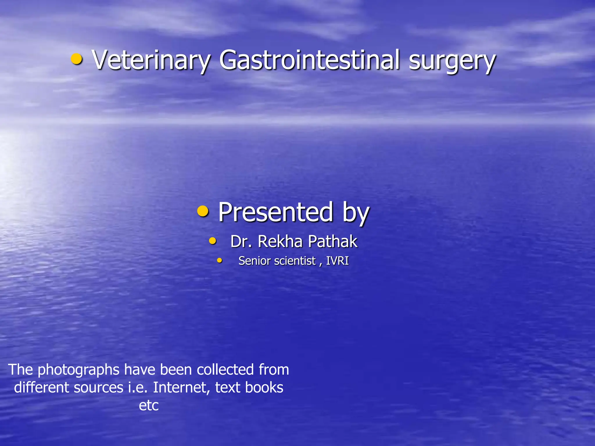
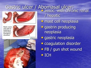
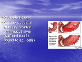
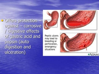
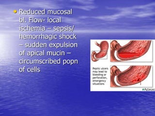
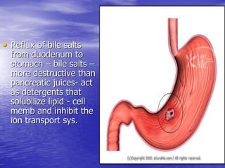
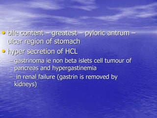
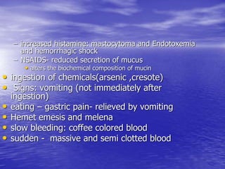
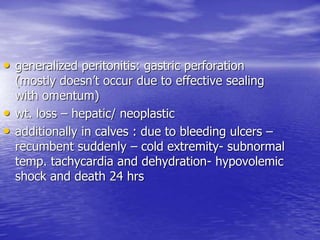
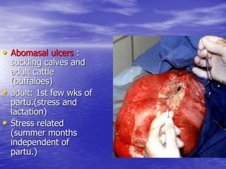
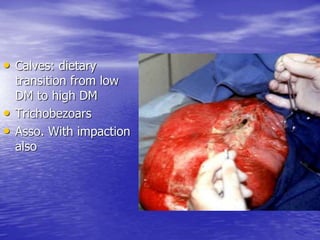
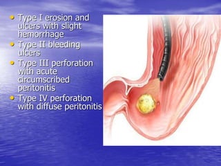
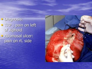
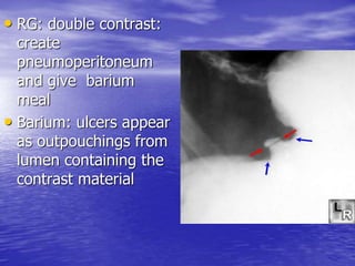
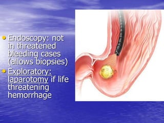
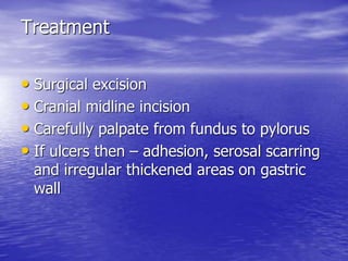
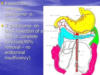
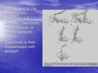
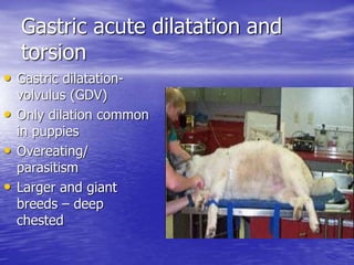
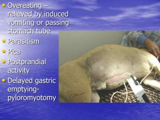
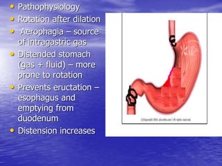
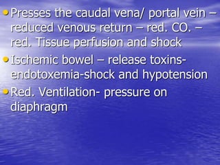
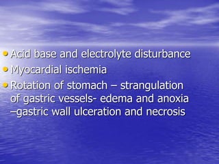
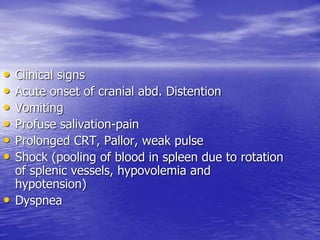
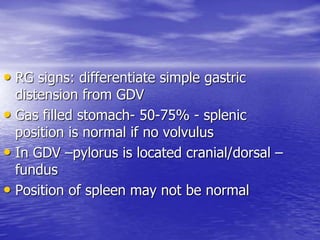
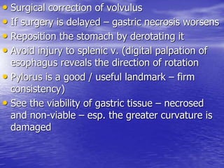
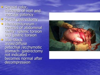
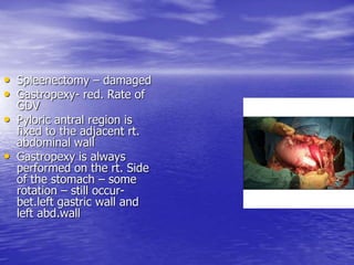
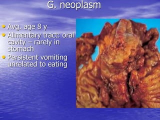
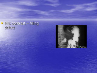
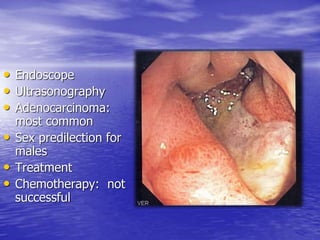
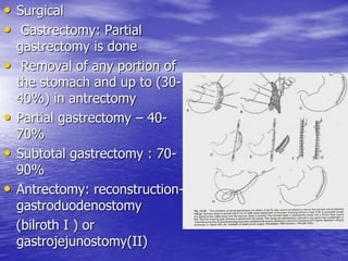
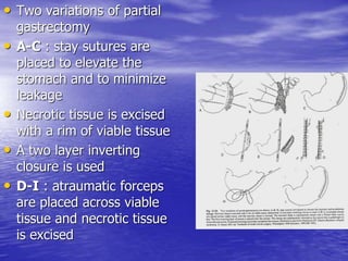
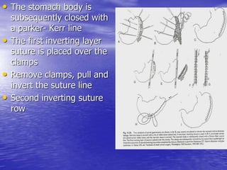
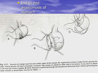
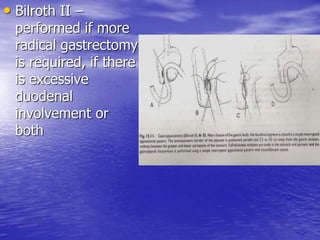
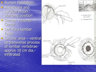
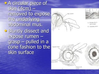
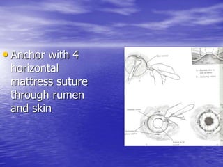
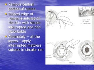
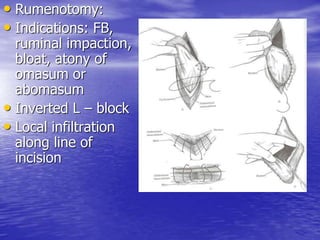
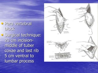
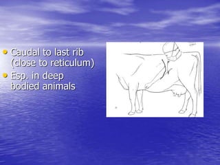
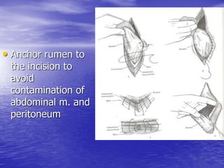
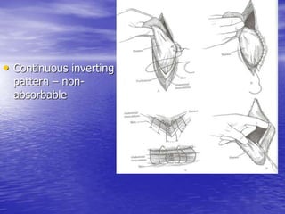
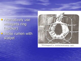
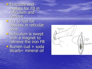
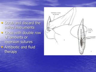
• Veterinary Gastrointestinal surgery
• Presented by
• Dr. Rekha Pathak
• Senior scientist , IVRI
The photographs have been collected from
different sources i.e. Internet, text books
etc
Gastric ulcer / Abomasal ulcer:
• assoc. with chronic renal
/ hepatic
• mast cell neoplasia
• gastrin producing
neoplasia
• gastric neoplasia
• coagulation disorder
• FB / gun shot wound
• ICH
• Uremia
• Poison
• Snakebite
• Primary ulcers are
less common
• Ulcers secondary –
common
• Aspirin:
experimentally to
produce ulcers
• Pathophysiology:
• Gastric/ duodenal
mucosa/ covered
with mucus layer
(sulfated mucin
bound to epi. cells)
• Offers protection –
against – corrosive
/ digestive effects
of gastric acid and
pepsin (auto
digestion and
ulceration)
• Reduced mucosal
bl. Flow- local
ischemia – sepsis/
hemorrhagic shock
– sudden expulsion
of apical mucin –
circumscribed popn
of cells
• Reflux of bile salts
from duodenum to
stomach – bile salts –
more destructive than
pancreatic juices- act
as detergents that
solubilize lipid - cell
memb and inhibit the
ion transport sys.
• bile content – greatest – pyloric antrum –
ulcer region of stomach
• hyper secretion of HCL
– gastrinoma ie non beta islets cell tumour of
pancreas and hypergastinemia
– in renal failure (gastrin is removed by
kidneys)
– increased histamine: mastocytoma and Endotoxemia
and hemorrhagic shock
– NSAIDS- reduced secretion of mucus
• alters the biochemical composition of mucin
• ingestion of chemicals(arsenic ,cresote)
• Signs: vomiting (not immediately after
ingestion)
• eating – gastric pain- relieved by vomiting
• Hemet emesis and melena
• slow bleeding: coffee colored blood
• sudden - massive and semi clotted blood
• generalized peritonitis: gastric perforation
(mostly doesn’t occur due to effective sealing
with omentum)
• wt. loss – hepatic/ neoplastic
• additionally in calves : due to bleeding ulcers –
recumbent suddenly – cold extremity- subnormal
temp. tachycardia and dehydration- hypovolemic
shock and death 24 hrs
• Abomasal ulcers :
suckling calves and
adult cattle
(buffaloes)
• adult: 1st few wks of
partu.(stress and
lactation)
• Stress related
(summer months
independent of
partu.)
• Calves: dietary
transition from low
DM to high DM
• Trichobezoars
• Asso. With impaction
also
• Type I erosion and
ulcers with slight
hemorrhage
• Type II bleeding
ulcers
• Type III perforation
with acute
circumscribed
peritonitis
• Type IV perforation
with diffuse peritonitis
• Diagnosis:
• TRP ; pain on left
of xiphoid
• Abomasal ulcer:
pain on rt. side
• RG: double contrast:
create
pneumoperitoneum
and give barium
meal
• Barium: ulcers appear
as outpouchings from
lumen containing the
contrast material
• Fluoroscopy: helps in variable
positioning and pin point the site
• Endoscopy: not
in threatened
bleeding cases
(allows biopsies)
• Exploratory:
laparotomy if life
threatening
hemorrhage
Treatment
• Surgical excision
• Cranial midline incision
• Carefully palpate from fundus to pylorus
• If ulcers then – adhesion, serosal scarring
and irregular thickened areas on gastric
wall
• Inspect the
pancreas-
gastrinoma- p.
nodules
• If gastrinoma- en
block resection of a
lobe or complete
pancreas(90%
removal – no
endocrinal
insufficiency)
• If no ulcers found
• Open stomach- find the bleeding site- also
on pyloric antrum(equidistant from lesser /
greater curvature)
• Extend to duodenum if necessary
• Small ulcers :
elliptical incision-
mucosa closed –
simple continuous
– 3/0 or 4/0
absorbable chromic
and interrupted
Lambert on serosa
and muscularis
• Multiple ulcers on
pyloric part –
bilroth I
gastrectomy
technique
• Bilroth technique I :
ligate the rt. Gastric
artery near pylorus on
the lesser curvature
• Rt. Gastroepiploic
vessels ligated
• Take care not to injure
the pancreas
• Pyloric and gastric
branches supplying the
area to be resected are
ligated
• 2 st. intestinal
clamps are placed
across the pyloric
antrum
• another 2 are
placed distal to the
pylorus and avoid
the common bile
duct.
• Excise the pyloric
sphincter and canal
• Gastric mucosa is
apposed with 3-0
synthetic
absorbable suture
in an Cushing
pattern starting
from the lesser
curvature and
continuing towards
the greater
curvature
• Equal in size to the
duodenal dia
• Apposed – 3-0 –
synthetic absorbable,
polypropylene, or
nylon – lamberts
pattern
• Duodenum is then
anastamosed with
stomach
Gastric acute dilatation and
torsion
• Gastric dilatation-
volvulus (GDV)
• Only dilation common
in puppies
• Overeating/
parasitism
• Larger and giant
breeds – deep
chested
• Overeating –
relieved by induced
vomiting or passing
stomach tube
• Parasitism
• Pica
• Postprandial
activity
• Delayed gastric
emptying-
pyloromyotomy
• Pathophysiology
• Rotation after dilation
• Aerophagia – source
of intragastric gas
• Distended stomach
(gas + fluid) – more
prone to rotation
• Prevents eructation –
esophagus and
emptying from
duodenum
• Distension increases
•Presses the caudal vena/ portal vein –
reduced venous return – red. CO. –
red. Tissue perfusion and shock
•Ischemic bowel – release toxins-
endotoxemia-shock and hypotension
•Red. Ventilation- pressure on
diaphragm
• Acid base and electrolyte disturbance
• Myocardial ischemia
• Rotation of stomach – strangulation
of gastric vessels- edema and anoxia
–gastric wall ulceration and necrosis
• Clinical signs
• Acute onset of cranial abd. Distention
• Vomiting
• Profuse salivation-pain
• Prolonged CRT, Pallor, weak pulse
• Shock (pooling of blood in spleen due to rotation
of splenic vessels, hypovolemia and
hypotension)
• Dyspnea
• RG signs: differentiate simple gastric
distension from GDV
• Gas filled stomach- 50-75% - splenic
position is normal if no volvulus
• In GDV –pylorus is located cranial/dorsal –
fundus
• Position of spleen may not be normal
• A tissue density line
dividing the gas filled
stomach into
compartments
• VD - pylorus is near
or near to the left of
the midline
• Gastric perforation-
pneumoperitoneum
• Clockwise 270
• Anticlockwise 90
• Preoperative care
• Gastric
decompression
• Needle
trocarization 18 G
needle
• Thrust on rt. Or
left wall – point of
greatest distension
• 2-3 needles – relieves
gas component of
distension
• Alternatively – if not
effectively reduced –
stomach
• Pass the s.tube
through mouth gag-
resistance is
encountered in gastro
esophageal junction –
rotate and advance
• Removal of
intragastric gas –
trocarization- corrects
the gastro esophageal
angle-allows passage
of S. tube
• Passage of st doesn’t
mean absence of g.
rotation
• Withdraw the tube
after decompression
• Sometimes for
decompression –
temporary Gastrotomy is
constructed
• Close the Gastrotomy
wound and proceed for
surgical correction of
rotation (Decompression
doesn’t always result in
normal gastric position)
• Shock therapy
• Surgical correction of volvulus
• If surgery is delayed – gastric necrosis worsens
• Reposition the stomach by derotating it
• Avoid injury to splenic v. (digital palpation of
esophagus reveals the direction of rotation
• Pylorus is a good / useful landmark – firm
consistency)
• See the viability of gastric tissue – necrosed
and non-viable – esp. the greater curvature is
damaged
• Serosal color,
thickness of wall and
vascular patency
• Partial gastrectomy
• Hemoperitoneum -
centesis of abdominal
cavity- splenic torsion
and gastric torsion
• Blue-black
areas/diffuse
petechial /ecchymotic
stomach- gastrectomy
not indicated –
becomes normal after
decompression
• Spleenectomy – damaged
• Gastropexy- red. Rate of
GDV
• Pyloric antral region is
fixed to the adjacent rt.
abdominal wall
• Gastropexy is always
performed on the rt. Side
of the stomach – some
rotation – still occur-
bet.left gastric wall and
left abd.wall
G. neoplasm
• Avg. age 8 y
• Alimentary tract: oral
cavity – rarely in
stomach
• Persistent vomiting
unrelated to eating
•Within the antrum on the lesser
curvature
•Metastasis: liver, lungs, spleen
•Leiomyoma/
rhabdomyosarcoma/polyps
(solitary or multiple)
• Polyps – due to
sharp fragment of
bones- resting for
long in antrum -
injure mucosa –
herniation of sub
mucosa
• Clinical signs;
anorexia
• Loss of wt.
Obstructing
gastric out flow
• Normal
peristalsis is
interfered
• Anemia
• Abd. Pain
• Emesis unrelated to
ingestion of food /water
• Melena
• palpation
• Exploratory laparotomy
• RG: contrast – filling
defect
• Endoscope
• Ultrasonography
• Adenocarcinoma:
most common
• Sex predilection for
males
• Treatment
• Chemotherapy: not
successful
• Surgical
• Gastrectomy: Partial
gastrectomy is done
• Removal of any portion of
the stomach and up to (30-
40%) in antrectomy
• Partial gastrectomy – 40-
70%
• Subtotal gastrectomy : 70-
90%
• Antrectomy: reconstruction-
gastroduodenostomy
(bilroth I ) or
gastrojejunostomy(II)
• Two variations of partial
gastrectomy
• A-C : stay sutures are
placed to elevate the
stomach and to minimize
leakage
• Necrotic tissue is excised
with a rim of viable tissue
• A two layer inverting
closure is used
• D-I : atraumatic forceps
are placed across viable
tissue and necrotic tissue
is excised
• The stomach body is
subsequently closed with
a parker- Kerr line
• The first inverting layer
suture is placed over the
clamps
• Remove clamps, pull and
invert the suture line
• Second inverting suture
row
• End to end
anastomosis of
stomach
• Bilroth II –
performed if more
radical gastrectomy
is required, if there
is excessive
duodenal
involvement or
both
Bloat
• Bloat : Major problems- GIT – cattle and
buffaloes
• Higher in buffaloes
• Acute/chronic
• Gaseous bloat – free gas - dorsal part of
rumen
• Frothy bloat – gas trapped with ingesta-
dispersed throughout the rumen content
• Acute: rapid feeding and sudden diet
change – large ruminants
• s. ruminants – large quantities of grain
ingestion/cereals
• More pressure on diaphragm –
hypoventilation and red. Venous return to
the heart
• signs: bulge on Para lumbar fossa
• Abdominal distension
• Cyanotic mm
• v. serious – lying down – asphyxiated –
open mouth- protruded tongue and
tachycardia
• Treatment: puncture wall – left side with
trocar and canula
• if frothy – antifoaming agents – turpentine
oil (80ml) + mustard oil (500-1000 ml)
• antifroth prepn. – bloatosil
• gives immediate relief to ailing animal
• avoid conc. – 2-3 days and leguminous
fodder
• Resort to rumenotomy / rumenostomy
• S. animal: IV- RL or oral soda bicarb
• Chronic bloat: TRP (FBS) – reticuloperitonitis/
fibrinous pneumonia – pleuritis involving the
vagus nerve
• Liver abscess, splenic cyst and abscess, enlarged
mediastinal lymph nodes, pyloric stenosis
• Rumen fistulation / rumenotomy can be done
• Rumen fistulation:
• Anesthesia and
surgical prepn.:
standing position
• Sternal recumb. –
Camel
• Left Para lumbar
fossa
• Circular area – ventral
to transverse process
of lumbar vertebrae-
approx 10 cm dia.-
infiltrated
• A circular piece of
skin (4cm) –
removed to expose
the underlying
abdominal mus.
• Bluntly dissect and
expose rumen –
grasp – pulled in a
cone fashion to the
skin surface
• Anchor with 4
horizontal
mattress suture
through rumen
and skin
• Remove central
portion of rumen
• Incised edge of
rumen is sutured to
the skin with simple
interrupted and non-
absorbable
• Alternately – all the
layers – apply
interrupted mattress
sutures in circular rim
• Rumenotomy:
• Indications: FB,
ruminal impaction,
bloat, atony of
omasum or
abomasum
• Inverted L – block
• Local infiltration
along line of
incision
• Para vertebral
block
• Surgical technique:
20 cm incision-
middle of tuber
coxae and last rib
5 cm ventral to
lumbar process
• Caudal to last rib
(close to reticulum)
• Esp. in deep
bodied animals
• Anchor rumen to
the incision to
avoid
contamination of
abdominal m. and
peritoneum
• Continuous inverting
pattern – non-
absorbable
• Alternatively use
weingarts ring
(quicker)
• Incise rumen with
scalpel
• Evacuate and
explore for FB in
reticulum and
remove
• Try to feel for
abscess in reticular
area
• Reticulum is swept
with a magnet to
retrieve the iron FB
• Rumen cud + soda
bicarb= mineral oil
• Scrub and discard the
soiled instruments
• Close with double row
of lamberts or
inversion sutures
• Antibiotic and fluid
therapy

dokumen.tips_veterinary-gastrointestinal-surgery.ppt

  • 1.
    • Veterinary Gastrointestinalsurgery • Presented by • Dr. Rekha Pathak • Senior scientist , IVRI The photographs have been collected from different sources i.e. Internet, text books etc
  • 2.
    Gastric ulcer /Abomasal ulcer: • assoc. with chronic renal / hepatic • mast cell neoplasia • gastrin producing neoplasia • gastric neoplasia • coagulation disorder • FB / gun shot wound • ICH
  • 3.
    • Uremia • Poison •Snakebite • Primary ulcers are less common • Ulcers secondary – common • Aspirin: experimentally to produce ulcers
  • 4.
    • Pathophysiology: • Gastric/duodenal mucosa/ covered with mucus layer (sulfated mucin bound to epi. cells)
  • 5.
    • Offers protection– against – corrosive / digestive effects of gastric acid and pepsin (auto digestion and ulceration)
  • 6.
    • Reduced mucosal bl.Flow- local ischemia – sepsis/ hemorrhagic shock – sudden expulsion of apical mucin – circumscribed popn of cells
  • 7.
    • Reflux ofbile salts from duodenum to stomach – bile salts – more destructive than pancreatic juices- act as detergents that solubilize lipid - cell memb and inhibit the ion transport sys.
  • 8.
    • bile content– greatest – pyloric antrum – ulcer region of stomach • hyper secretion of HCL – gastrinoma ie non beta islets cell tumour of pancreas and hypergastinemia – in renal failure (gastrin is removed by kidneys)
  • 9.
    – increased histamine:mastocytoma and Endotoxemia and hemorrhagic shock – NSAIDS- reduced secretion of mucus • alters the biochemical composition of mucin • ingestion of chemicals(arsenic ,cresote) • Signs: vomiting (not immediately after ingestion) • eating – gastric pain- relieved by vomiting • Hemet emesis and melena • slow bleeding: coffee colored blood • sudden - massive and semi clotted blood
  • 10.
    • generalized peritonitis:gastric perforation (mostly doesn’t occur due to effective sealing with omentum) • wt. loss – hepatic/ neoplastic • additionally in calves : due to bleeding ulcers – recumbent suddenly – cold extremity- subnormal temp. tachycardia and dehydration- hypovolemic shock and death 24 hrs
  • 11.
    • Abomasal ulcers: suckling calves and adult cattle (buffaloes) • adult: 1st few wks of partu.(stress and lactation) • Stress related (summer months independent of partu.)
  • 12.
    • Calves: dietary transitionfrom low DM to high DM • Trichobezoars • Asso. With impaction also
  • 13.
    • Type Ierosion and ulcers with slight hemorrhage • Type II bleeding ulcers • Type III perforation with acute circumscribed peritonitis • Type IV perforation with diffuse peritonitis
  • 14.
    • Diagnosis: • TRP; pain on left of xiphoid • Abomasal ulcer: pain on rt. side
  • 15.
    • RG: doublecontrast: create pneumoperitoneum and give barium meal • Barium: ulcers appear as outpouchings from lumen containing the contrast material
  • 16.
    • Fluoroscopy: helpsin variable positioning and pin point the site
  • 17.
    • Endoscopy: not inthreatened bleeding cases (allows biopsies) • Exploratory: laparotomy if life threatening hemorrhage
  • 18.
    Treatment • Surgical excision •Cranial midline incision • Carefully palpate from fundus to pylorus • If ulcers then – adhesion, serosal scarring and irregular thickened areas on gastric wall
  • 19.
    • Inspect the pancreas- gastrinoma-p. nodules • If gastrinoma- en block resection of a lobe or complete pancreas(90% removal – no endocrinal insufficiency)
  • 20.
    • If noulcers found • Open stomach- find the bleeding site- also on pyloric antrum(equidistant from lesser / greater curvature) • Extend to duodenum if necessary
  • 21.
    • Small ulcers: elliptical incision- mucosa closed – simple continuous – 3/0 or 4/0 absorbable chromic and interrupted Lambert on serosa and muscularis • Multiple ulcers on pyloric part – bilroth I gastrectomy technique
  • 22.
    • Bilroth techniqueI : ligate the rt. Gastric artery near pylorus on the lesser curvature • Rt. Gastroepiploic vessels ligated • Take care not to injure the pancreas • Pyloric and gastric branches supplying the area to be resected are ligated
  • 23.
    • 2 st.intestinal clamps are placed across the pyloric antrum • another 2 are placed distal to the pylorus and avoid the common bile duct. • Excise the pyloric sphincter and canal
  • 24.
    • Gastric mucosais apposed with 3-0 synthetic absorbable suture in an Cushing pattern starting from the lesser curvature and continuing towards the greater curvature
  • 25.
    • Equal insize to the duodenal dia • Apposed – 3-0 – synthetic absorbable, polypropylene, or nylon – lamberts pattern • Duodenum is then anastamosed with stomach
  • 26.
    Gastric acute dilatationand torsion • Gastric dilatation- volvulus (GDV) • Only dilation common in puppies • Overeating/ parasitism • Larger and giant breeds – deep chested
  • 27.
    • Overeating – relievedby induced vomiting or passing stomach tube • Parasitism • Pica • Postprandial activity • Delayed gastric emptying- pyloromyotomy
  • 28.
    • Pathophysiology • Rotationafter dilation • Aerophagia – source of intragastric gas • Distended stomach (gas + fluid) – more prone to rotation • Prevents eructation – esophagus and emptying from duodenum • Distension increases
  • 29.
    •Presses the caudalvena/ portal vein – reduced venous return – red. CO. – red. Tissue perfusion and shock •Ischemic bowel – release toxins- endotoxemia-shock and hypotension •Red. Ventilation- pressure on diaphragm
  • 30.
    • Acid baseand electrolyte disturbance • Myocardial ischemia • Rotation of stomach – strangulation of gastric vessels- edema and anoxia –gastric wall ulceration and necrosis
  • 31.
    • Clinical signs •Acute onset of cranial abd. Distention • Vomiting • Profuse salivation-pain • Prolonged CRT, Pallor, weak pulse • Shock (pooling of blood in spleen due to rotation of splenic vessels, hypovolemia and hypotension) • Dyspnea
  • 32.
    • RG signs:differentiate simple gastric distension from GDV • Gas filled stomach- 50-75% - splenic position is normal if no volvulus • In GDV –pylorus is located cranial/dorsal – fundus • Position of spleen may not be normal
  • 33.
    • A tissuedensity line dividing the gas filled stomach into compartments • VD - pylorus is near or near to the left of the midline • Gastric perforation- pneumoperitoneum • Clockwise 270 • Anticlockwise 90
  • 34.
    • Preoperative care •Gastric decompression • Needle trocarization 18 G needle • Thrust on rt. Or left wall – point of greatest distension
  • 35.
    • 2-3 needles– relieves gas component of distension • Alternatively – if not effectively reduced – stomach • Pass the s.tube through mouth gag- resistance is encountered in gastro esophageal junction – rotate and advance
  • 36.
    • Removal of intragastricgas – trocarization- corrects the gastro esophageal angle-allows passage of S. tube • Passage of st doesn’t mean absence of g. rotation • Withdraw the tube after decompression
  • 37.
    • Sometimes for decompression– temporary Gastrotomy is constructed • Close the Gastrotomy wound and proceed for surgical correction of rotation (Decompression doesn’t always result in normal gastric position) • Shock therapy
  • 38.
    • Surgical correctionof volvulus • If surgery is delayed – gastric necrosis worsens • Reposition the stomach by derotating it • Avoid injury to splenic v. (digital palpation of esophagus reveals the direction of rotation • Pylorus is a good / useful landmark – firm consistency) • See the viability of gastric tissue – necrosed and non-viable – esp. the greater curvature is damaged
  • 39.
    • Serosal color, thicknessof wall and vascular patency • Partial gastrectomy • Hemoperitoneum - centesis of abdominal cavity- splenic torsion and gastric torsion • Blue-black areas/diffuse petechial /ecchymotic stomach- gastrectomy not indicated – becomes normal after decompression
  • 40.
    • Spleenectomy –damaged • Gastropexy- red. Rate of GDV • Pyloric antral region is fixed to the adjacent rt. abdominal wall • Gastropexy is always performed on the rt. Side of the stomach – some rotation – still occur- bet.left gastric wall and left abd.wall
  • 41.
    G. neoplasm • Avg.age 8 y • Alimentary tract: oral cavity – rarely in stomach • Persistent vomiting unrelated to eating
  • 42.
    •Within the antrumon the lesser curvature •Metastasis: liver, lungs, spleen •Leiomyoma/ rhabdomyosarcoma/polyps (solitary or multiple)
  • 43.
    • Polyps –due to sharp fragment of bones- resting for long in antrum - injure mucosa – herniation of sub mucosa • Clinical signs; anorexia
  • 44.
    • Loss ofwt. Obstructing gastric out flow • Normal peristalsis is interfered • Anemia • Abd. Pain
  • 45.
    • Emesis unrelatedto ingestion of food /water • Melena • palpation • Exploratory laparotomy
  • 46.
    • RG: contrast– filling defect
  • 47.
    • Endoscope • Ultrasonography •Adenocarcinoma: most common • Sex predilection for males • Treatment • Chemotherapy: not successful
  • 48.
    • Surgical • Gastrectomy:Partial gastrectomy is done • Removal of any portion of the stomach and up to (30- 40%) in antrectomy • Partial gastrectomy – 40- 70% • Subtotal gastrectomy : 70- 90% • Antrectomy: reconstruction- gastroduodenostomy (bilroth I ) or gastrojejunostomy(II)
  • 49.
    • Two variationsof partial gastrectomy • A-C : stay sutures are placed to elevate the stomach and to minimize leakage • Necrotic tissue is excised with a rim of viable tissue • A two layer inverting closure is used • D-I : atraumatic forceps are placed across viable tissue and necrotic tissue is excised
  • 50.
    • The stomachbody is subsequently closed with a parker- Kerr line • The first inverting layer suture is placed over the clamps • Remove clamps, pull and invert the suture line • Second inverting suture row
  • 51.
    • End toend anastomosis of stomach
  • 52.
    • Bilroth II– performed if more radical gastrectomy is required, if there is excessive duodenal involvement or both
  • 53.
    Bloat • Bloat :Major problems- GIT – cattle and buffaloes • Higher in buffaloes • Acute/chronic • Gaseous bloat – free gas - dorsal part of rumen • Frothy bloat – gas trapped with ingesta- dispersed throughout the rumen content
  • 54.
    • Acute: rapidfeeding and sudden diet change – large ruminants • s. ruminants – large quantities of grain ingestion/cereals • More pressure on diaphragm – hypoventilation and red. Venous return to the heart
  • 55.
    • signs: bulgeon Para lumbar fossa • Abdominal distension • Cyanotic mm • v. serious – lying down – asphyxiated – open mouth- protruded tongue and tachycardia
  • 56.
    • Treatment: puncturewall – left side with trocar and canula • if frothy – antifoaming agents – turpentine oil (80ml) + mustard oil (500-1000 ml) • antifroth prepn. – bloatosil • gives immediate relief to ailing animal • avoid conc. – 2-3 days and leguminous fodder
  • 57.
    • Resort torumenotomy / rumenostomy • S. animal: IV- RL or oral soda bicarb • Chronic bloat: TRP (FBS) – reticuloperitonitis/ fibrinous pneumonia – pleuritis involving the vagus nerve • Liver abscess, splenic cyst and abscess, enlarged mediastinal lymph nodes, pyloric stenosis • Rumen fistulation / rumenotomy can be done
  • 58.
    • Rumen fistulation: •Anesthesia and surgical prepn.: standing position • Sternal recumb. – Camel • Left Para lumbar fossa • Circular area – ventral to transverse process of lumbar vertebrae- approx 10 cm dia.- infiltrated
  • 59.
    • A circularpiece of skin (4cm) – removed to expose the underlying abdominal mus. • Bluntly dissect and expose rumen – grasp – pulled in a cone fashion to the skin surface
  • 60.
    • Anchor with4 horizontal mattress suture through rumen and skin
  • 61.
    • Remove central portionof rumen • Incised edge of rumen is sutured to the skin with simple interrupted and non- absorbable • Alternately – all the layers – apply interrupted mattress sutures in circular rim
  • 62.
    • Rumenotomy: • Indications:FB, ruminal impaction, bloat, atony of omasum or abomasum • Inverted L – block • Local infiltration along line of incision
  • 63.
    • Para vertebral block •Surgical technique: 20 cm incision- middle of tuber coxae and last rib 5 cm ventral to lumbar process
  • 64.
    • Caudal tolast rib (close to reticulum) • Esp. in deep bodied animals
  • 65.
    • Anchor rumento the incision to avoid contamination of abdominal m. and peritoneum
  • 66.
  • 67.
    • Alternatively use weingartsring (quicker) • Incise rumen with scalpel
  • 68.
    • Evacuate and explorefor FB in reticulum and remove • Try to feel for abscess in reticular area • Reticulum is swept with a magnet to retrieve the iron FB • Rumen cud + soda bicarb= mineral oil
  • 69.
    • Scrub anddiscard the soiled instruments • Close with double row of lamberts or inversion sutures • Antibiotic and fluid therapy