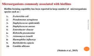
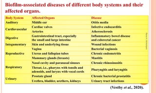
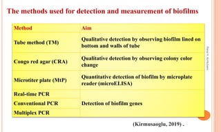
BIOFILM
preparation by :
Rana Hamad saleh
Rana
H.
Al-Hichami
 Introduction.
 Development of biofilm.
 Biofilm formation .
 What are the Factors affecting
biofilm formation?
 Why Biofilms Complicate
Infections ?
 Microorganisms commonly
associated with biofilms.
 Biofilm-associated diseases of
different body systems and their
affected organs.
 The methods used for detection and
measurement of biofilms.
 Antibiotic Tolerance and Resistance
in Biofilms and treatment .
 Reference .
Objectives
Rana
H.
Al-Hichami
 Bacterial growth is characterized by two life forms, one being as single cells
(planktonic) and the other being in sessile aggregates. The later is commonly
referred to as the biofilm mode of growth (Bjarnsholt et al., 2011).
 So, biofilm can be defined is an association of micro-organisms in which cells stick to
each other on a surface encased within matrix of extracellular polymeric
substance(EPS) produced by bacteria themselves (Muhsin et al., 2015) .
 The biofilm matrix is composed of cell wall teichoic acids, DNA, N-acetyl-
glucosamine and host products (Nandakumar et al., 2013) .
Introduction
Rana
H.
Al-Hichami
 Biofilms harbor considerable microbial diversity. Bacteria, archaea, algae,
fungi, protozoa and viruses all form important components of the biofilm
matrix and contribute to the biodiversity (Bjarnsholt et al., 2011).
 Biofilms are present everywhere in nature and can be found in industrial
places, hotels, waste water channels, bathrooms, labs, hospital settings and
commonly occur on hard surfaces submerged in or exposed to an aqueous
solution (Muhsin et al., 2015) .
 The ability of bacteria to adhere and colonize on to the surface is an important
feature in the pathogenesis of infection. (Nandakumar et al., 2013) .
Rana
H.
Al-Hichami
Development of biofilm
 The first description dates back to the 17th century, when Anton Von
Leeuwenhoek - the inventor of the Microscope, saw microbial aggregates (now
known to be Biofilms) on scrapings of plaque from his teeth (Chandki et al.,
2011) .
 In 1978 Costerton et al defined the term biofilm for the first time, and the
phenomenon was reviewed and described in 1987 by Costerton et al, as a
matrix-enclosed mode of growth (Bjarnsholt et al., 2011) .
Rana
H.
Al-Hichami
Biofilms formation
Biofilm formation on any surface involves mainly 3 stages. The first stage involve
attachment of free-floating microorganisms onto the surface, followed by secretion of
extracellular polymeric substance (EPS), rapid proliferation and aggregation of
bacteria in the slime layer. These aggregates further differentiate into characteristic
biofilms (Nandakumar et al., 2013) .
(Heidi, 2018) .
Rana
H.
Al-Hichami
What are the Factors affecting biofilm formation?
(Nandakumar et al., 2013)
Biofilm
Implant
Bacteria Host
• Surface
hydrophobicity
• Roughness
• Charge
• Porosity
• Surface chemistry
• Cell Receptor
• PH
• Proteins
• Platelets
• Cell surface
hydrophobicity
• Adhesins
• Quorum sensing
molecules
• Extracellular
polysscharride (EPS)
Rana
H.
Al-Hichami
Why Biofilms Complicate Infections ?
 Biofilms have serious medical implications. Often, microbes will accumulate on
damaged tissue such as heart valves or hard surfaces such as teeth, bacteria also
have an affinity for implanted medical devices such as catheters, gastrostomy
tubes, and urinary catheters, and readily form biofilms on these surfaces, Treating
these types of infections is extremely difficult due to antibiotics being unable to
penetrate the thick glycocalyx of the biofilm (Heidi, 2018) .
Rana
H.
Al-Hichami
Microorganisms commonly associated with biofilms
1- Escherichia coli
2- Pseudomonas aeruginosa
3- Staphylococcus epidermidis
4- Staphylococcus aureus
5- Enterobacter cloacae
6- Klebsiella pneumoniae
7- Actenomyces israelii
8- Haemophilus influenza
9- Burkholderia cepacia
10- Candida albicans
Biofilm forming capability has been reported in large number of microorganisms
species such as :
(Muhsin et al., 2015)
Rana
H.
Al-Hichami
Biofilm-associated diseases of different body systems and their
affected organs.
(Vestby et al., 2020).
Body System Affected Organs Disease
Auditory Middle ear Otitis media
Cardiovascular
Cardiac valves Infective endocarditis
Arteries Atherosclerosis
Digestive
Gastrointestinal tract, especially
the small and large intestine
Inflammatory bowel disease
and colorectal cancer
Integumentary Skin and underlying tissue Wound infections
Reproductive
Vagina Bacterial vaginosis
Uterus and fallopian tubes Chronic endometritis
Mammary glands (breasts) Mastitis
Respiratory
Nasal cavity and paranasal sinuses Chronic rhinosinusitis
Throat, i.e., pharynx with tonsils and
adenoids, and larynx with vocal cords
Pharyngitis and laryngitis
Urinary
Prostate gland Chronic bacterial prostatitis
Urethra, bladder, urethers, kidneys Urinary tract infections
Rana
H.
Al-Hichami
Method Aim
Tube method (TM)
Qualitative detection by observing biofilm lined on
bottom and walls of tube
Congo red agar (CRA)
Qualitative detection by observing colony color
change
Microtiter plate (MtP)
Quantitative detection of biofilm by microplate
reader (microELISA)
Real-time PCR
Detection of biofilm genes
Conventional PCR
Multiplex PCR
(Kirmusaoglu, 2019) .
The methods used for detection and measurement of biofilms
Rana
H.
Al-Hichami
 The resistance of bacteria against antibiotics and other components of
antimicrobial agents are supported by glycocalyx matrix. (Singh et al ., 2017) .
 When bacteria that survived antibiotic treatment of biofilms are susceptible to
the antibiotics during planktonic growth, the recalcitrance to antibiotic
treatment displayed by the biofilm is often referred to as tolerance (Bjarnsholt
et al., 2011) .
 The best possible treatment for biofilm-based infections is to inhibit the initial
attachment stage thus preventing the infection from starting (Khatoon et al.,
2018) .
Antibiotic tolerance and resistance in biofilms and treatment
Rana
H.
Al-Hichami
1. Bjarnsholt, T., Jensen, P.O., Moser, C. and Hoiby, N. eds., 2011. Biofilm
infections (Vol. 17). New York, NY: Springer.
2. Chandki, R., Banthia, P. and Banthia, R., 2011. Biofilms: A microbial home.
Journal of Indian Society of Periodontology, 15(2), p.111.
3. Heidi, S., 2018. Microbiology: a systems approach Fifth edition.
4. Khatoon, Z., McTiernan, C.D., Suuronen, E.J., Mah, T.F. and Alarcon, E.I., 2018.
Bacterial biofilm formation on implantable devices and approaches to its
treatment and prevention. Heliyon, 4(12), p.e01067.
5. Kırmusaoglu, S., 2019. The methods for detection of biofilm and screening
antibiofilm activity of agents. In Antimicrobials, Antibiotic Resistance, Antibiofilm
Strategies and Activity Methods. Intech Open.
Reference
Rana
H.
Al-Hichami
6. Muhsin, J., Ufaq, T., Tahir, H. and Saadia, A., 2015. Bacterial biofilm: its
composition, formation and role in human infections. J Microbiol Biotechnol, 4,
pp.1-14.
7. Nandakumar, V., Chittaranjan, S., Kurian, V.M. and Doble, M., 2013.
Characteristics of bacterial biofilm associated with implant material in clinical
practice. Polymer journal, 45(2), pp.137-152.
8. Singh, S., Singh, S.K., Chowdhury, I. and Singh, R., 2017. Understanding the
mechanism of bacterial biofilms resistance to antimicrobial agents. The open
microbiology journal, 11, p.53.
9. Vestby, L.K., Grønseth, T., Simm, R. and Nesse, L.L., 2020. Bacterial Biofilm and
its Role in the Pathogenesis of Disease. Antibiotics, 9(2), p.59.
Rana
H.
Al-Hichami
THANKS FOR LESSENING
Rana
H.
Al-Hichami

Biofilm

  • 1.
    BIOFILM preparation by : RanaHamad saleh Rana H. Al-Hichami
  • 2.
     Introduction.  Developmentof biofilm.  Biofilm formation .  What are the Factors affecting biofilm formation?  Why Biofilms Complicate Infections ?  Microorganisms commonly associated with biofilms.  Biofilm-associated diseases of different body systems and their affected organs.  The methods used for detection and measurement of biofilms.  Antibiotic Tolerance and Resistance in Biofilms and treatment .  Reference . Objectives Rana H. Al-Hichami
  • 3.
     Bacterial growthis characterized by two life forms, one being as single cells (planktonic) and the other being in sessile aggregates. The later is commonly referred to as the biofilm mode of growth (Bjarnsholt et al., 2011).  So, biofilm can be defined is an association of micro-organisms in which cells stick to each other on a surface encased within matrix of extracellular polymeric substance(EPS) produced by bacteria themselves (Muhsin et al., 2015) .  The biofilm matrix is composed of cell wall teichoic acids, DNA, N-acetyl- glucosamine and host products (Nandakumar et al., 2013) . Introduction Rana H. Al-Hichami
  • 4.
     Biofilms harborconsiderable microbial diversity. Bacteria, archaea, algae, fungi, protozoa and viruses all form important components of the biofilm matrix and contribute to the biodiversity (Bjarnsholt et al., 2011).  Biofilms are present everywhere in nature and can be found in industrial places, hotels, waste water channels, bathrooms, labs, hospital settings and commonly occur on hard surfaces submerged in or exposed to an aqueous solution (Muhsin et al., 2015) .  The ability of bacteria to adhere and colonize on to the surface is an important feature in the pathogenesis of infection. (Nandakumar et al., 2013) . Rana H. Al-Hichami
  • 5.
    Development of biofilm The first description dates back to the 17th century, when Anton Von Leeuwenhoek - the inventor of the Microscope, saw microbial aggregates (now known to be Biofilms) on scrapings of plaque from his teeth (Chandki et al., 2011) .  In 1978 Costerton et al defined the term biofilm for the first time, and the phenomenon was reviewed and described in 1987 by Costerton et al, as a matrix-enclosed mode of growth (Bjarnsholt et al., 2011) . Rana H. Al-Hichami
  • 6.
    Biofilms formation Biofilm formationon any surface involves mainly 3 stages. The first stage involve attachment of free-floating microorganisms onto the surface, followed by secretion of extracellular polymeric substance (EPS), rapid proliferation and aggregation of bacteria in the slime layer. These aggregates further differentiate into characteristic biofilms (Nandakumar et al., 2013) . (Heidi, 2018) . Rana H. Al-Hichami
  • 7.
    What are theFactors affecting biofilm formation? (Nandakumar et al., 2013) Biofilm Implant Bacteria Host • Surface hydrophobicity • Roughness • Charge • Porosity • Surface chemistry • Cell Receptor • PH • Proteins • Platelets • Cell surface hydrophobicity • Adhesins • Quorum sensing molecules • Extracellular polysscharride (EPS) Rana H. Al-Hichami
  • 8.
    Why Biofilms ComplicateInfections ?  Biofilms have serious medical implications. Often, microbes will accumulate on damaged tissue such as heart valves or hard surfaces such as teeth, bacteria also have an affinity for implanted medical devices such as catheters, gastrostomy tubes, and urinary catheters, and readily form biofilms on these surfaces, Treating these types of infections is extremely difficult due to antibiotics being unable to penetrate the thick glycocalyx of the biofilm (Heidi, 2018) . Rana H. Al-Hichami
  • 9.
    Microorganisms commonly associatedwith biofilms 1- Escherichia coli 2- Pseudomonas aeruginosa 3- Staphylococcus epidermidis 4- Staphylococcus aureus 5- Enterobacter cloacae 6- Klebsiella pneumoniae 7- Actenomyces israelii 8- Haemophilus influenza 9- Burkholderia cepacia 10- Candida albicans Biofilm forming capability has been reported in large number of microorganisms species such as : (Muhsin et al., 2015) Rana H. Al-Hichami
  • 10.
    Biofilm-associated diseases ofdifferent body systems and their affected organs. (Vestby et al., 2020). Body System Affected Organs Disease Auditory Middle ear Otitis media Cardiovascular Cardiac valves Infective endocarditis Arteries Atherosclerosis Digestive Gastrointestinal tract, especially the small and large intestine Inflammatory bowel disease and colorectal cancer Integumentary Skin and underlying tissue Wound infections Reproductive Vagina Bacterial vaginosis Uterus and fallopian tubes Chronic endometritis Mammary glands (breasts) Mastitis Respiratory Nasal cavity and paranasal sinuses Chronic rhinosinusitis Throat, i.e., pharynx with tonsils and adenoids, and larynx with vocal cords Pharyngitis and laryngitis Urinary Prostate gland Chronic bacterial prostatitis Urethra, bladder, urethers, kidneys Urinary tract infections Rana H. Al-Hichami
  • 11.
    Method Aim Tube method(TM) Qualitative detection by observing biofilm lined on bottom and walls of tube Congo red agar (CRA) Qualitative detection by observing colony color change Microtiter plate (MtP) Quantitative detection of biofilm by microplate reader (microELISA) Real-time PCR Detection of biofilm genes Conventional PCR Multiplex PCR (Kirmusaoglu, 2019) . The methods used for detection and measurement of biofilms Rana H. Al-Hichami
  • 12.
     The resistanceof bacteria against antibiotics and other components of antimicrobial agents are supported by glycocalyx matrix. (Singh et al ., 2017) .  When bacteria that survived antibiotic treatment of biofilms are susceptible to the antibiotics during planktonic growth, the recalcitrance to antibiotic treatment displayed by the biofilm is often referred to as tolerance (Bjarnsholt et al., 2011) .  The best possible treatment for biofilm-based infections is to inhibit the initial attachment stage thus preventing the infection from starting (Khatoon et al., 2018) . Antibiotic tolerance and resistance in biofilms and treatment Rana H. Al-Hichami
  • 13.
    1. Bjarnsholt, T.,Jensen, P.O., Moser, C. and Hoiby, N. eds., 2011. Biofilm infections (Vol. 17). New York, NY: Springer. 2. Chandki, R., Banthia, P. and Banthia, R., 2011. Biofilms: A microbial home. Journal of Indian Society of Periodontology, 15(2), p.111. 3. Heidi, S., 2018. Microbiology: a systems approach Fifth edition. 4. Khatoon, Z., McTiernan, C.D., Suuronen, E.J., Mah, T.F. and Alarcon, E.I., 2018. Bacterial biofilm formation on implantable devices and approaches to its treatment and prevention. Heliyon, 4(12), p.e01067. 5. Kırmusaoglu, S., 2019. The methods for detection of biofilm and screening antibiofilm activity of agents. In Antimicrobials, Antibiotic Resistance, Antibiofilm Strategies and Activity Methods. Intech Open. Reference Rana H. Al-Hichami
  • 14.
    6. Muhsin, J.,Ufaq, T., Tahir, H. and Saadia, A., 2015. Bacterial biofilm: its composition, formation and role in human infections. J Microbiol Biotechnol, 4, pp.1-14. 7. Nandakumar, V., Chittaranjan, S., Kurian, V.M. and Doble, M., 2013. Characteristics of bacterial biofilm associated with implant material in clinical practice. Polymer journal, 45(2), pp.137-152. 8. Singh, S., Singh, S.K., Chowdhury, I. and Singh, R., 2017. Understanding the mechanism of bacterial biofilms resistance to antimicrobial agents. The open microbiology journal, 11, p.53. 9. Vestby, L.K., Grønseth, T., Simm, R. and Nesse, L.L., 2020. Bacterial Biofilm and its Role in the Pathogenesis of Disease. Antibiotics, 9(2), p.59. Rana H. Al-Hichami
  • 15.