Full Set of Slides.ppt
- 1. Maslak, P. ASH Image Bank 2008;2008:8-00044. Copyright ©2008 American Society of Hematology.
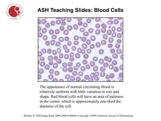
ASH Teaching Slides: Blood Cells
The appearance of normal circulating blood is
relatively uniform with little variation in size and
shape. Red blood cells will have an area of paleness
in the center, which is approximately one-third the
diameter of the cell.
- 2. Maslak, P. ASH Image Bank 2008;2008:8-00067. Copyright ©2008 American Society of Hematology.
ASH Teaching Slides: Blood Cells
Normal blood smear. The four larger cells shown are called
granulocytes, a type of white blood cell.
- 3. ASH Teaching Slides: Blood Cells
Schrier, S. ASH Image Bank 202;2002:100345. Copyright ©2002 American Society of Hematology.
Iron-deficiency anemia is indicated by red blood cells that are
paler and of a smaller size than normal.
- 4. Schrier, S. ASH Image Bank 2001;2001:100248. Copyright ©2001 American Society of Hematology.
ASH Teaching Slides: Blood Cells
Blood smear; arrows indicate sickled cells.
- 5. Maslak, P. ASH Image Bank 2001;2001:100202. Copyright ©2001 American Society of Hematology.
ASH Teaching Slides: Blood Cells
Chronic myelogenous leukemia. The blood smear shows an
increased number of neutrophils, a type of white blood cell.
- 6. Maslak, P. ASH Image Bank 2007;2007:7-00011. Copyright ©2007 American Society of Hematology.
ASH Teaching Slides: Blood Cells
Hairy cell leukemia. The characteristic cell of this type
of leukemia has projections uniformly distributed around
its border that give it a hairy appearance.
- 7. Kadin, M. ASH Image Bank 2002;2002:100484. Copyright ©2002 American Society of Hematology.
ASH Teaching Slides: Blood Cells
Hodgkin lymphoma. The large cells with an owl-like
appearance are called Reed-Sternberg cells and are a sign of
Hodgkin lymphoma.