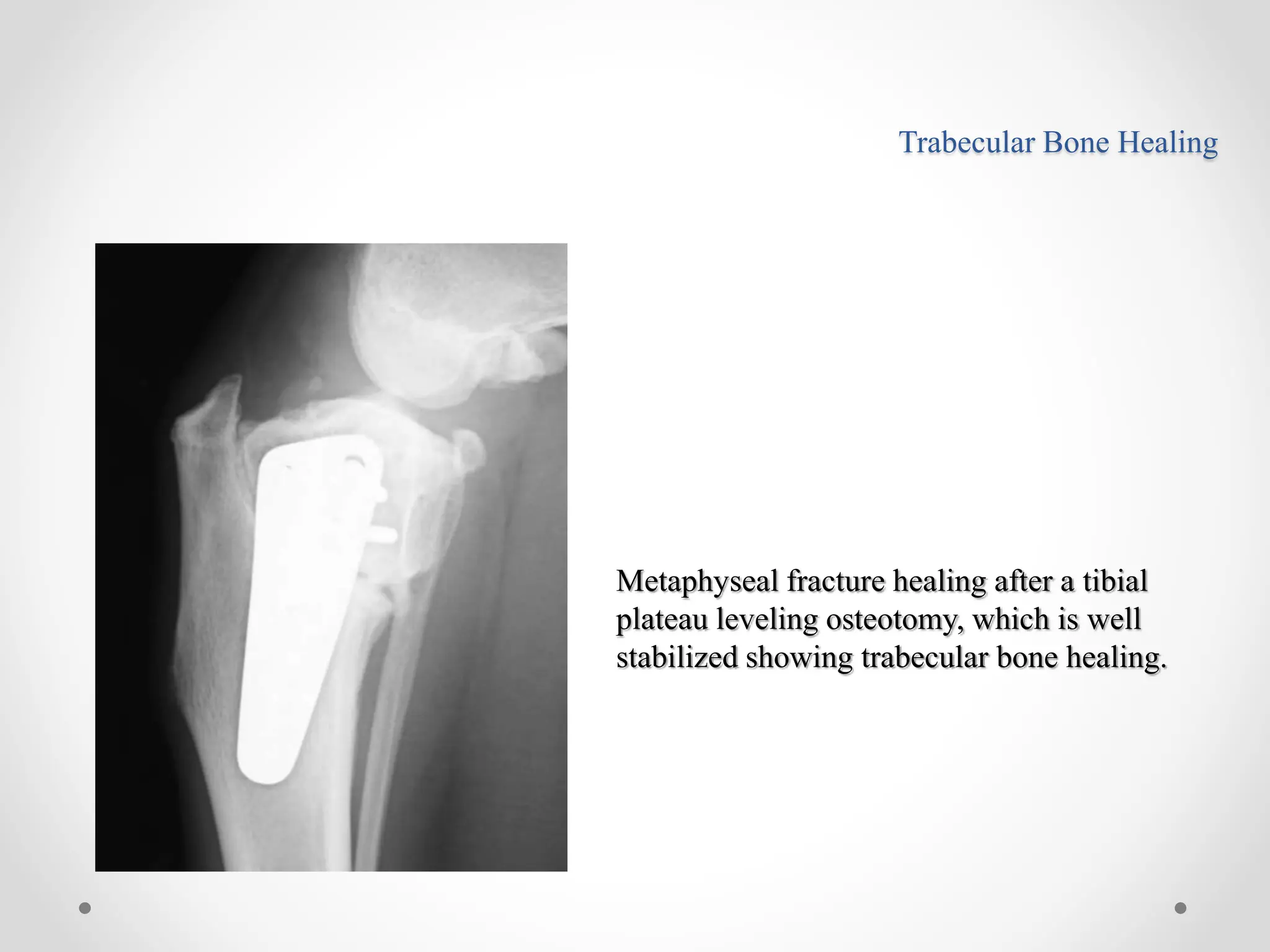
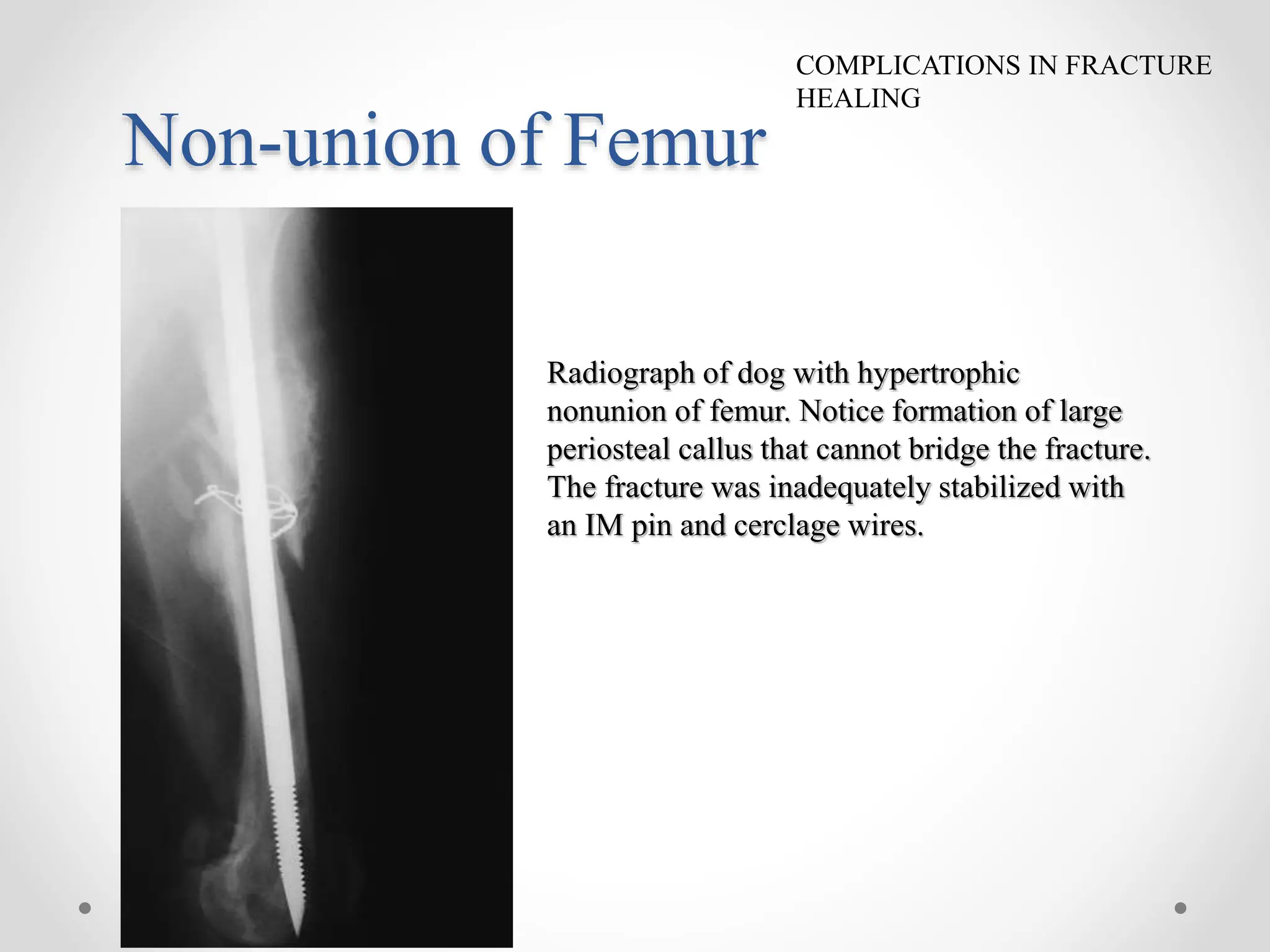
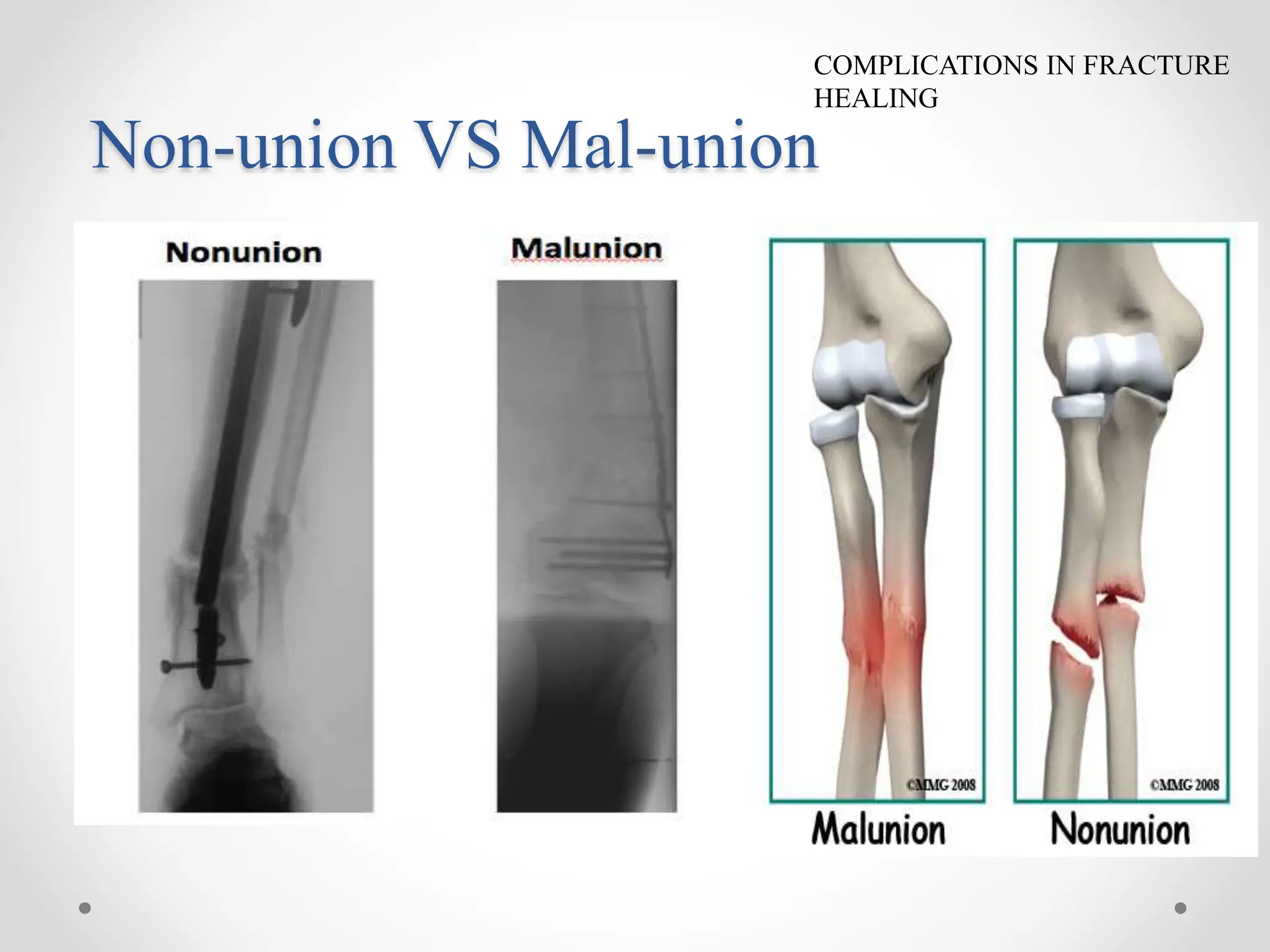
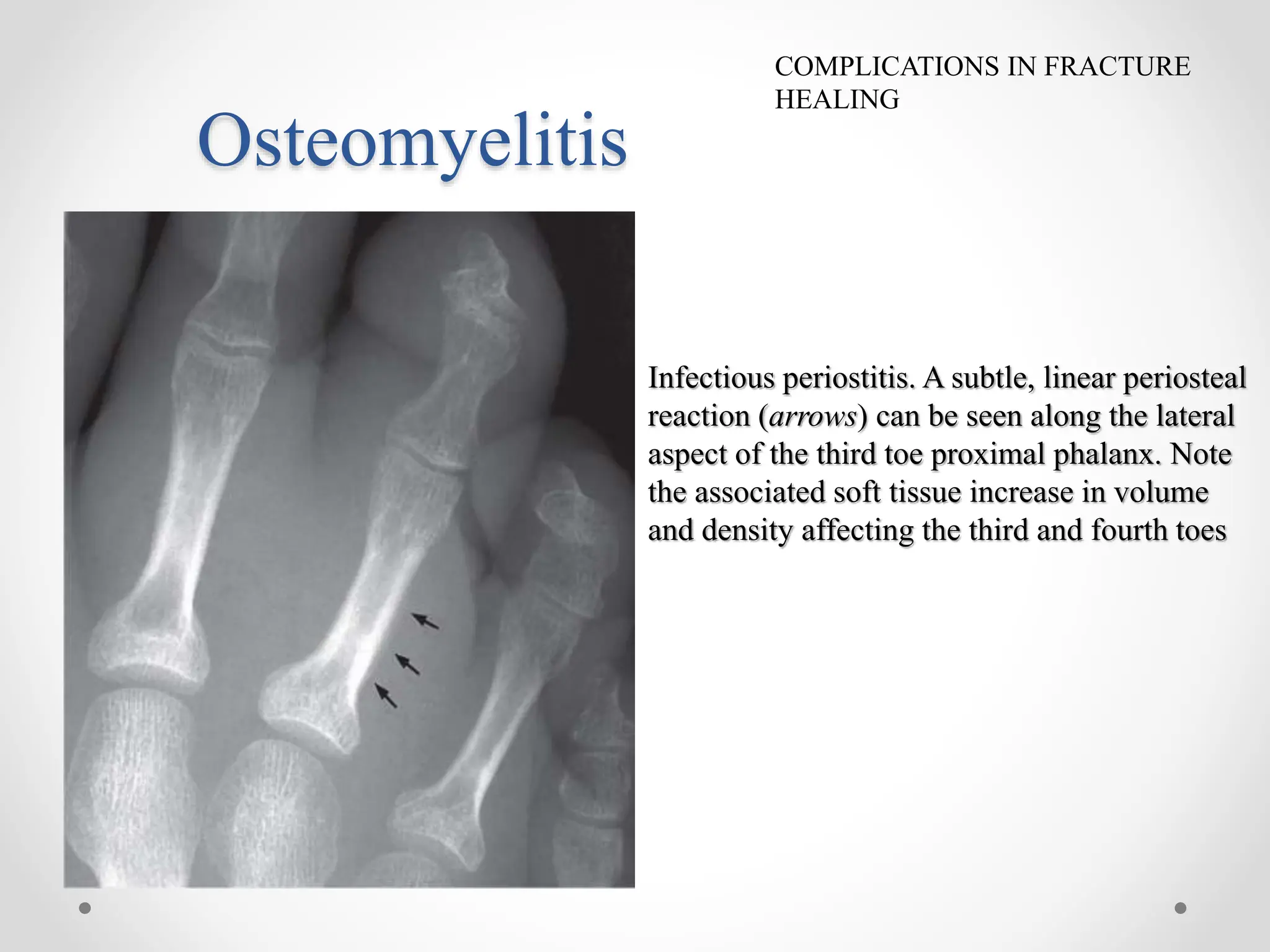
Fracture Healing, Complications
& factors promotes the fracture
healing
Presented By:
Surgery Group 2
Presented To:
Dr.Asad Manzoor
 Fracture healing
 Goals of fracture healing
Encourage Healing
Restore function
Cosmetically acceptable appearance
 Factors influence the fracture healing
Biologic Events
Mechanical Events
Blood supply of healing long bones
Blood supply to bone. A, Normal bone.
B, Immature bone. C, Fractured bone
(extraosseous blood
supply). D, Healing bone.
Effect of different fixation devices on circulation to fracture
bone
Fracture Healing
Phases of fracture healing
 Reactive phase
Inflammatory phase
Granulation tissue formation
 Reparative phase
Cartilage callus formation
Lamellar bone deposition
Trabecular bone formation
 Remodelling phase
Trabecular bone  compact bone
Time Distribution of different
phases of fracture healing
Inflammatory phase
Hematoma formation soon after the fracture
Reparative phase
Reparative + Remodeling phase
Collagen & muco-polyscharides level
during the fracture healing
Indirect bone healing
 Hematoma & granulation tissue formation
 Granulation tissue replaced by fibrous tissue & fibrocartilage
 Local resorption of mineralized tissue
 Vascularization of resorption cavity
 Formation of lamellar bone in this cavity
 Remodeling
Indirect bone healing
Indirect bone healing A-Bone resorption B-Periosteal callus formation C-
Mineralization of fibrocartilage D-Bone remodeling
Direct bone healing
 Fracture having small gaps (150-300µm) between fractured
ends
 Gaps filled with the network of fibrous tissue
 Remodeling of bone union within 7-8 weeks + longitudinal
reconstruction of fracture site
 Radiographicaly slowly increasing density of fracture line
without bridging periosteal & endosteal callus
Direct Bone Healing
Direct bone healing
Simultaneous union and reconstruction(A, B)
Gap fills with fibrous bone (C)Haversian
remodeling(D).
Direct Bone Healing
Fracture healing in a transverse
radial fracture in mature dog treated with bone plate.
Intra-membranous Bone Healing
 Direct differentiation of mesenchymal cells in to osteoblasts
 Bone bridging between comminuted bone fragments after
biologic fixation technique
 Periosteal callus is smaller in comminuted fracture
 Resorption of woven bone & formation of lamellar bone
 Remodeling
Intra-membranous Bone Healing
A, Comminuted fractures fixed with
biologic techniques of indirect
reduction, major segment alignment,
and optimal stabilization appear to heal
with a combination
of direct differentiation of
mesenchymal cells to osteoblasts
and endochondral ossification. B, The
fracture site fills with endosteal and
bridging callus. C, Resorption of
woven bone and formation of lamellar
bone at the fracture sites result in
remodeling of bony callus to cortical
bone.
Intra-membranous Bone Healing
A, Fracture healing in comminuted nonreducible radial fracture treated
with closed reduction and external fixation. B and C,
Radiographically endosteal bone appears first, followed by bone
bridging between fragments with minimal periosteal callus formation.
D, Bone remodeling occurs after the fracture lines are bridged.
Trabecular Bone Healing
 Increase osteoblastic activity on either side of fracture
 New bone deposit on existing trabeculae
 Fracture gap filled with woven bone
 Radiographicaly formation of one or two dense band at
fracture site
Trabecular Bone Healing
Metaphyseal fracture healing after a tibial
plateau leveling osteotomy, which is well
stabilized showing trabecular bone healing.
COMPLICATIONS IN FRACTURE
HEALING
 Delayed union (healing slower than normal)
 Non-union (arrested fracture repair process)
 Osteomyelitis (inflammatory condition of bone & medullary
canal)
 Mal-union (anatomic bone alignment not achieved)
Delayed Union
Delayed healing after 6 weeks of
bone marrow injection
COMPLICATIONS IN FRACTURE
HEALING
Non-union of Femur
Radiograph of dog with hypertrophic
nonunion of femur. Notice formation of large
periosteal callus that cannot bridge the fracture.
The fracture was inadequately stabilized with
an IM pin and cerclage wires.
COMPLICATIONS IN FRACTURE
HEALING
Non-union VS Mal-union
COMPLICATIONS IN FRACTURE
HEALING
Osteomyelitis
Infectious periostitis. A subtle, linear periosteal
reaction (arrows) can be seen along the lateral
aspect of the third toe proximal phalanx. Note
the associated soft tissue increase in volume
and density affecting the third and fourth toes
COMPLICATIONS IN FRACTURE
HEALING
Factors promotes the fracture healing
The factors that promotes fracture healing are:
 Growth hormones
 Thyroid hormone
 Calcitonin
 Insulin
 Vitamin K
 Vitamin C
 Vitamin D
 Immobilization
Factors promotes the fracture healing
 Anabolic steroids
 Young age
 Nutrition status
 Cerament
 Electric current
 Physical exercise
 Minerals
 High protein diet
Suggested Reading
 Griffon DJ: Fracture healing. In Johnson AL, Houlton
JEF, Vannini R, editors: AO principles of fracture
management in the dog and cat, Thieme, NY, 2005, AO
Publishing, pp. 73-97.
 Johnson AL, Egger EL, Eurell JC, et al: Biomechanics
and biology of fracture healing with external skeletal
fixation, Compend Cont Educ Pract Vet 20:487, 1998.
 Wilson JW: Blood supply to developing, mature and
healing bone. In Sumner-Smith G, (editor): Bone in
clinical orthopedics, ed 2, Thieme, New York NY, 2002,
AO Publishing, p. 23.
Suggested Reading
 Budsberg SC: Osteomyelitis, In Johnson AL, Houlton
JEF, Vannini R, editors: AO principles of fracture
management in the dog and cat, Thieme NY, 2005, AO
Publishing.
 Johnson AL: Corrective osteotomies, In Johnson AL,
Houlton JEF, and Vannini R, editors: AO principles of
fracture management in the dog and cat, Thieme, NY,
2005, AO Publishing.
Any question??????
Topic for Performance:
Pinning of long bone

Fracture Healing, Complications & factors promotes the fracture healing.pptx

  • 1.
    Fracture Healing, Complications &factors promotes the fracture healing Presented By: Surgery Group 2 Presented To: Dr.Asad Manzoor
  • 2.
     Fracture healing Goals of fracture healing Encourage Healing Restore function Cosmetically acceptable appearance  Factors influence the fracture healing Biologic Events Mechanical Events
  • 3.
    Blood supply ofhealing long bones Blood supply to bone. A, Normal bone. B, Immature bone. C, Fractured bone (extraosseous blood supply). D, Healing bone.
  • 4.
    Effect of differentfixation devices on circulation to fracture bone
  • 5.
  • 6.
    Phases of fracturehealing  Reactive phase Inflammatory phase Granulation tissue formation  Reparative phase Cartilage callus formation Lamellar bone deposition Trabecular bone formation  Remodelling phase Trabecular bone  compact bone
  • 7.
    Time Distribution ofdifferent phases of fracture healing
  • 8.
  • 9.
  • 10.
  • 11.
    Collagen & muco-polyscharideslevel during the fracture healing
  • 12.
    Indirect bone healing Hematoma & granulation tissue formation  Granulation tissue replaced by fibrous tissue & fibrocartilage  Local resorption of mineralized tissue  Vascularization of resorption cavity  Formation of lamellar bone in this cavity  Remodeling
  • 13.
  • 14.
    Indirect bone healing Indirectbone healing A-Bone resorption B-Periosteal callus formation C- Mineralization of fibrocartilage D-Bone remodeling
  • 15.
    Direct bone healing Fracture having small gaps (150-300µm) between fractured ends  Gaps filled with the network of fibrous tissue  Remodeling of bone union within 7-8 weeks + longitudinal reconstruction of fracture site  Radiographicaly slowly increasing density of fracture line without bridging periosteal & endosteal callus
  • 16.
    Direct Bone Healing Directbone healing Simultaneous union and reconstruction(A, B) Gap fills with fibrous bone (C)Haversian remodeling(D).
  • 17.
    Direct Bone Healing Fracturehealing in a transverse radial fracture in mature dog treated with bone plate.
  • 18.
    Intra-membranous Bone Healing Direct differentiation of mesenchymal cells in to osteoblasts  Bone bridging between comminuted bone fragments after biologic fixation technique  Periosteal callus is smaller in comminuted fracture  Resorption of woven bone & formation of lamellar bone  Remodeling
  • 19.
    Intra-membranous Bone Healing A,Comminuted fractures fixed with biologic techniques of indirect reduction, major segment alignment, and optimal stabilization appear to heal with a combination of direct differentiation of mesenchymal cells to osteoblasts and endochondral ossification. B, The fracture site fills with endosteal and bridging callus. C, Resorption of woven bone and formation of lamellar bone at the fracture sites result in remodeling of bony callus to cortical bone.
  • 20.
    Intra-membranous Bone Healing A,Fracture healing in comminuted nonreducible radial fracture treated with closed reduction and external fixation. B and C, Radiographically endosteal bone appears first, followed by bone bridging between fragments with minimal periosteal callus formation. D, Bone remodeling occurs after the fracture lines are bridged.
  • 21.
    Trabecular Bone Healing Increase osteoblastic activity on either side of fracture  New bone deposit on existing trabeculae  Fracture gap filled with woven bone  Radiographicaly formation of one or two dense band at fracture site
  • 22.
    Trabecular Bone Healing Metaphysealfracture healing after a tibial plateau leveling osteotomy, which is well stabilized showing trabecular bone healing.
  • 23.
    COMPLICATIONS IN FRACTURE HEALING Delayed union (healing slower than normal)  Non-union (arrested fracture repair process)  Osteomyelitis (inflammatory condition of bone & medullary canal)  Mal-union (anatomic bone alignment not achieved)
  • 24.
    Delayed Union Delayed healingafter 6 weeks of bone marrow injection COMPLICATIONS IN FRACTURE HEALING
  • 25.
    Non-union of Femur Radiographof dog with hypertrophic nonunion of femur. Notice formation of large periosteal callus that cannot bridge the fracture. The fracture was inadequately stabilized with an IM pin and cerclage wires. COMPLICATIONS IN FRACTURE HEALING
  • 26.
  • 27.
    Osteomyelitis Infectious periostitis. Asubtle, linear periosteal reaction (arrows) can be seen along the lateral aspect of the third toe proximal phalanx. Note the associated soft tissue increase in volume and density affecting the third and fourth toes COMPLICATIONS IN FRACTURE HEALING
  • 28.
    Factors promotes thefracture healing The factors that promotes fracture healing are:  Growth hormones  Thyroid hormone  Calcitonin  Insulin  Vitamin K  Vitamin C  Vitamin D  Immobilization
  • 29.
    Factors promotes thefracture healing  Anabolic steroids  Young age  Nutrition status  Cerament  Electric current  Physical exercise  Minerals  High protein diet
  • 30.
    Suggested Reading  GriffonDJ: Fracture healing. In Johnson AL, Houlton JEF, Vannini R, editors: AO principles of fracture management in the dog and cat, Thieme, NY, 2005, AO Publishing, pp. 73-97.  Johnson AL, Egger EL, Eurell JC, et al: Biomechanics and biology of fracture healing with external skeletal fixation, Compend Cont Educ Pract Vet 20:487, 1998.  Wilson JW: Blood supply to developing, mature and healing bone. In Sumner-Smith G, (editor): Bone in clinical orthopedics, ed 2, Thieme, New York NY, 2002, AO Publishing, p. 23.
  • 31.
    Suggested Reading  BudsbergSC: Osteomyelitis, In Johnson AL, Houlton JEF, Vannini R, editors: AO principles of fracture management in the dog and cat, Thieme NY, 2005, AO Publishing.  Johnson AL: Corrective osteotomies, In Johnson AL, Houlton JEF, and Vannini R, editors: AO principles of fracture management in the dog and cat, Thieme, NY, 2005, AO Publishing.
  • 32.
  • 33.