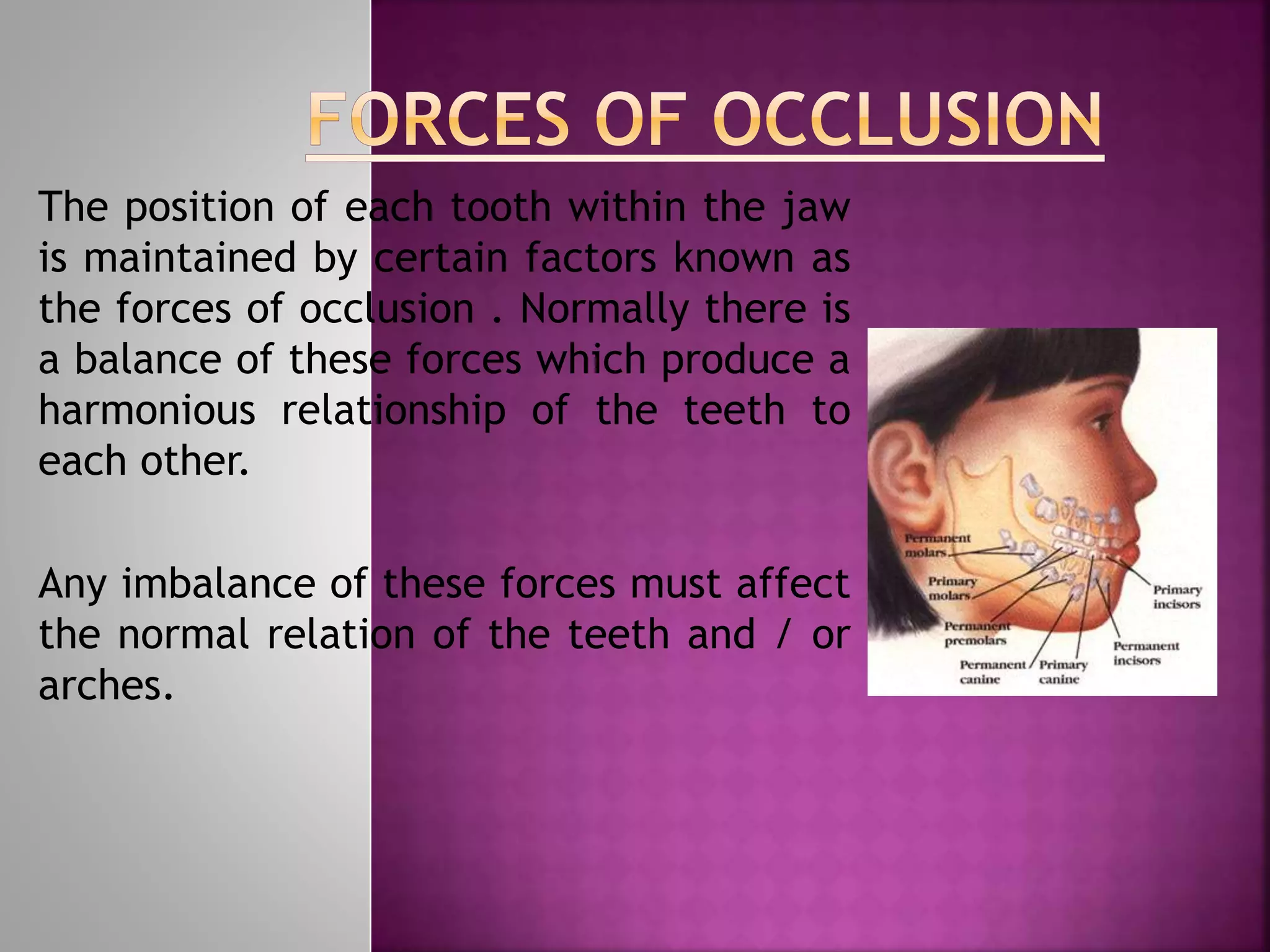
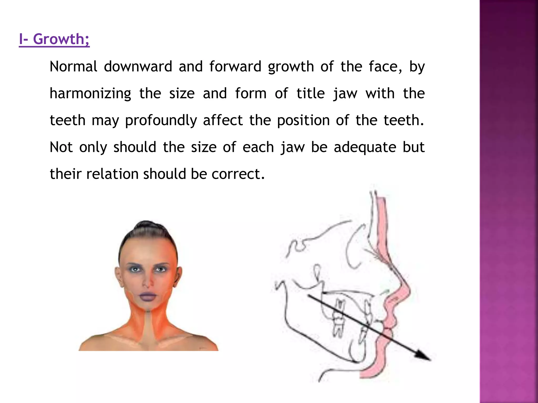
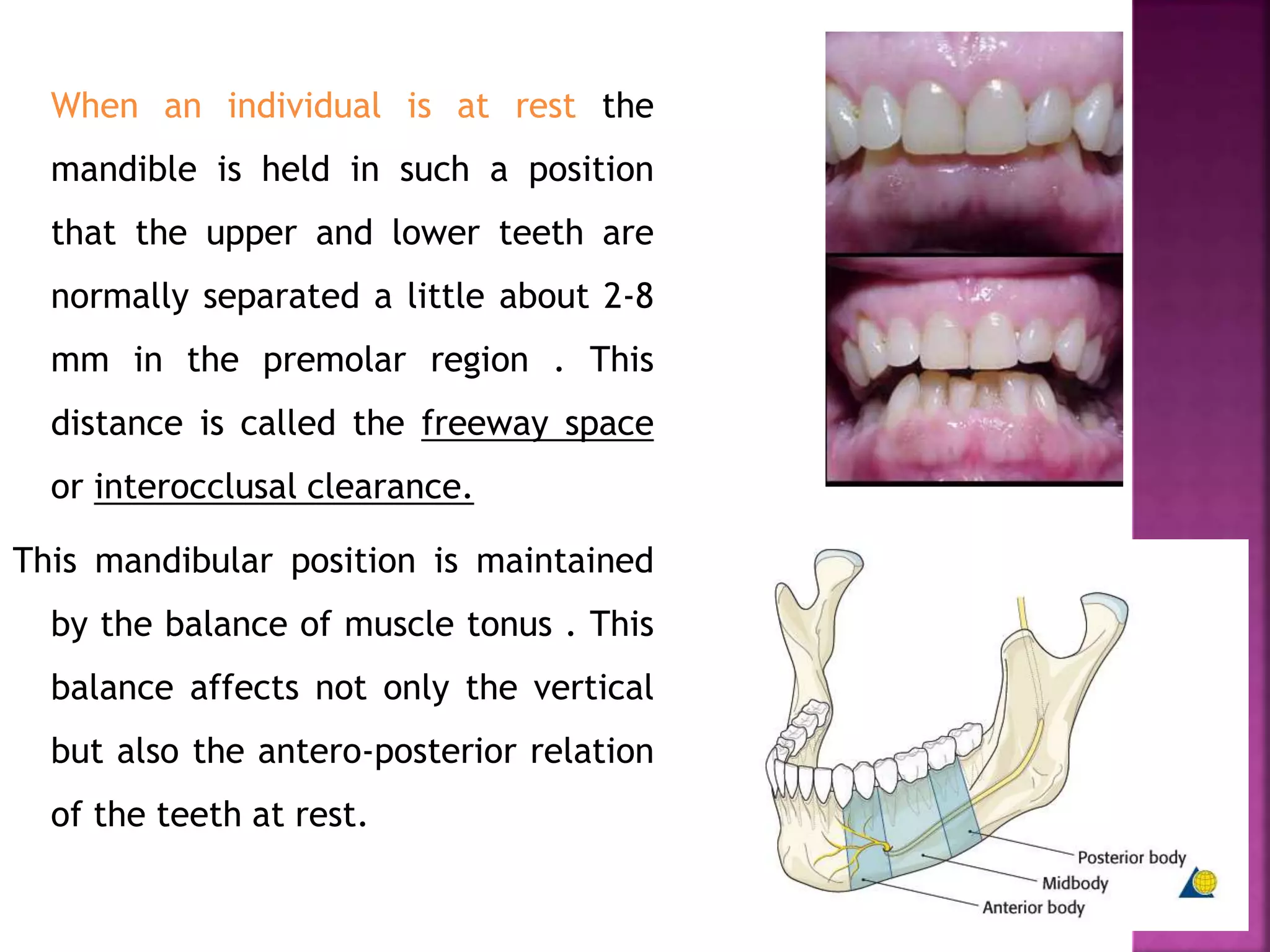
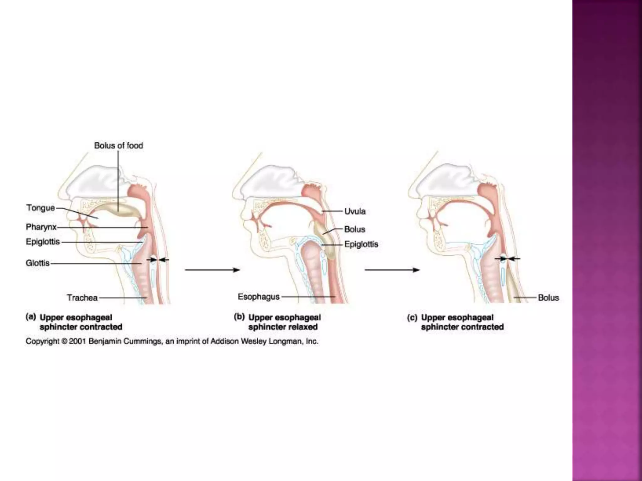
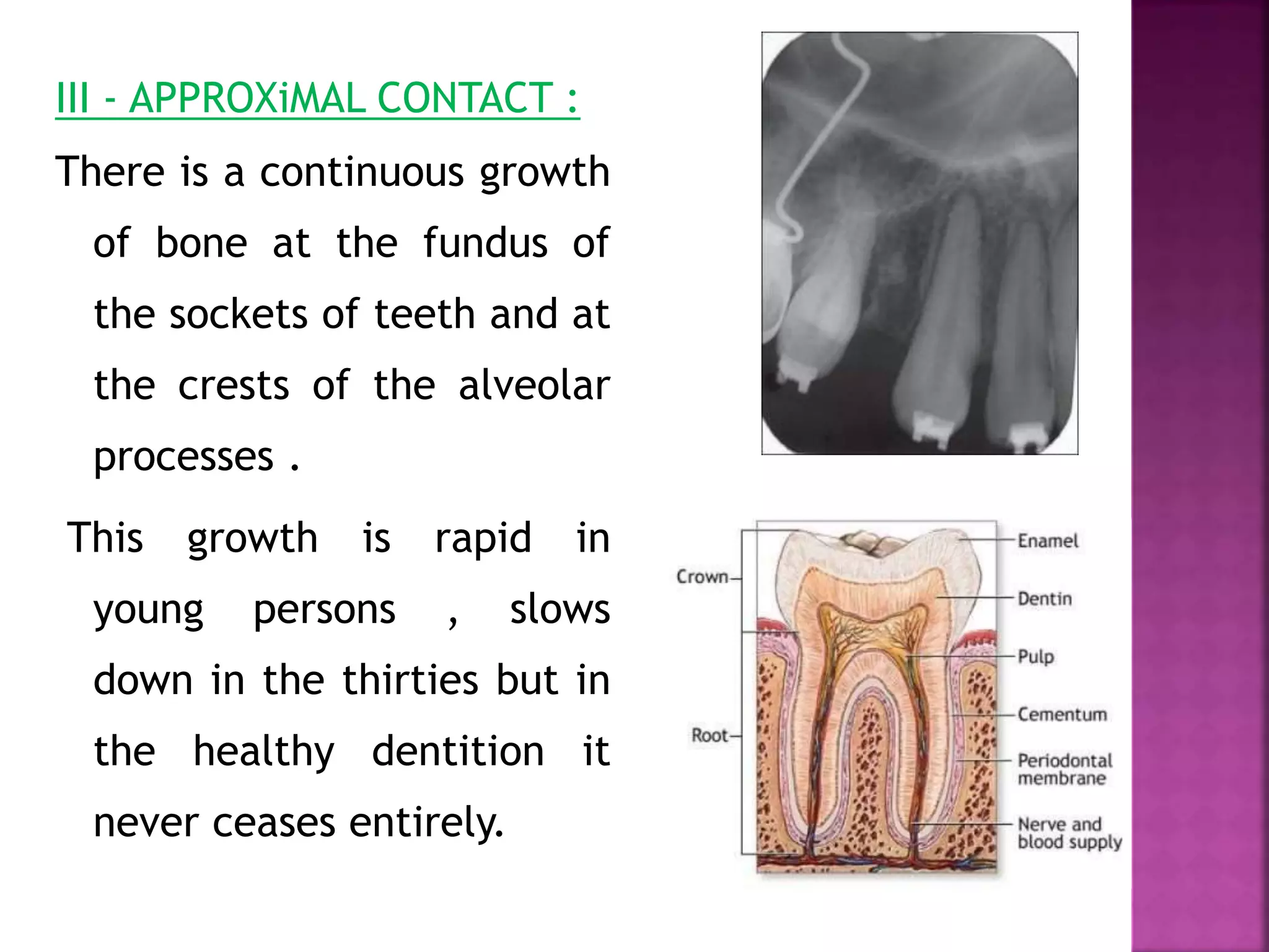
Dr. Ahmed Maher Fouda
Assistant professor of
Orthodontics
Faculty of Dentistry- Mansoura
University
by
The position of each tooth within the jaw
is maintained by certain factors known as
the forces of occlusion . Normally there is
a balance of these forces which produce a
harmonious relationship of the teeth to
each other.
Any imbalance of these forces must affect
the normal relation of the teeth and / or
arches.
The forces of occlusion are:
 1- Growth
 2- Muscle tonus
 3- Approximal contact
 4- Mastication
I- Growth;
Normal downward and forward growth of the face, by
harmonizing the size and form of title jaw with the
teeth may profoundly affect the position of the teeth.
Not only should the size of each jaw be adequate but
their relation should be correct.
 To achieve this , it is necessary that the growth of all those
bones that contribute to the formation of the face should
be proportionate. Growth of the soft tissues, particularly
muscles, should also be harmonious .
II - Muscle tonus:
is the continuous and passive partial contraction of the
muscles
Forces generated by the muscles may be either:
1-passive due to muscle tonus which is continuous but
very light.
2-active associated with muscle activity which is always
greater and intermittent.
passive muscle forces :
Certain muscles exert a constant tension (muscle tonus) upon
the jaws. At rest a muscle is in a state of tonus , In this
state of tonus a small proportion of fibers are contracted,
the proportion of fibers is constant but they are not always
the same fibers .
Successive groups taking over the function of maintaining
tension. It is known that the number of fibers contracted is
proportional to the amount of stretch put upon the muscle.
 Muscles that elevate the mandible:
The masseter : elevation and retraction of
mandible.
The temporalis : elevation and retraction of
mandible.
The medial pterygoid : elevates mandible, closes
jaw, helps lateral pterygoids in moving the jaw
from side to side.
 Muscle that depress the mandible:
The lateral pterygoid : depresses mandible
 Temporalis muscle
 Medial pterygoid muscle
 Masseter muscle
 The muscles which have a direct effect on the jaws or
the dento-alveolar structures are those of deglutition,
expression and mastication.
 MUSCLES OF DEGLUTATION:
 muscle of the mouth
 Muscle of soft palate
 Muscles of pharynx
 MUSCLES OF EXPRESSION:
 Buccinator , orbicularis oris and mentalis
 Muscles of mastication:
 The masseter
 The temporalis
 The lateral Pterygoid
 The medial pterygoid
 The tongue within the vestibule is applied
to the lingual surfaces of the teeth and
the hard and soft palates. The lips and
cheeks are applied to the labial and
buccal surfaces of the teeth.
The upper incisors are prevented from moving labially by
the lip pressure and the lower incisors by the overbite
of the upper incisors.
There is a tension from the orbicularis oris muscle on the
upper incisors and it should be remembered that
orbicularis oris muscle derives some of its fibers from
the buccinator muscle which blends with it at the angle
of the mouth.
The buccinator passes
backwards and inwards
around the maxillary
tuberosity to be inserted
into the pterygomandibular
raphae. From the posterior
aspect of this ligament arise
some of the fibres of the
superior constrictor muscle
which other fibres are
inserted into prevertebral
fascia and base of the
cranium. There is thus a
continuous chain of muscle
encircling the pharynx and
dental arches .
 The buccinator muscle, often limits
buccal movement of upper third molar,
causing the upper dental arch to assume a
horse-shoe shape .
Three of the muscles of mastcation (temporalis , masseter and
medial pterygoid ) support the mandible against gravity and the
action of the submandibular muscles . The former exert an upward
pull and the latter downward pull.
Masseter
Temporalis
Medial PterygoidThe masseter attaches superiorly to the medial and inferior aspect of the zygomatic arch and inferiorly to the lower border
on the lateral surface of the ramus of the mandible.
The temporalis attaches superiorly to the floor of the temporal fossa to the fascia that overlies it. Inferiorly it attaches to the
coronoid process and to the anterior surface of the ramus.
The medial pterygoid runs from the medial surface of the lateral pterygoid plate and the palatine bone to the lower edge of
the medial surface of the ramus of the mandible.
The lateral pterygoid attaches to the lateral surface of the lateral pterygoid plate and the greater wing of the sphenoid bone.
It runs to the pterygoid fovea at the neck of the mandible and the capsule of the temperomandibular joint.
Lateral
Pterygoid
When an individual is at rest the
mandible is held in such a position
that the upper and lower teeth are
normally separated a little about 2-8
mm in the premolar region . This
distance is called the freeway space
or interocclusal clearance.
This mandibular position is maintained
by the balance of muscle tonus . This
balance affects not only the vertical
but also the antero-posterior relation
of the teeth at rest.
active muscle forces :
 This kind of force exert pressure
intermittently. The degree of force is larger
than that of muscle tonus. Also the
frequency varies.
 One of the activities is the deglutition that
concern the orthodontist very much, also
speech , mastication and expression should
not be ignored.
III - APPROXiMAL CONTACT :
There is a continuous growth
of bone at the fundus of
the sockets of teeth and at
the crests of the alveolar
processes .
This growth is rapid in
young persons , slows
down in the thirties but in
the healthy dentition it
never ceases entirely.
 There is also
constant growth of
bone on the distal
wall of each socket,
whereas the mesial
wall shows
resorption of bone
alternating with
localized reparative
apposition .
The roots of the teeth also
enlarge and this enlargement
does not cease when the root
is fully formed. By continuous
apposition of cementum the
roots grow slightly in its
transverse diameters and more
rapidly in length.
 Elongation of the
root and growth of
bone at the
fundus, of the
socket are
correlated with a
continued vertical
eruption of the
tooth during its
functional period.
The changes on the mesial and distal alveolar walls are
correlated to a movement of all teeth toward the
midline, i.e., mesial drift .
The continued occluso-mesial
movement is necessary to
compensate for the loss of tooth
substance at the occlusal and
contact surface .
it is well known clinically that the
contact between neighboring
teeth in a complete adult
dentition is maintained by a
tendency for the posterior teeth
to move forward.
The deciduous teeth are more or less vertical in the
alveolus, so that when occlude, there is little
dislodging force ..
 When the roots of a complete permanent
dentition are examined, they show that
the degree of root curvature increase
towards the back of the mouth, i.e.,
distal inclination of the teeth axes
increases towards the end of the series
 These teeth, therefore, erupt slightly
mesially inclined, and when they come into
occlusion, they exert upon each other an
equal and opposite force.
As a result of their axial inclinations, this force will be divided
into vertical and horizontal compenents of force. The vertical
components will cancel each other and leaving just a mesial force
on all the teeth .This mesial force is called the anterior
component of force which transmits to the teeth by approximal
contact.
IV : Mastication:
The forces of mastication exerted on the teeth
can be divide into those which applied in the
following direction:
 1-vertically
 2-antero-posteriorly
 3-transversely
The mandible moves from its position of rest vertically into
occlusion and then applies direct vertical pressure to the upper
teeth. In the mesiodistai direction,there is a forward resultant,
most marked in the third molar region. In the bucco-lingual
direction, the upper and lower molars are directly opposed
although their axes are not vertical.
The axes of the incisors,
however, are not directly
opposed labio-lingually; the
upper incisors being inclined
labially. The forward resultant
of occlusal pressure is
absorbed partly by the lips and
partly by the palatal curvature
of the upper incisor roots.
 The roots of the lower incisors resist
lingual pressure because they are
flattened mesio-distally.
Movements of the mandible in this direction are not used
frequently in mastication because the overbite of the incisors
causes the molars and premolars to be disengaged if the smallest
excursions are made. However, for some action the mandible is
protruded sufficiently to bring the incisors into occlusion.
The movement is at first an incising the lateral margin of the orbit
to the junction of the temporal ridge and the supra-orbital ridge.
 The pterygoid plates
which lie behind the
tuberosity of the
maxilla and pass
upwards and backwards
to the inferior surface
of the great wing of the
sphenoid bone.
In the molar region, lateral excursions of the mandible at first cause
the large mesio-palatal cusps of the upper molars to glide up the
buccal cusps of the lower molars and if movement is continued, the
cusp to cusp contact of the buccal cusps of the opposite side
disengages the molars. Shearing actions may be performed by the
premolars and canines, the buccal cusps of the lower premolars
engaging those of the upper premolars.
Bone, unlike other connective tissue responds to mild
degree of pressure and tension by changes in form.
Those changes are accomplished by resorption of
existing bone and deposition of new bone. In this
respect bone is more plastic than any other connective
tissue.
During mastication, certain stresses and strains are exerted not
only upon the alveolar bone but also upon the whole face and
anterior part of the base of the cranium. In order to withstand
these, the bones which are made up of thin plates are thickened
into well defined ridges. Most of these ridges pass upwards from
the alveolar bone towards the base of the cranium. Some, however,
lie transversely or antero-posteriorly to withstand forces in these
directions .
1 - The canine eminence which is a thickened ridge arises
from the region
of the upper canine . It extends upwarde on the facial
surface of the maxilla to the medial end of the infraorbital
margin and continues up the lateral wall of the nose to the
mesial end of the supraorbital ridge .
2 - The key ridge or the root of the Zygomatic process which arises
on the lateral wall of the maxilla over the roots of the first
permanent molars and becomes the zygomatic process . It
continues upwards to approximal contact .The anterior coinpenent
of force on one side of the arch cancels out that of the other side
in the midline provided that there is an intact dental arch. If the
interproximal contacts are destroyed by loss of teeth, those behind
the gap tend to drift forward, whilst those in front will lack
forward development
The supraorbital ridges and infraorbital margins .
 The arched vault of the palate.
 The zygomatic arch which lies
horizontally and it is the continuation of
the zygomatic process . It is continued
posteriorly with the temporal ridge.
The mylohyoid ridge and its continuation upwards and backwards to
the condyle .
 The external oblique ridge winch is a
thickening of bone passing backwards and
upwards on the external surface to the
root of the coronoid process and up its
anterior border.
external oblique ridge
 The inferior border of the mandible is
thickened and the gonial angle is slightly
everted and thickened where the
masseter muscle is inserted .
Forces of occlusion.ppt

Forces of occlusion.ppt

  • 1.
    Dr. Ahmed MaherFouda Assistant professor of Orthodontics Faculty of Dentistry- Mansoura University by
  • 2.
    The position ofeach tooth within the jaw is maintained by certain factors known as the forces of occlusion . Normally there is a balance of these forces which produce a harmonious relationship of the teeth to each other. Any imbalance of these forces must affect the normal relation of the teeth and / or arches.
  • 3.
    The forces ofocclusion are:  1- Growth  2- Muscle tonus  3- Approximal contact  4- Mastication
  • 4.
    I- Growth; Normal downwardand forward growth of the face, by harmonizing the size and form of title jaw with the teeth may profoundly affect the position of the teeth. Not only should the size of each jaw be adequate but their relation should be correct.
  • 7.
     To achievethis , it is necessary that the growth of all those bones that contribute to the formation of the face should be proportionate. Growth of the soft tissues, particularly muscles, should also be harmonious .
  • 9.
    II - Muscletonus: is the continuous and passive partial contraction of the muscles Forces generated by the muscles may be either: 1-passive due to muscle tonus which is continuous but very light. 2-active associated with muscle activity which is always greater and intermittent.
  • 10.
    passive muscle forces: Certain muscles exert a constant tension (muscle tonus) upon the jaws. At rest a muscle is in a state of tonus , In this state of tonus a small proportion of fibers are contracted, the proportion of fibers is constant but they are not always the same fibers . Successive groups taking over the function of maintaining tension. It is known that the number of fibers contracted is proportional to the amount of stretch put upon the muscle.
  • 12.
     Muscles thatelevate the mandible: The masseter : elevation and retraction of mandible. The temporalis : elevation and retraction of mandible. The medial pterygoid : elevates mandible, closes jaw, helps lateral pterygoids in moving the jaw from side to side.  Muscle that depress the mandible: The lateral pterygoid : depresses mandible
  • 13.
  • 14.
  • 15.
  • 17.
     The muscleswhich have a direct effect on the jaws or the dento-alveolar structures are those of deglutition, expression and mastication.  MUSCLES OF DEGLUTATION:  muscle of the mouth  Muscle of soft palate  Muscles of pharynx  MUSCLES OF EXPRESSION:  Buccinator , orbicularis oris and mentalis
  • 18.
     Muscles ofmastication:  The masseter  The temporalis  The lateral Pterygoid  The medial pterygoid
  • 22.
     The tonguewithin the vestibule is applied to the lingual surfaces of the teeth and the hard and soft palates. The lips and cheeks are applied to the labial and buccal surfaces of the teeth.
  • 23.
    The upper incisorsare prevented from moving labially by the lip pressure and the lower incisors by the overbite of the upper incisors. There is a tension from the orbicularis oris muscle on the upper incisors and it should be remembered that orbicularis oris muscle derives some of its fibers from the buccinator muscle which blends with it at the angle of the mouth.
  • 25.
    The buccinator passes backwardsand inwards around the maxillary tuberosity to be inserted into the pterygomandibular raphae. From the posterior aspect of this ligament arise some of the fibres of the superior constrictor muscle which other fibres are inserted into prevertebral fascia and base of the cranium. There is thus a continuous chain of muscle encircling the pharynx and dental arches .
  • 26.
     The buccinatormuscle, often limits buccal movement of upper third molar, causing the upper dental arch to assume a horse-shoe shape .
  • 29.
    Three of themuscles of mastcation (temporalis , masseter and medial pterygoid ) support the mandible against gravity and the action of the submandibular muscles . The former exert an upward pull and the latter downward pull.
  • 31.
    Masseter Temporalis Medial PterygoidThe masseterattaches superiorly to the medial and inferior aspect of the zygomatic arch and inferiorly to the lower border on the lateral surface of the ramus of the mandible. The temporalis attaches superiorly to the floor of the temporal fossa to the fascia that overlies it. Inferiorly it attaches to the coronoid process and to the anterior surface of the ramus. The medial pterygoid runs from the medial surface of the lateral pterygoid plate and the palatine bone to the lower edge of the medial surface of the ramus of the mandible. The lateral pterygoid attaches to the lateral surface of the lateral pterygoid plate and the greater wing of the sphenoid bone. It runs to the pterygoid fovea at the neck of the mandible and the capsule of the temperomandibular joint. Lateral Pterygoid
  • 33.
    When an individualis at rest the mandible is held in such a position that the upper and lower teeth are normally separated a little about 2-8 mm in the premolar region . This distance is called the freeway space or interocclusal clearance. This mandibular position is maintained by the balance of muscle tonus . This balance affects not only the vertical but also the antero-posterior relation of the teeth at rest.
  • 34.
    active muscle forces:  This kind of force exert pressure intermittently. The degree of force is larger than that of muscle tonus. Also the frequency varies.  One of the activities is the deglutition that concern the orthodontist very much, also speech , mastication and expression should not be ignored.
  • 36.
    III - APPROXiMALCONTACT : There is a continuous growth of bone at the fundus of the sockets of teeth and at the crests of the alveolar processes . This growth is rapid in young persons , slows down in the thirties but in the healthy dentition it never ceases entirely.
  • 38.
     There isalso constant growth of bone on the distal wall of each socket, whereas the mesial wall shows resorption of bone alternating with localized reparative apposition .
  • 39.
    The roots ofthe teeth also enlarge and this enlargement does not cease when the root is fully formed. By continuous apposition of cementum the roots grow slightly in its transverse diameters and more rapidly in length.
  • 40.
     Elongation ofthe root and growth of bone at the fundus, of the socket are correlated with a continued vertical eruption of the tooth during its functional period.
  • 43.
    The changes onthe mesial and distal alveolar walls are correlated to a movement of all teeth toward the midline, i.e., mesial drift .
  • 45.
    The continued occluso-mesial movementis necessary to compensate for the loss of tooth substance at the occlusal and contact surface . it is well known clinically that the contact between neighboring teeth in a complete adult dentition is maintained by a tendency for the posterior teeth to move forward.
  • 46.
    The deciduous teethare more or less vertical in the alveolus, so that when occlude, there is little dislodging force ..
  • 49.
     When theroots of a complete permanent dentition are examined, they show that the degree of root curvature increase towards the back of the mouth, i.e., distal inclination of the teeth axes increases towards the end of the series
  • 52.
     These teeth,therefore, erupt slightly mesially inclined, and when they come into occlusion, they exert upon each other an equal and opposite force.
  • 54.
    As a resultof their axial inclinations, this force will be divided into vertical and horizontal compenents of force. The vertical components will cancel each other and leaving just a mesial force on all the teeth .This mesial force is called the anterior component of force which transmits to the teeth by approximal contact.
  • 55.
    IV : Mastication: Theforces of mastication exerted on the teeth can be divide into those which applied in the following direction:  1-vertically  2-antero-posteriorly  3-transversely
  • 56.
    The mandible movesfrom its position of rest vertically into occlusion and then applies direct vertical pressure to the upper teeth. In the mesiodistai direction,there is a forward resultant, most marked in the third molar region. In the bucco-lingual direction, the upper and lower molars are directly opposed although their axes are not vertical.
  • 57.
    The axes ofthe incisors, however, are not directly opposed labio-lingually; the upper incisors being inclined labially. The forward resultant of occlusal pressure is absorbed partly by the lips and partly by the palatal curvature of the upper incisor roots.
  • 58.
     The rootsof the lower incisors resist lingual pressure because they are flattened mesio-distally.
  • 59.
    Movements of themandible in this direction are not used frequently in mastication because the overbite of the incisors causes the molars and premolars to be disengaged if the smallest excursions are made. However, for some action the mandible is protruded sufficiently to bring the incisors into occlusion.
  • 60.
    The movement isat first an incising the lateral margin of the orbit to the junction of the temporal ridge and the supra-orbital ridge.
  • 61.
     The pterygoidplates which lie behind the tuberosity of the maxilla and pass upwards and backwards to the inferior surface of the great wing of the sphenoid bone.
  • 62.
    In the molarregion, lateral excursions of the mandible at first cause the large mesio-palatal cusps of the upper molars to glide up the buccal cusps of the lower molars and if movement is continued, the cusp to cusp contact of the buccal cusps of the opposite side disengages the molars. Shearing actions may be performed by the premolars and canines, the buccal cusps of the lower premolars engaging those of the upper premolars.
  • 63.
    Bone, unlike otherconnective tissue responds to mild degree of pressure and tension by changes in form. Those changes are accomplished by resorption of existing bone and deposition of new bone. In this respect bone is more plastic than any other connective tissue.
  • 64.
    During mastication, certainstresses and strains are exerted not only upon the alveolar bone but also upon the whole face and anterior part of the base of the cranium. In order to withstand these, the bones which are made up of thin plates are thickened into well defined ridges. Most of these ridges pass upwards from the alveolar bone towards the base of the cranium. Some, however, lie transversely or antero-posteriorly to withstand forces in these directions .
  • 67.
    1 - Thecanine eminence which is a thickened ridge arises from the region of the upper canine . It extends upwarde on the facial surface of the maxilla to the medial end of the infraorbital margin and continues up the lateral wall of the nose to the mesial end of the supraorbital ridge .
  • 68.
    2 - Thekey ridge or the root of the Zygomatic process which arises on the lateral wall of the maxilla over the roots of the first permanent molars and becomes the zygomatic process . It continues upwards to approximal contact .The anterior coinpenent of force on one side of the arch cancels out that of the other side in the midline provided that there is an intact dental arch. If the interproximal contacts are destroyed by loss of teeth, those behind the gap tend to drift forward, whilst those in front will lack forward development
  • 70.
    The supraorbital ridgesand infraorbital margins .
  • 71.
     The archedvault of the palate.
  • 72.
     The zygomaticarch which lies horizontally and it is the continuation of the zygomatic process . It is continued posteriorly with the temporal ridge.
  • 73.
    The mylohyoid ridgeand its continuation upwards and backwards to the condyle .
  • 75.
     The externaloblique ridge winch is a thickening of bone passing backwards and upwards on the external surface to the root of the coronoid process and up its anterior border. external oblique ridge
  • 76.
     The inferiorborder of the mandible is thickened and the gonial angle is slightly everted and thickened where the masseter muscle is inserted .