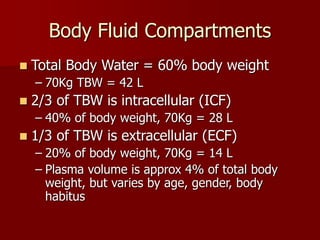
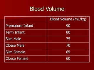
Fluid Management and
Shock Resuscitation
Kallie Honeywood
UBC Anaesthesia PGY-3
Outline
 Normal Fluid Requirements
 Definition of Shock
 Types of Shock
– Hypovolemic
– Cardiogenic
– Distributive
– Obstructive
 Resuscitation Fluids
 Goals of Resuscitation
Body Fluid Compartments
 Total Body Water = 60% body weight
– 70Kg TBW = 42 L
 2/3 of TBW is intracellular (ICF)
– 40% of body weight, 70Kg = 28 L
 1/3 of TBW is extracellular (ECF)
– 20% of body weight, 70Kg = 14 L
– Plasma volume is approx 4% of total body
weight, but varies by age, gender, body
habitus
Blood Volume
Blood Volume (mL/kg)
Premature Infant 90
Term Infant 80
Slim Male 75
Obese Male 70
Slim Female 65
Obese Female 60
Peri-operative Maintenance Fluids
 Water
 Sodium
 Potassium replacement can be omitted for
short periods of time
 Chloride, Mg, Ca, trace minerals and
supplementation needed only for chronic
IV maintenance
 Most commonly Saline, Lactated Ringers,
Plasmalyte
4 – 2 – 1 Rule
 100 – 50 – 20 Rule for daily fluid
requirements
 4 mL/kg for 1st 10 kg
 2 mL/kg for 2nd 10 kg
 1 mL/kg for each additional kg
Maintenance Fluids: Example
 60 kg female
 1st 10 kg: 4 mL/kg x 10 kg = 40 mL
 2nd 10 kg: 2 mL/kg x 10 kg = 20 mL
 Remaining: 60 kg – 20 kg = 40 kg
1 mL/kg x 40 kg = 40 mL
 Maintenance Rate = 120 mL/hr
Fluid Deficits
 Fasting
 Bowel Loss (Bowel Prep, vomiting, diarrhea)
 Blood Loss
– Trauma
– Fractures
 Burns
 Sepsis
 Pancreatitis
Insensible Fluid Loss
 Evaporative
 Exudative
 Tissue Edema (surgical manipulation)
 Fluid Sequestration (bowel, lung)
 Extent of fluid loss or redistribution (the
“Third Space”) dependent on type of
surgical procedure
 Mobilization of Third Space Fluid POD#3
Insensible Fluid Loss
 4 – 6 – 8 Rule
 Replace with Crystalloid (NS, LR,
Plasmalyte)
 Minor: 4 mL/kg/hr
 Moderate: 6 mL/kg/hr
 Major: 8 mL/kg/hr
Example
 68 kg female for laparoscopic
cholecystectomy
 Fasted since midnight, OR start at 8am
 Maintenance = 40 + 20 + 48 = 108 mL/hr
 Deficit = 108 mL/hr x 8hr
= 864 mL
 3rd Space (4mL/kg/hr) = 272 mL/hr
Example
 Intra-operative Fluid Replacement of:
– Fluid Deficit 864 mL
– Maintenance Fluid 108 mL/hr
– 3rd Space Loss 272 mL/hr
– Ongoing blood loss (crystalloid vs. colloid)
Shock
 Circulatory failure leading to inadequate
perfusion and delivery of oxygen to vital
organs
 Blood Pressure is often used as an indirect
estimator of tissue perfusion
 Oxygen delivery is an interaction of
Cardiac Output, Blood Volume, Systemic
Vascular Resistance
DO2
CaO2
CO
Sat %
PaO2
Hgb
HR
SV
Preload
Contractility
Afterload
Types of Shock
 Hypovolemic – most common
 Hemorrhagic, occult fluid loss
 Cardiogenic
 Ischemia, arrhythmia, valvular, myocardial
depression
 Distributive
 Anaphylaxis, sepsis, neurogenic
 Obstructive
 Tension pneumo, pericardial tamponade, PE
Shock States
BP CVP PCWP CO SVR
Hypovolemia
Cardiogenic -
LV
- RV
Distributive
Obstructive
DO2
CaO2
CO
Sat %
PaO2
Hgb
HR
SV
Preload
Contractility
Afterload
Hypovolemic Shock
 Most common
 Trauma
 Blood Loss
 Occult fluid loss (GI)
 Burns
 Pancreatitis
 Sepsis (distributive, relative
hypovolemia)
Assessment of Stages of Shock
% Blood
Volume loss
< 15% 15 – 30% 30 – 40% >40%
HR <100 >100 >120 >140
SBP N N, DBP,
postural drop
Pulse
Pressure
N or
Cap Refill < 3 sec > 3 sec >3 sec or
absent
absent
Resp 14 - 20 20 - 30 30 - 40 >35
CNS anxious v. anxious confused lethargic
Treatment 1 – 2 L
crystalloid, +
maintenance
2 L
crystalloid, re-
evaluate
2 L crystalloid, re-evaluate,
replace blood loss 1:3
crystalloid, 1:1 colloid or blood
products. Urine output >0.5
mL/kg/hr
Fluid Resuscitation of Shock
 Crystalloid Solutions
– Normal saline
– Ringers Lactate solution
– Plasmalyte
 Colloid Solutions
– Pentastarch
– Blood products (albumin, RBC, plasma)
Crystalloid Solutions
 Normal Saline
 Lactated Ringers Solution
 Plasmalyte
 Require 3:1 replacement of volume loss
 e.g. estimate 1 L blood loss, require 3 L of
crystalloid to replace volume
Colloid Solutions
 Pentaspan
 Albumin 5%
 Red Blood Cells
 Fresh Frozen Plasma
 Replacement of lost volume in 1:1 ratio
Oxygen Carrying Capacity
 Only RBC contribute to oxygen carrying
capacity (hemoglobin)
 Replacement with all other solutions will
– support volume
– Improve end organ perfusion
– Will NOT provide additional oxygen carrying
capacity
RBC Transfusion
 BC Red Cell Transfusion Guidelines
recommend transfusion only to keep Hgb
>70 g/dL unless
– Comorbid disease necessitating higher
transfusion trigger (CAD, pulmonary disease,
sepsis)
– Hemodynamic instability despite adequate
fluid resuscitation
Crystalloid vs. Colloid
 SAFE study (Saline vs. Albumin Fluid
Evaluation)
– Critically ill patients in ICU
– Randomized to Saline vs. 4% Albumin for fluid
resuscitation
– No difference in 28 day all cause mortality
– No difference in length of ICU stay,
mechanical ventilation, RRT, other organ
failure
NEJM 2004; 350 (22), 2247- 2256
Goals of Fluid Resuscitation
 Easily measured
– Mentation
– Blood Pressure
– Heart Rate
– Jugular Venous Pressure
– Urine Output
Goals of Fluid Resuscitation
 A little less easily measured
– Central Venous Pressure (CVP)
– Left Atrial Pressure
– Central Venous Oxygen Saturation SCVO2
Goals of Fluid Resuscitation
 A bit more of a pain to measure
– Pulmonary Capillary Wedge Pressure (PCWP)
– Systemic Vascular Resistance (SVR)
– Cardiac Output / Cardiac Index
Mixed Venous Oxygenation
 Used as a surrogate marker of end organ
perfusion and oxygen delivery
 Should be interpreted in context of other clinical
information
 True mixed venous is drawn from the pulmonary
artery (mixing of venous blood from upper and
lower body)
 Often sample will be drawn from central venous
catheter (superior vena cava, R atrium)
Mixed Venous Oxygenation
 Normal oxygen saturation of venous blood
68% – 77%
 Low SCVO2
– Tissues are extracting far more oxygen than
usual, reflecting sub-optimal tissue perfusion
(and oxygenation)
 Following trends of SCVO2 to guide
resuscitation (fluids, RBC, inotropes,
vasopressors)
Goals of Resuscitation
 Rivers Study- Early Goal Directed Therapy
in Sepsis and Septic Shock
– Emergency department with severe sepsis or
septic shock, randomized to goal directed
protocol vs standard therapy prior to
admission to ICU
– Early goal directed therapy conferred lower
APACHE scores, incidating less severe organ
dysfunction
DO2
CaO2
CO
Sat %
PaO2
Hgb
HR
SV
Preload
Contractility
Afterload
Bottom Line
 Resuscitation of Shock is all about getting
oxygen to the tissues
 Initial assessment of volume deficit, replace that
(with crystalloid), and reassess
 Continue volume resuscitation to target
endpoints
 Can use mixed venous oxygen saturation to
estimate tissue perfusion and oxygenation
References
 Clinical Anesthesia 3rd Ed. Morgan et al.
Lange Medical / McGraw Hill, 2002
 Anesthesiology Review 3rd Ed. Faust, R.
Churchill-Livingstone, 2002
 Rivers, E. et al. NEJM 2001; 345 (19):
1368 – 77
 SAFE Investigators. NEJM 2004; 350:
2247 - 56

Fluid resuscitation and shock management

  • 1.
    Fluid Management and ShockResuscitation Kallie Honeywood UBC Anaesthesia PGY-3
  • 2.
    Outline  Normal FluidRequirements  Definition of Shock  Types of Shock – Hypovolemic – Cardiogenic – Distributive – Obstructive  Resuscitation Fluids  Goals of Resuscitation
  • 3.
    Body Fluid Compartments Total Body Water = 60% body weight – 70Kg TBW = 42 L  2/3 of TBW is intracellular (ICF) – 40% of body weight, 70Kg = 28 L  1/3 of TBW is extracellular (ECF) – 20% of body weight, 70Kg = 14 L – Plasma volume is approx 4% of total body weight, but varies by age, gender, body habitus
  • 4.
    Blood Volume Blood Volume(mL/kg) Premature Infant 90 Term Infant 80 Slim Male 75 Obese Male 70 Slim Female 65 Obese Female 60
  • 5.
    Peri-operative Maintenance Fluids Water  Sodium  Potassium replacement can be omitted for short periods of time  Chloride, Mg, Ca, trace minerals and supplementation needed only for chronic IV maintenance  Most commonly Saline, Lactated Ringers, Plasmalyte
  • 6.
    4 – 2– 1 Rule  100 – 50 – 20 Rule for daily fluid requirements  4 mL/kg for 1st 10 kg  2 mL/kg for 2nd 10 kg  1 mL/kg for each additional kg
  • 7.
    Maintenance Fluids: Example 60 kg female  1st 10 kg: 4 mL/kg x 10 kg = 40 mL  2nd 10 kg: 2 mL/kg x 10 kg = 20 mL  Remaining: 60 kg – 20 kg = 40 kg 1 mL/kg x 40 kg = 40 mL  Maintenance Rate = 120 mL/hr
  • 8.
    Fluid Deficits  Fasting Bowel Loss (Bowel Prep, vomiting, diarrhea)  Blood Loss – Trauma – Fractures  Burns  Sepsis  Pancreatitis
  • 9.
    Insensible Fluid Loss Evaporative  Exudative  Tissue Edema (surgical manipulation)  Fluid Sequestration (bowel, lung)  Extent of fluid loss or redistribution (the “Third Space”) dependent on type of surgical procedure  Mobilization of Third Space Fluid POD#3
  • 10.
    Insensible Fluid Loss 4 – 6 – 8 Rule  Replace with Crystalloid (NS, LR, Plasmalyte)  Minor: 4 mL/kg/hr  Moderate: 6 mL/kg/hr  Major: 8 mL/kg/hr
  • 11.
    Example  68 kgfemale for laparoscopic cholecystectomy  Fasted since midnight, OR start at 8am  Maintenance = 40 + 20 + 48 = 108 mL/hr  Deficit = 108 mL/hr x 8hr = 864 mL  3rd Space (4mL/kg/hr) = 272 mL/hr
  • 12.
    Example  Intra-operative FluidReplacement of: – Fluid Deficit 864 mL – Maintenance Fluid 108 mL/hr – 3rd Space Loss 272 mL/hr – Ongoing blood loss (crystalloid vs. colloid)
  • 13.
    Shock  Circulatory failureleading to inadequate perfusion and delivery of oxygen to vital organs  Blood Pressure is often used as an indirect estimator of tissue perfusion  Oxygen delivery is an interaction of Cardiac Output, Blood Volume, Systemic Vascular Resistance
  • 14.
  • 15.
    Types of Shock Hypovolemic – most common  Hemorrhagic, occult fluid loss  Cardiogenic  Ischemia, arrhythmia, valvular, myocardial depression  Distributive  Anaphylaxis, sepsis, neurogenic  Obstructive  Tension pneumo, pericardial tamponade, PE
  • 16.
    Shock States BP CVPPCWP CO SVR Hypovolemia Cardiogenic - LV - RV Distributive Obstructive
  • 17.
  • 18.
    Hypovolemic Shock  Mostcommon  Trauma  Blood Loss  Occult fluid loss (GI)  Burns  Pancreatitis  Sepsis (distributive, relative hypovolemia)
  • 19.
    Assessment of Stagesof Shock % Blood Volume loss < 15% 15 – 30% 30 – 40% >40% HR <100 >100 >120 >140 SBP N N, DBP, postural drop Pulse Pressure N or Cap Refill < 3 sec > 3 sec >3 sec or absent absent Resp 14 - 20 20 - 30 30 - 40 >35 CNS anxious v. anxious confused lethargic Treatment 1 – 2 L crystalloid, + maintenance 2 L crystalloid, re- evaluate 2 L crystalloid, re-evaluate, replace blood loss 1:3 crystalloid, 1:1 colloid or blood products. Urine output >0.5 mL/kg/hr
  • 20.
    Fluid Resuscitation ofShock  Crystalloid Solutions – Normal saline – Ringers Lactate solution – Plasmalyte  Colloid Solutions – Pentastarch – Blood products (albumin, RBC, plasma)
  • 21.
    Crystalloid Solutions  NormalSaline  Lactated Ringers Solution  Plasmalyte  Require 3:1 replacement of volume loss  e.g. estimate 1 L blood loss, require 3 L of crystalloid to replace volume
  • 22.
    Colloid Solutions  Pentaspan Albumin 5%  Red Blood Cells  Fresh Frozen Plasma  Replacement of lost volume in 1:1 ratio
  • 23.
    Oxygen Carrying Capacity Only RBC contribute to oxygen carrying capacity (hemoglobin)  Replacement with all other solutions will – support volume – Improve end organ perfusion – Will NOT provide additional oxygen carrying capacity
  • 24.
    RBC Transfusion  BCRed Cell Transfusion Guidelines recommend transfusion only to keep Hgb >70 g/dL unless – Comorbid disease necessitating higher transfusion trigger (CAD, pulmonary disease, sepsis) – Hemodynamic instability despite adequate fluid resuscitation
  • 25.
    Crystalloid vs. Colloid SAFE study (Saline vs. Albumin Fluid Evaluation) – Critically ill patients in ICU – Randomized to Saline vs. 4% Albumin for fluid resuscitation – No difference in 28 day all cause mortality – No difference in length of ICU stay, mechanical ventilation, RRT, other organ failure NEJM 2004; 350 (22), 2247- 2256
  • 26.
    Goals of FluidResuscitation  Easily measured – Mentation – Blood Pressure – Heart Rate – Jugular Venous Pressure – Urine Output
  • 27.
    Goals of FluidResuscitation  A little less easily measured – Central Venous Pressure (CVP) – Left Atrial Pressure – Central Venous Oxygen Saturation SCVO2
  • 28.
    Goals of FluidResuscitation  A bit more of a pain to measure – Pulmonary Capillary Wedge Pressure (PCWP) – Systemic Vascular Resistance (SVR) – Cardiac Output / Cardiac Index
  • 29.
    Mixed Venous Oxygenation Used as a surrogate marker of end organ perfusion and oxygen delivery  Should be interpreted in context of other clinical information  True mixed venous is drawn from the pulmonary artery (mixing of venous blood from upper and lower body)  Often sample will be drawn from central venous catheter (superior vena cava, R atrium)
  • 30.
    Mixed Venous Oxygenation Normal oxygen saturation of venous blood 68% – 77%  Low SCVO2 – Tissues are extracting far more oxygen than usual, reflecting sub-optimal tissue perfusion (and oxygenation)  Following trends of SCVO2 to guide resuscitation (fluids, RBC, inotropes, vasopressors)
  • 31.
    Goals of Resuscitation Rivers Study- Early Goal Directed Therapy in Sepsis and Septic Shock – Emergency department with severe sepsis or septic shock, randomized to goal directed protocol vs standard therapy prior to admission to ICU – Early goal directed therapy conferred lower APACHE scores, incidating less severe organ dysfunction
  • 33.
  • 34.
    Bottom Line  Resuscitationof Shock is all about getting oxygen to the tissues  Initial assessment of volume deficit, replace that (with crystalloid), and reassess  Continue volume resuscitation to target endpoints  Can use mixed venous oxygen saturation to estimate tissue perfusion and oxygenation
  • 35.
    References  Clinical Anesthesia3rd Ed. Morgan et al. Lange Medical / McGraw Hill, 2002  Anesthesiology Review 3rd Ed. Faust, R. Churchill-Livingstone, 2002  Rivers, E. et al. NEJM 2001; 345 (19): 1368 – 77  SAFE Investigators. NEJM 2004; 350: 2247 - 56