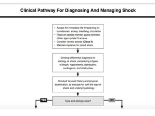
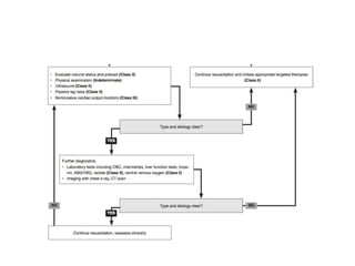
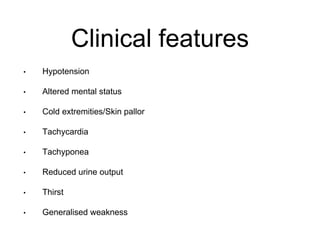
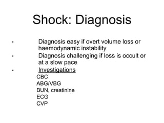
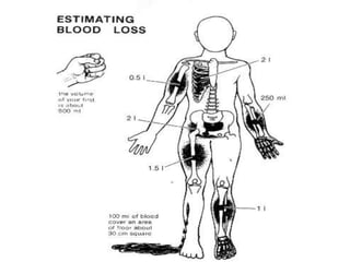
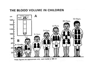
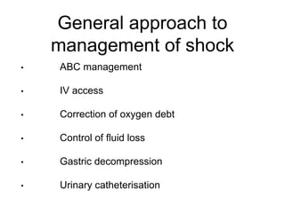
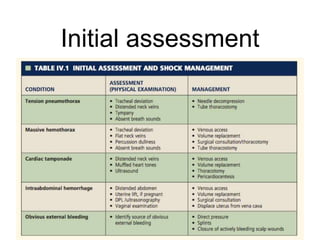
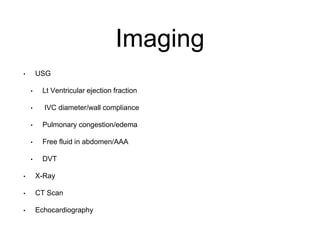
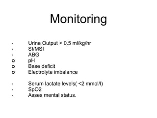
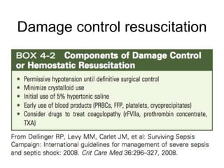
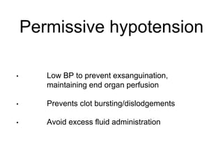
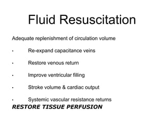
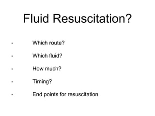
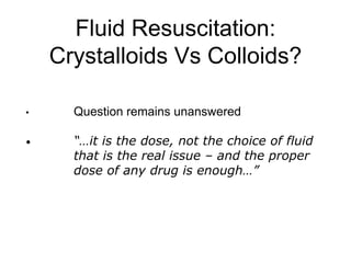
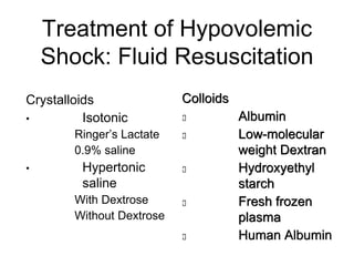
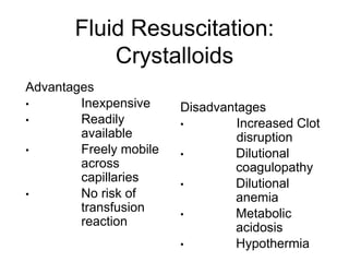
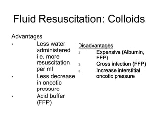
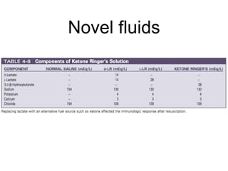
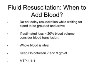
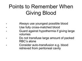
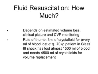
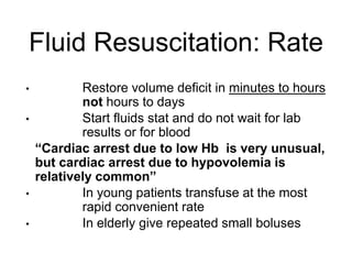
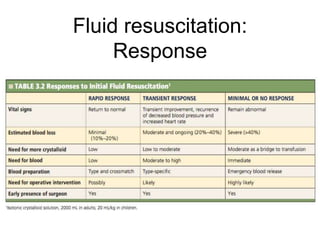
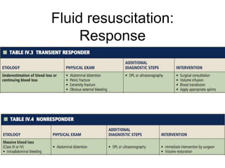
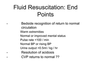
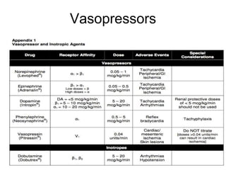
EVALUATION AND
MANAGEMENT OF
SHOCK
Presented By: Dr. MK Tiwari
Clinical features
• Hypotension
• Altered mental status
• Cold extremities/Skin pallor
• Tachycardia
• Tachyponea
• Reduced urine output
• Thirst
• Generalised weakness
Shock: Diagnosis
• Diagnosis easy if overt volume loss or
haemodynamic instability
• Diagnosis challenging if loss is occult or
at a slow pace
• Investigations
• CBC
• ABG/VBG
• BUN, creatinine
• ECG
• CVP
General approach to
management of shock
• ABC management
• IV access
• Correction of oxygen debt
• Control of fluid loss
• Gastric decompression
• Urinary catheterisation
Initial assessment
Imaging
• USG
• Lt Ventricular ejection fraction
• IVC diameter/wall compliance
• Pulmonary congestion/edema
• Free fluid in abdomen/AAA
• DVT
• X-Ray
• CT Scan
• Echocardiography
Monitoring
• Urine Output > 0.5 ml/kg/hr
• SI/MSI
• ABG
 pH
 Base deficit
 Electrolyte imbalance
• Serum lactate levels( <2 mmol/l)
• SpO2
• Asses mental status.
Damage control resuscitation
Permissive hypotension
• Low BP to prevent exsanguination,
maintaining end organ perfusion
• Prevents clot bursting/dislodgements
• Avoid excess fluid administration
Fluid Resuscitation
Adequate replenishment of circulation volume
• Re-expand capacitance veins
• Restore venous return
• Improve ventricular filling
• Stroke volume & cardiac output
• Systemic vascular resistance returns
RESTORE TISSUE PERFUSION
Fluid Resuscitation?
• Which route?
• Which fluid?
• How much?
• Timing?
• End points for resuscitation
Fluid Resuscitation:
Crystalloids Vs Colloids?
• Question remains unanswered
• “…it is the dose, not the choice of fluid
that is the real issue – and the proper
dose of any drug is enough…”
Treatment of Hypovolemic
Shock: Fluid Resuscitation
Crystalloids
• Isotonic
• Ringer’s Lactate
• 0.9% saline
• Hypertonic
saline
• With Dextrose
• Without Dextrose
Colloids
 Albumin
 Low-molecular
weight Dextran
 Hydroxyethyl
starch
 Fresh frozen
plasma
 Human Albumin
Fluid Resuscitation:
Crystalloids
Advantages
• Inexpensive
• Readily
available
• Freely mobile
across
capillaries
• No risk of
transfusion
reaction
Disadvantages
• Increased Clot
disruption
• Dilutional
coagulopathy
• Dilutional
anemia
• Metabolic
acidosis
• Hypothermia
Fluid Resuscitation: Colloids
Advantages
• Less water
administered
i.e. more
resuscitation
per ml
• Less decrease
in oncotic
pressure
• Acid buffer
(FFP)
Disadvantages
 Expensive (Albumin,
FFP)
 Cross infection (FFP)
 Increase interstitial
oncotic pressure
Types of fluid available
Novel fluids
Fluid Resuscitation: When to
Add Blood?
• Do not delay resuscitation while waiting for
blood to be grouped and arrive
• If estimated loss > 20% blood volume
consider blood transfusion.
• Whole blood is ideal
• Keep Hb between 7 and 9 gm/dL
• MTP-1:1:1
Points to Remember When
Giving Blood
• Always use youngest possible blood
• Use fully cross-matched blood
• Guard against hypothermia if giving large
volumes
• Do not transfuse large amount of packed
RBC’s alone
• Consider auto-transfusion e.g. blood
retrieved from peritoneal cavity
Fluid Resuscitation: How
Much?
• Depends on estimated volume loss,
clinical picture and CVP monitoring
• Rule of thumb: 3ml of crystalloid for every
ml of blood lost e.g. 70kg patient in Class
III shock has lost almost 1500 ml of blood
and needs 4500 ml of crystalloids for
volume replacement
Fluid Resuscitation: Rate
• Restore volume deficit in minutes to hours
not hours to days
• Start fluids stat and do not wait for lab
results or for blood
“Cardiac arrest due to low Hb is very unusual,
but cardiac arrest due to hypovolemia is
relatively common”
• In young patients transfuse at the most
rapid convenient rate
• In elderly give repeated small boluses
Fluid resuscitation:
Response
Fluid resuscitation:
Response
Fluid Resuscitation: End
Points
• Bedside recognition of return to normal
circulation
• Warm extremities
• Normal or improved mental status
• Pulse rate <100 / min
• Normal BP or rising BP
• Urine output >0.5ml / kg / hr
• Resolution of acidosis
• CVP returns to normal ??
Lethal Triad
• Acidosis
• Hypothermia
• Coagulopathy
Acidosis
• Treat underlying cause of shock
• Correction of PH
• THAM (tromethamine tris hydrodymethyl
aminomethane)- lean body weight (kgs)xbase
deficit(mmol/l)
Acidosis
• Treat underlying cause of shock
• Correction of PH
• THAM (tromethamine tris hydrodymethyl
aminomethane)- lean body weight (kgs)xbase
deficit(mmol/l)
Hypothermia
Hypothermia
Hypothermia
Coagulopathy
• rFVIIa
• Factor IX/Prothrombin complex concentrate
• Trenaxemic Acid
• Cryoprecipitate
Coagulopathy
• rFVIIa
• Factor IX/Prothrombin complex concentrate
• Trenaxemic Acid
• Cryoprecipitate
Oxygen delivery
• DO2= COxCaO2
• CO= HRx Stroke volume
• CaO2= (1.34xHbxSpO2)+(0.003xPaO2)
• VO2=COx(CaO2-CvO2)
Adjuvant Therapy
• Vasopressors
• Epinephrine
• Nor-epinephrine
• Dopamine
• Dobutamine
• Positioning
• Not head-down but elevate only both legs
• Pulmonary support (O2 by face mask)
• Analgesics – small doses by intravenous
route
Vasopressors
• Vasopressor therapy initially to target a mean arterial
pressure (MAP) of 65 mm Hg Norepinephrine as the
first choice vasopressor
• Epinephrine (added to and potentially substituted for
norepinephrine) when an additional agent is needed to
maintain adequate blood pressure
• Vasopressin 0.03 units/minute can be added to
norepinephrine (NE) with intent of either raising MAP
or decreasing NE dosage
Vasopressors
Damage control surgery
• Principles
• Initial homeostasis and packing
• Control of contamination
• Correction of acidosis, coagulopathy and
hypothermia
• Definitive surgery within 24 hrs
Phases of DCS
• Stage I- Prehospital & Hospital
• Rapid evaluation
• CXR, Pelvis X-Ray,ICD Insertion
• Damage control resuscitation
• Permissive hypotension
• 20-30 mins
Phases of DCS
• Stage II-Damage control laparotomy
• Control hemorrhage
• Prevent contamination
• Avoid further injuries
Phases of DCS
• Stage III-Resuscitation
• Reverse the sequel of hypotension
• Physiological and biochemical restoration
• Adequate Oxygen delivery
• Agressive core rewarming
• Correction of coagulopathy
• Phase IV- Definitive surgery
Complications of DCS
• Abdominal compartment syndrome
• Enterocutaneous fistula
• Sepsis
Detrimental effect of
resuscitation
• RL induced inflammatory process
• Ischemic-reperfusion injury
• Abdominal compartment syndrome
Future Of Resuscitation
The ideal blood substitute would do the following:
• Deliver oxygen
• Require no compatibility testing
• Have few side effects
• Have prolonged storage capabilities
• Persist in the circulation
• Be cost-effective
Future Of Resuscitation
• HBOC
• Perflurocarbons(PFC)
• Pyruvate ringer solutions
• Freeze dried plasma
• Suspended animation
PITFALLS: DIAGNOSIS AND
MANAGEMENT OF SHOCK
Normal BP: Occult shock
Inadequate volume resuscitation
Volume to vasopressor mismatch
Resuscitation fluid replaces volume but no inherent pro
survival property
THANK YOU

Shock

  • 1.
  • 4.
    Clinical features • Hypotension •Altered mental status • Cold extremities/Skin pallor • Tachycardia • Tachyponea • Reduced urine output • Thirst • Generalised weakness
  • 5.
    Shock: Diagnosis • Diagnosiseasy if overt volume loss or haemodynamic instability • Diagnosis challenging if loss is occult or at a slow pace • Investigations • CBC • ABG/VBG • BUN, creatinine • ECG • CVP
  • 8.
    General approach to managementof shock • ABC management • IV access • Correction of oxygen debt • Control of fluid loss • Gastric decompression • Urinary catheterisation
  • 9.
  • 10.
    Imaging • USG • LtVentricular ejection fraction • IVC diameter/wall compliance • Pulmonary congestion/edema • Free fluid in abdomen/AAA • DVT • X-Ray • CT Scan • Echocardiography
  • 11.
    Monitoring • Urine Output> 0.5 ml/kg/hr • SI/MSI • ABG  pH  Base deficit  Electrolyte imbalance • Serum lactate levels( <2 mmol/l) • SpO2 • Asses mental status.
  • 12.
  • 13.
    Permissive hypotension • LowBP to prevent exsanguination, maintaining end organ perfusion • Prevents clot bursting/dislodgements • Avoid excess fluid administration
  • 14.
    Fluid Resuscitation Adequate replenishmentof circulation volume • Re-expand capacitance veins • Restore venous return • Improve ventricular filling • Stroke volume & cardiac output • Systemic vascular resistance returns RESTORE TISSUE PERFUSION
  • 15.
    Fluid Resuscitation? • Whichroute? • Which fluid? • How much? • Timing? • End points for resuscitation
  • 16.
    Fluid Resuscitation: Crystalloids VsColloids? • Question remains unanswered • “…it is the dose, not the choice of fluid that is the real issue – and the proper dose of any drug is enough…”
  • 17.
    Treatment of Hypovolemic Shock:Fluid Resuscitation Crystalloids • Isotonic • Ringer’s Lactate • 0.9% saline • Hypertonic saline • With Dextrose • Without Dextrose Colloids  Albumin  Low-molecular weight Dextran  Hydroxyethyl starch  Fresh frozen plasma  Human Albumin
  • 18.
    Fluid Resuscitation: Crystalloids Advantages • Inexpensive •Readily available • Freely mobile across capillaries • No risk of transfusion reaction Disadvantages • Increased Clot disruption • Dilutional coagulopathy • Dilutional anemia • Metabolic acidosis • Hypothermia
  • 19.
    Fluid Resuscitation: Colloids Advantages •Less water administered i.e. more resuscitation per ml • Less decrease in oncotic pressure • Acid buffer (FFP) Disadvantages  Expensive (Albumin, FFP)  Cross infection (FFP)  Increase interstitial oncotic pressure
  • 20.
    Types of fluidavailable
  • 21.
  • 22.
    Fluid Resuscitation: Whento Add Blood? • Do not delay resuscitation while waiting for blood to be grouped and arrive • If estimated loss > 20% blood volume consider blood transfusion. • Whole blood is ideal • Keep Hb between 7 and 9 gm/dL • MTP-1:1:1
  • 23.
    Points to RememberWhen Giving Blood • Always use youngest possible blood • Use fully cross-matched blood • Guard against hypothermia if giving large volumes • Do not transfuse large amount of packed RBC’s alone • Consider auto-transfusion e.g. blood retrieved from peritoneal cavity
  • 24.
    Fluid Resuscitation: How Much? •Depends on estimated volume loss, clinical picture and CVP monitoring • Rule of thumb: 3ml of crystalloid for every ml of blood lost e.g. 70kg patient in Class III shock has lost almost 1500 ml of blood and needs 4500 ml of crystalloids for volume replacement
  • 25.
    Fluid Resuscitation: Rate •Restore volume deficit in minutes to hours not hours to days • Start fluids stat and do not wait for lab results or for blood “Cardiac arrest due to low Hb is very unusual, but cardiac arrest due to hypovolemia is relatively common” • In young patients transfuse at the most rapid convenient rate • In elderly give repeated small boluses
  • 26.
  • 27.
  • 28.
    Fluid Resuscitation: End Points •Bedside recognition of return to normal circulation • Warm extremities • Normal or improved mental status • Pulse rate <100 / min • Normal BP or rising BP • Urine output >0.5ml / kg / hr • Resolution of acidosis • CVP returns to normal ??
  • 29.
    Lethal Triad • Acidosis •Hypothermia • Coagulopathy
  • 30.
    Acidosis • Treat underlyingcause of shock • Correction of PH • THAM (tromethamine tris hydrodymethyl aminomethane)- lean body weight (kgs)xbase deficit(mmol/l)
  • 31.
    Acidosis • Treat underlyingcause of shock • Correction of PH • THAM (tromethamine tris hydrodymethyl aminomethane)- lean body weight (kgs)xbase deficit(mmol/l)
  • 32.
  • 33.
  • 34.
  • 35.
    Coagulopathy • rFVIIa • FactorIX/Prothrombin complex concentrate • Trenaxemic Acid • Cryoprecipitate
  • 36.
    Coagulopathy • rFVIIa • FactorIX/Prothrombin complex concentrate • Trenaxemic Acid • Cryoprecipitate
  • 37.
    Oxygen delivery • DO2=COxCaO2 • CO= HRx Stroke volume • CaO2= (1.34xHbxSpO2)+(0.003xPaO2) • VO2=COx(CaO2-CvO2)
  • 38.
    Adjuvant Therapy • Vasopressors •Epinephrine • Nor-epinephrine • Dopamine • Dobutamine • Positioning • Not head-down but elevate only both legs • Pulmonary support (O2 by face mask) • Analgesics – small doses by intravenous route
  • 39.
    Vasopressors • Vasopressor therapyinitially to target a mean arterial pressure (MAP) of 65 mm Hg Norepinephrine as the first choice vasopressor • Epinephrine (added to and potentially substituted for norepinephrine) when an additional agent is needed to maintain adequate blood pressure • Vasopressin 0.03 units/minute can be added to norepinephrine (NE) with intent of either raising MAP or decreasing NE dosage
  • 40.
  • 41.
    Damage control surgery •Principles • Initial homeostasis and packing • Control of contamination • Correction of acidosis, coagulopathy and hypothermia • Definitive surgery within 24 hrs
  • 42.
    Phases of DCS •Stage I- Prehospital & Hospital • Rapid evaluation • CXR, Pelvis X-Ray,ICD Insertion • Damage control resuscitation • Permissive hypotension • 20-30 mins
  • 43.
    Phases of DCS •Stage II-Damage control laparotomy • Control hemorrhage • Prevent contamination • Avoid further injuries
  • 44.
    Phases of DCS •Stage III-Resuscitation • Reverse the sequel of hypotension • Physiological and biochemical restoration • Adequate Oxygen delivery • Agressive core rewarming • Correction of coagulopathy • Phase IV- Definitive surgery
  • 45.
    Complications of DCS •Abdominal compartment syndrome • Enterocutaneous fistula • Sepsis
  • 46.
    Detrimental effect of resuscitation •RL induced inflammatory process • Ischemic-reperfusion injury • Abdominal compartment syndrome
  • 47.
    Future Of Resuscitation Theideal blood substitute would do the following: • Deliver oxygen • Require no compatibility testing • Have few side effects • Have prolonged storage capabilities • Persist in the circulation • Be cost-effective
  • 48.
    Future Of Resuscitation •HBOC • Perflurocarbons(PFC) • Pyruvate ringer solutions • Freeze dried plasma • Suspended animation
  • 49.
    PITFALLS: DIAGNOSIS AND MANAGEMENTOF SHOCK Normal BP: Occult shock Inadequate volume resuscitation Volume to vasopressor mismatch Resuscitation fluid replaces volume but no inherent pro survival property
  • 50.

Editor's Notes

  • #2 Shock is defined as inadequate perfusion to maintain normal organ function. With prolonged anaerobic metabolism, tissue acidosis and O2 debt accumulate. Thus, the goal in the treatment of shock is restoration of adequate organ perfusion and tissue oxygenation. Resuscitation is complete when O2 debt is repaid, tissue acidosis is corrected, and aerobic metabolism is restored. Shock in a trauma patient or postoperative patient should be presumed to be due to hemorrhagic until proven otherwise. The clinical signs of shock may be evidenced by agitation, cool clammy extremities, tachycardia, weak or absent peripheral pulses, and hypotension.
  • #5 * Hypotension (due to hypovolaemia, perhaps followed by myocardial insufficiency) * Skin pallor (vasoconstriction due to catecholamine release) * Tachycardia (due to catecholamine release) * Confusion, aggression, drowsiness, and coma (due to cerebral hypoxia and acidosis) * Tachypnoea (due to hypoxia and acidosis) * General weakness (due to hypoxia and acidosis) * Thirst (due to hypovolaemia) * Reduced urine output (due to reduced perfusion).
  • #6 The diagnosis and treatment of shock must occur almost simultaneously. For most trauma patients, treatment is instituted as if the patient has hypovolemic shock, unless there is clear evidence that the shock state has a different cause. base deficit is associated with hypovolemia and impaired tissue perfusion, a base deficit does not accurately identify an elevated serum lactate concentration or shock. However, worsening base deficit is associated with increasing morbidity. Cultures as clinically appropriate before antimicrobial therapy if no significant delay (> 45 mins) in the start of antimicrobial(s) At least 2 sets of blood cultures (both aerobic and anaerobic bottles) be obtained before antimicrobial therapy with at least 1 drawn percutaneously and 1 drawn through each vascular access device, unless the device was recently (<48 hrs) inserted
  • #11 inferior vena cava diameter, inferior vena cava inspiratory collapse, and the ratio of internal jugular vein height to width and found that maxi- mal inferior vena cava diameter was better able to differentiate a central venous pressure < 10 mm Hg from a central venous pressure > 10 mm Hg versus the other 2 measures.
  • #14 when to use: uncontrolled bleeding small bolus of 250ml crystalloid to maintain SBP-90mmhg, MAP-65mmhg, Controversy-TBI, target SPB - 110mmhg
  • #19 Proinflammatory- RL NS- abdominal compartment Metabolic acidocis ( dilution of bicarbonate buffers) , hyperchloremic non anion gap metabolic acidosis
  • #21 plasmalyte- low Cl and additional gluconate and mg(renal failure)
  • #22 Further investigations showed that when the lactate in LR was replaced with other sources of energy that could be better used by the mitochondria, the inflammatory aspects were attenuated. One such novel fluid was ketone Ringer’s solution L lactate- intermediate in mammalian tissue D lactate- methylyoxal glycosylate to lactic acid D lactate- highly pro inflammatory, neuropsychotic disturbances HTS- reduces inflammatory response and imunosupression (immunomodulator), less volume compared to NS
  • #24 Use fully cross-matched blood unless impending lethal exsanguination; then use type ‘O’ blood; in girls & women of childbearing age give ‘O’ negative blood Whole blood- no pulmonary problems,or coagulation problem in lack of blood aggressive use of FFP,
  • #31 decreased perfusion causing lactic acidosis, and consumptive coagulopathy. the resuscitation injury from the amount and type of fluid infused contributing to hypothermia if the fluid is not warmed and dilutional coagulopathy. The 50-mL ampule of sodium bicarbonate has 1 mEq/ mL—in essence, similar to giving a hypertonic concentration of sodium, which quickly draws fluid into the vascular space. Given its high sodium concentration, a 50-mL bolus of sodium bicarbonate has physiologic results similar to 325 mL of normal saline or 385 mL of LR. Essentially, it is like giving small doses of HTS. Sodium bicarbonate quickly increases CO2 levels by its conversion in the liver, so if the minute ventilation is not increased, respiratory acidosis can result.
  • #32 THAM (trometamol; tris-hydroxymethyl aminomethane) is a biologically inert amino alcohol of low toxicity, which buffers carbon dioxide and acids in vitro and in vivo. decreased co2 production At 37 degrees C, the pK (the pH at which the weak conjugate acid or base in the solution is 50% ionised) of THAM is 7.8, making it a more effective buffer than bicarbonate in the physiological range of blood pH. accepting a proton, generating bicarbonate and decreasing the partial pressure of carbon dioxide in arterial blood (paCO2)
  • #33 Hypothermia Coagulopathy due to reduced enzymatic activity Enhancing fibrinolytic activity Platelet aggregation dysfunction( inhibition of TXB2 inhibition factor IX and thromboplastin is most affected Induced hypothermia is vastly different from spontaneous hypothermia, which is typically from shock, inadequate tissue perfusion, or cold fluid infusion. It takes 1 kcal to raise the temperature of 1 liter of water by 1°C. However, we are not made of pure water, and blood has a specific heat coefficient of 0.87. Thus, the human body as a whole has a specific heat coefficient of 0.83. Therefore, it actually takes 62.25 kcal (75 kg × 0.83) to raise body temperature by 1° C.
  • #34 Induced hypothermia is vastly different from spontaneous hypothermia, which is typically from shock, inadequate tissue perfusion, or cold fluid infusion. It takes 1 kcal to raise the temperature of 1 liter of water by 1°C. However, we are not made of pure water, and blood has a specific heat coefficient of 0.87. Thus, the human body as a whole has a specific heat coefficient of 0.83. Therefore, it actually takes 62.25 kcal (75 kg × 0.83) to raise body temperature by 1° C.
  • #36 It works with tissue factor and activated platelets. with tissue injury, TF is released at high levels at the site of injury, as are activated platelets. Theoretically, because rFVIIa targets the site of tissue injury, it is ideal in surgery. Reduces need for massive blood transfusion For example, if a patient had blunt injuries to the spleen and a femur fracture and was infused rFVIIa, the drug would work predominantly at the site of injury but would not create a systemic state of throm- botic or embolic problems. PCC actually has many factors (factors II, VII, IX, X) in it, including variable amounts of factor VIIa,
  • #37 TXA- Tranexamic acid (TXA) is a synthetic analogue of the amino acid lysine. Antifibrinolytic Time dependent action- survival advantage when given early CRASH 2 trial(Clinical Randomization of an Antifibrinolytic in Significant Haemorrhage) Placebo Vs 1gm/10min+1gm/hr -reduced death due to haemorrhage -No significant increase in clots - reduced 30 days hospital mortality Fibrinogen + platelet primary substitute for clot formation Reduced fibrinogen associated with increased mortality consider if Fibrinogen <1gm/L
  • #38 The definition of shock is inadequate tissue perfusion, Blood delivers oxygen by red cells, which contain hemoglobin. The simple calculation of oxygen delivery (DO2) is the cardiac output (CO) multiplied by the content of oxygen carried by a volume of blood (CaO2): The average hemoglobin carries 1.34 mL of oxygen per gram, depending on the arterial hemoglobin (Hgb) oxygen saturation (SaO2) of the red cell. In addition, a minor amount of oxygen is dissolved in plasma. This amount is calculated by multiplying the solubility constant 0.003 times the partial pressure of oxygen in the arterial blood (PaO2). The CaO2 of arterial blood is calculated as follows: In states of hemorrhage and resuscitation, the stroke volume is affected by infusion of fluids. As blood volume is decreased, it will ultimately affect stroke volume and is compensated by an increase in heart rate. Oxygen consumption (VO2) by cells is calculated by subtract- ing the content of oxygen in the venous system (CvO2) from delivered oxygen content in the arterial blood (CaO2):
  • #39 To optimize oxygen delivery, one of the most efficient ways, according to past calculations, was to add hemoglobin. If the hemoglobin level increased from 8.0 to 10 g/dL, by transfusing 2 units of blood, oxygen delivery would increase by 25%. Blood transfusions were part of the optimization process because they also increased wedge pressure and LVED volume and thus cardiac output, but it was rarely noted that transfusions placed patients on the flat part of the consumption curve.
  • #40 Vasopressors should only be initiated with/after adequate resuscitation is provided with crystalloids, colloids, and/or blood products. Vasopressors are not recommended in the initial stabilization of hemorrhagic shock. Permissive hypotension may be employed until bleeding is controlled in patients requiring emergent surgical intervention. Epinephrin-a,b1,b2-2-10ug/min, CO & SVR increses NE- a,b1, SVR markdly increses increasing CO, 2-20ug/min Dop- 0.3-5ug/kg/min dop rec,5-10ug/kg/min- positive ionotropic b1, 10ug/kg/min a1mediated vasoconstriction
  • #41 High dose of pressor agents worsen flow at the capillary bed and tends to increase lactate level
  • #42 High dose of pressor agents worsen flow at the capillary bed and tends to increase lactate level
  • #45 Aims to restore physiology at the expense of anatomical reconstruction On going Damage control resuscitation Peri hepatic packing baloon catheter temponad Splenectomy pancreatic packing and drainage Bile drainage Cieliac axix and spleenic artery ligation Common hepatic artery ligation Aorta,SMA, common iliac and internal iliac - intraluminal shunting/ grafting renal artery nephrectomy
  • #47 Tensly disdended abdomen inadequate ventilation, elevated peak expiratory airway pressure, oliguria anuria,hypoxia IAP >20 mmhg Sudden release of pressure lead to ischemic repercussion injury,acidosis, vasodilatation,
  • #48 It was shown that neutrophils are activated after a 40% blood volume hemorrhage when followed by resuscitation with LR. The type and amount of fluids directly caused inflammation. All the artificial fluids used to raise BP could cause the inflammatory sequelae of shock.
  • #49 In contrast to volume expanders, blood substitutes refer to fluids that can carry oxygen.
  • #50 In contrast to volume expanders, blood substitutes refer to fluids that can carry oxygen. HBOC- source of HB- discarded human blood, bovine, transgenic E.colino communicable pathogen,no abo rh antigen, universally compatible, short half life, free radical generation, immunosuppression, repercussion injury PFC-biologically inert, dissolves large quantity on oxygen and co2 than plasma. immiscible in water, linear relationship to o2 partial pressure