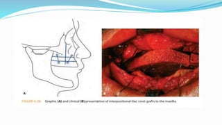
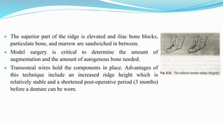
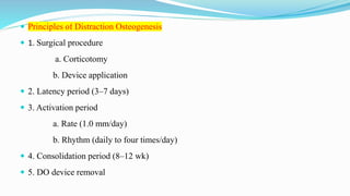
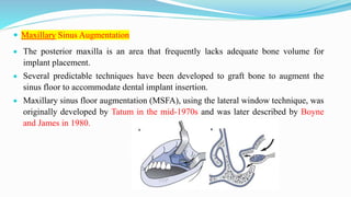
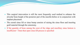
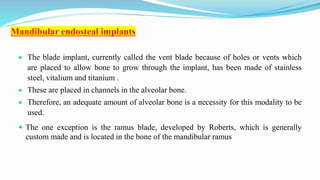
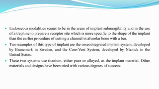
3. Introduction :
With evolution human masticatory system which is functionally efficient using natural teeth
and an omnivorous diet.
This system would be designed in a totally different fashion if it were selected based on the
model of the complete denture wearer.
Dentures are rigid pieces of acrylic resin which are shaped to fit the soft tissue covering of
the jaws and to be compatible with the functioning and ever-changing oral environment.
4. Dentures are subject to the physical laws which form the realities of this oral
environment.
No denture, regardless of how well it is constructed, can be better than the foundation
on which it is placed.
The main goal of pre-prosthetic surgery is to modify oral environment to render it
free of disease and make its form more compatible with requirements of complete
denture wearing
5. Characteristics of Ideal form:
Ridges – broad & flat
A Mucosa which is Firm & Resilient
with nicely shaped buccal and lingual
sulci which are uninterrupted by
frenae scars or redundant tissue folds.
An inter-arch distance (minimum 20
- 24 mm) and relationship which
allows room for the denture and its
components
• Vertical height minimum
5mm
• Nearly parallel
• Non-undercut, bony walls.
6. Preoperative Examination
It must first be determined that no mental or physical condition exists which would
contraindicate the wearing and use of the denture itself.
The next goal of the preoperative examination then becomes one of determining
realistic treatment goals based on the age of the patient.
The success of complete denture prosthodontics frequently depends heavily on the
patient’s attitude toward his treatment. The patient must understand and accept his
treatment if he is to cooperate postoperatively and, post prosthetically.
7. Complete radiographic evaluation using cephalometrics as well as lateral and
anteroposterior head films may be required.
Frontal and profile photographs are invaluable in evaluating and discussing potential
facial changes.
All radiographs and photographs may be obtained with correctly shaped trial bases and
wax rims in place so that the ridge relationships and facial esthetics after surgery may
be assessed.
SIGNIFICANCE OF RADIOGRAPHS
8. A complete understanding and acceptance of the limitations of denture
performance must be accomplished before treatment is begun.
Explanations before treatment are diagnostic, those after treatment are
excuse
9. SECONDARY PREOPERATIVE EXAMINATION PROCEDURES
Patients frequently have oral tissues which have been abused and distorted by their existing
malfitting prosthesis.
Often correcting the soft-tissue problems to a point where the need for surgery to these
tissues is eliminated.
The simplest procedure can be is by removal of prosthesis from oral cavity for a short
period of time accompanied by soft tissue stimulation with a soft tooth brush or damp cloth.
Patient cooperation may be difficult or impossible to obtain if the patient is socially active
and refuses to remove the dentures for an extended period of time.
10. Because of this, the procedure is often augmented by the tissue conditioning materials.
This material will more correctly extend-soft tissue coverage and will absorb some of the
stresses of function.
It allows the deformed tissue to recover its-normal form.
11. Secondary Hard Tissue Procedures
These procedures, which involve ridge augmentation and ridge relationship alterations
may require hospitalization of the patient and special pre and post-surgical care.
They are 1. Ridge augmentation procedures
2. Ridge relationship procedures
3. Implant procedures.
12. RIDGE AUGMENTATION PROCEDURES:
With increase in life expectancy there is an increase in the severely resorbed alveolar
ridge is becoming more the rule than the exception in modern day prosthodontic practice.
The goal of this procedures is to recreate an edentulous ridge with characteristics
which are compatible with requirements of denture wearing.
Many variables combine to effect the success of these techniques.
Among these are: the materials used for the augmentation, the augmentation site, surgical
design, prosthodontic design, patient interest, prosthodontic follow up and the skill of the
surgeon and the prosthodontist.
13. The grafts can be
Autografts : Graft of same person from different site.
Homografts : Graft taken from different individual of same species.
Alloplastic : artificial synthethic grafts
Xenografts : grafts taken from different species.
14. No one procedure has proven eminently successful in augmenting the ridge to an ideal
height and width with predictable stability for long periods of time.
In many instance
the rate of resorption of the augmentation > than the rate of resorption of the original
bony site with resultant minimal benefit to the patient.
15.
16. The amount of bone selected and used for the augmentation should, to some degree,
compensate for the expected loss due to resorption but should not impinge on the inter-arch
distance.
For a prosthodontic point of view, both final ridge height and width are equally important
in judging the success of the procedure.
17. In many procedures several major operations must occur:
The collection of the graft material and the oral surgical placement procedure .
Soft tissue revisions to restore the vestibule of the augmented ridge and prepare it for
dentures.
In patients with an adequate amount of bony mandible remaining, simpler and less
demanding soft tissue may be needed
Severe ridge resorption : mandible > maxilla.
However, the demands of esthetics, phonetics, retention and comfort frequently
demand a maxillary ridge augmentation procedure.
18. Osteogenesis with heamtopoeitic bone marrow
One procedure which has shown some success in the past takes advantage of the
osteogenic potential of hematopoetic bone marrow through the use of particulate bone
marrow and cancellous bone chips contained within a metal crib (mesh).
The mesh is left in place over the graft and acts to protect and contain the bone graft
(which is usually of iliac crest origin) for a period of 10 to 14 weeks.
Following removal of the mesh, soft tissue procedures are instituted to reconstruct the
vestibules which were obliterated to supply adequate soft tissue to cover the graft.
Complications include a high rate of dehiscences over the metal crib and a high
resorption rate of the graft under the prosthesis.
19. Other procedures currently used for maxillary ridge augmentation are the classic LeFort I
maxillary osteotomy with down fracture of the maxillae and the total alveolar maxillary
osteotomy which leaves the palate in place but allows downward movement of the alveolar
ridge segments.
Interpositional or inlay bone grafting with iliac crest bone used as blocks along with
particulate bone and marrow is frequently used in both of these procedures.
These two techniques might be called composite or combination procedures as they combine
a number of separate techniques.
20. Such procedures will alter the spacial relations of the ridges as well as
the ridge height and must be used with caution only after careful pre-
operative evaluation.
Vestibuloplasties are often required following the procedures.
This possibility must be discussed with the patient before surgery.
21. A maxillary splint which is constructed on the mounted diagnostic casts following cast
surgery may be used to keep the graft and soft tissue adapted during healing, to prevent
hematoma formation, and for fixation of the maxillae during the healing period.
This splint will help to ensure the proper spatial repositioning after the maxillae which is
disarticulated from the cranial base.
In the edentulous patient an opposing mandibular splint is required to interface with the
maxillary splint.
22. The splints are usually worn for a period of 6 to 8 weeks.
Dentures are not constructed before the graft has been allowed to heal
and mature for approximately 3 to 4 months.
23. Mandibular ridge augmentation
It is performed more frequently than maxillary augmentation.
In addition to restoring ridge shape, these procedures may also be
used in an attempt to strengthen the severely atrophic mandible
which is in danger of spontaneous pathologic fracture.
A delay of 6 to 12 months is frequently encountered before the
final prosthesis is delivered.
Mandibular augmentations 1. Onlay grafting
2. Inlay grafting
24. The results of these procedures have proven to be far from perfect, with success depending
on graft material and site.
The use of the rib for mandibular augmentation has fallen into disfavor due to the high
initial resorption rate of 50 per cent or more in the first 2 years after placement.
In this procedure two ribs or one rib and bone particles from the iliac crest are used.
The rib is onlayed over the ridge and the spaces remaining filled with bone particles from
the second rib or the iliac crest.
A minimum of 4 months is allowed for graft maturity before vestibuloplasties are used to
recreate the vestibules.
26. This long postoperative healing period is necessary as the functional shape of the
alveolar ridge must remodel before dentures can be constructed. This remodeling
takes place continuously.
Sanders and Cox and others have suggested inferior border rib grafting as a possible
solution to these problems.
This procedure was developed initially for the reduction of fractures of the
mandibular body.
When used on the inferior border the graft does not have to bear the weight of the
denture, the vestibules are preserved, no splint is needed and the healing period before
a denture can be worn is reduced.
27. Disadvantages
It include sensory or motor nerve deficiencies which lead to lip biting and changes in
facial appearance..
The use of the iliac crest for onlay or inlay grafting either through an extraoral or intra-
oral approach was once the procedure of choice.
This procedure has been supplanted by osteotomy techniques with interpositional
grafting.
Donor blocks of iliac crest provide the cortical bone framework and cancellous marrow
is used to fill the spaces between the segments of cut cortical bone.
28. In the horizontal osteotomy technique an adequate vertical height of mandible must
exist so that the mandible can be cut horizontally.
This cut is, placed below the level of the mandibular canal and below the mental
foramen to avoid injury to the mandibular nerve.
29. The superior part of the ridge is elevated and iliac bone blocks,
particulate bone, and marrow are sandwiched in between.
Model surgery is critical to determine the amount of
augmentation and the amount of autogenous bone needed.
Transosteal wires hold the components in place. Advantages of
this technique include an increased ridge height which is
relatively stable and a shortened post-operative period (3 months)
before a denture can be worn.
30. Disadvantages include nerve trauma, paresthesia, mandibular fracture,
and flap dehiscence.
The procedure also can not be performed if other surgical procedures
such as previous bone grafting have been done.
The vertical osteotomy was originated by Harle and modified by
Peterson and Slade.
It is used where insufficient vertical mandibular bone height is present
for the horizontal osteotomy technique but adequate bone width
(approximately 10 mm) is present.
31. In this technique, the mandible is split vertically and the lingual section is elevated to
increase the mandibular height.
Cancellous bone or particulate bone and marrow is placed to correct the contours and fill
in the gaps on the facial side of the elevated segment.
Trans-osteal wires hold the segments in place for a period of 3 to 4 months before
vestibuloplasties are performed.
32. Great care must be exercised to avoid nerve trauma.
It should be noted that this technique has lost favor recently due to the
problem of unavoidable nerve trauma and the resultant paresthesia.
33. The most recent and exciting advances in the area of ridge augmentation, both
maxillary and mandibular, involves the use of alloplastic materials either alone or in
combination with autogenous bone.
Hydroxylapatite, a bone substitute, is similar to the mineral structure of both bone
and tooth. The nonporous form is in use at this time.
It has proven to be biocompatible, nonresorbable and nonosteogenic
34. The graft material produces a bond with the bone due sometimes to a deposition of new
bone mineral on the supporting matrix of hydroxylapatite and other times to a fibrous
ingrowth.
In some patients lack of bond to the ridge and fibrous ingrowth leads to the production of
a mobile but firm and rigid body beneath the tissue.
This material also, unless mixed with cancellous bone, remains somewhat compressible,
sometimes necessitating refitting of the prosthesis over time.
When used for ridge augmentation this synthetic material is placed by tunnelling beneath
the mucosa and periosteum.
35. Ridge relationship procedures:
It is procedure in which arch size &
arch relationship to each other in
space is corrected for any
discrepancies.
Improper relationship of jaws
create problems both in function
and esthetics.
This malrelated jaws cannot be
corrected by extraction of teeth
,which can be corrected with ridge
relationship surgeries resulting
from skeletal related disharmonius
.
Malrelated jaws should be
analyzed early in the diagnostic
procedures, and surgical
corrections should be performed
prior to removal of all teeth.
36. Secondary plastic surgical procedures are sometimes required to shorten sagging muscles
left following the repositioning of their bony attachments.
Relapse tendencies in both the vertical and horizontal directions are common due to
muscle pull.
This may be decreased with the use of skeletal fixation but can never be completely and
predictably eliminated.
The final results, even after careful planning and execution, often include minor errors
which need to be corrected prosthetically.
In some instances, orthognathic surgery seem like a radical form of treatment, but for many
patients it is the only hope
37.
38. Maxillary advancement procedure
It is used to correct problem of maxillary retrusion in complete denture patients who
might have skeletal maxillary retrusion and pseudo-retrusion due to resorption of small
maxilla.
Lateral relationships are also important along with antero-posterior & vertical
relationship of arches.
The procedure of choice is the lefort I osteotomy with or without interpositional
grafting which is both stable and reliable.
Procedures producing maxillary retrusion (retropositioning) are less commonly
performed because the problem of true skeletal maxillary protrusion is less common
than retrusion.
39. In the past, a radical alveolectomy was used to correct the problem of maxillary
protrusion.
The result was a more correct bony relationship with a severe and prosthetically
uncorrectable denture base deformity.
Fortunately, this procedure rarely, if ever, used today.
40. Mandibular advancement
It involves the retrognathic mandible are not commonly performed because there is less
bone and soft tissue for the surgeon to work with.
The retrognathic mandible, however, does produce a great prosthodontic problem and
sometimes demands this procedure.
The problems of a smaller bony mandible with a smaller denture bearing area makes
control of the mechanics of complete denture prosthodontics and occlusion problematic.
Performance of this procedure has the potential to greatly improve the situation.
41. Although several procedures are currently used for mandibular advancement the sagittal
osteotomy or several of its variants performed from an intra-oral approach seems to be the
procedure of choice.
Once again, this demanding surgical technique requires an equally demanding presurgical
workup.
A thin ramus or severe mandibular atrophy in the posterior region of the mandible
contraindicates this procedure
42. If a movement greater than approximately 8 mm is required the use of bone grafts and one
of the variants techniques is necessitated.
Skeletal fixation for 6 to 8 weeks is required. Following this, prosthodontic procedures may
be performed.
Mandibular prognathism may be of an acquired nature, due to ridge resorption, or a true
skeletal abnormality which requires a mandibular retrusion procedure to correct.
While prognathism is a common finding which can, in many instances, be aided
prosthetically by careful tooth placement and control of the occlusion, the control of
esthetics is a definite and often uncontrollable problem in the severe situation.
43. The currently accepted techniques involve an osteotomy procedure in the subcondylar
region or an osteotomy in the ramus.
The body of the mandible is most often avoided in osteotomy procedures of this -nature.
The subcondylar (oblique) osteotomy involves either an extra-oral or an intra-oral
approach
44. The intra-oral approach may be used when less than 10 mm of movement is required.
With this approach, access is limited, making greater modification of the segments
difficult.
An extra-oral approach which allows good access for bone and muscle surgery can be
used when movement of 10 to 12 mm is required.
The sagittal or ramus osteotomy is similar to that discussed for mandibular
advancement procedures but is usually performed by an intra-oral approach.
It may be performed and is preferred when extreme movement (10mm) is required and
when symmetry of the segments to be moved is present
45. Rarely, an additional procedure performed in the body of the mandible may be
required when symmetry is lacking or a greater amount of movement is required of
one segment.
Skeletally based intermaxillary fixation of at least 4 to 6 weeks is required.
Surgical splints are a necessity and should be worn until definitive complete dentures
are inserted. In some instances, an accompanying procedure to the coronoid is
necessary to allow free movement of the mandible.
Relapse tendencies due to muscle imbalances are a real prosthodontic problem.
46. Even slight relapse changes may produce great changes in the occlusion and may
lead to uncontrollable forces on the denture bearing foundation.
Close follow-up is necessary, and consideration must be given to delaying definitive
prosthodontic treatment when relapse is anticipated.
47. Distraction osteogenesis (DO),
Osteodistraction, or callatosis, is a biologic process of new bone formation that
occurs between bone segments that are separated by gradual incremental traction.
This process commences when distraction forces are applied to the callous tissues
that connect the divided bone segments and continues as long as these tissues are
stretched by the applied forces.
The traction generates tension that stimulates new bone formation in the gap parallel
to the vector of distraction.
48. These factors are based upon the law of tension-stress that states that gradual traction on
living tissues creates stresses that stimulate and maintain the regeneration and continued
growth of both hard and soft tissues.
DO is a surgical-orthopedic process, rather than an isolated surgical procedure,
involving gradual separation of a fracture callous, which produces an unlimited quantity
of new bone, with associated adaptive soft tissues changes, referred to as distraction
histogenesis.
50. Principles of Distraction Osteogenesis
1. Surgical procedure
a. Corticotomy
b. Device application
2. Latency period (3–7 days)
3. Activation period
a. Rate (1.0 mm/day)
b. Rhythm (daily to four times/day)
4. Consolidation period (8–12 wk)
5. DO device removal
51. Secondary Soft Tissue Procedures
The simple soft tissue problems are often seen following tooth extraction.
These procedures are often made necessary by years of denture wearing and neglect which
injure and modify the denture basal seat area and make successful denture wearing difficult
or impossible.
These techniques are useful and are preferred over the hard tissue procedures and when
adequate bone exists beneath the soft tissue and muscle covering.
52. This bone must somehow be uncovered by modifying the relationships
of hard and soft tissues.
Thus, existing bone is made available for use in support and retention of
the denture.
53. The diagnostic work up includes pantographic and cephalometric radiographs.
These are valuable in order to observe structures which could limit the extent and success of
the extension procedure.
In the maxilla these structures are the anterior nasal spine and the malar buttresses.
In the mandible they are the mental foramina, the genial tubercles, and the inferior border of
the mandible.
54. In some instances, the surgeon will require a splint to be placed immediately following the
surgery.
This splint may be made on an over-extended impression of the involved ridge and
modified with the placement of a soft lining material at the time of surgery.
Such splints may be maintained by relining during the healing period and worn till
definitive dentures are ready for insertion.
Sometimes the existing denture may be used for this purpose.
Quite frequently however, the occlusion on the existing denture needs severe correction.
55. MAXILLARY RIDGE EXTENSION PROCEDURES
The procedures currently used to uncover existing maxillary bone and make it available for
denture retention and support involve one of the various techniques for vestibuloplasty.
These include 1. Submucous resection vestibuloplasty,
2. Secondary epithelialization vestibuloplasty
3. Soft tissue graft vestibuloplasty with oral mucosal tissue or skin.
A cut-off point of 10 mm of available maxillary bone has been set as a necessity for the
success of these procedures.
56. This procedure was originally designed as a closed procedure by' Obwegeser.
Adequate mucosal length must be available for this procedure to be successful without
disproportionate alteration of the upper lip.
If a tongue blade or mouth mirror is placed to the height of the maxillary vestibule
without distortion or inversion of the upper lip, adequate labiovestibular depth is
present.
If distortion occurs, then maxillary vestibuloplasty using split-thickness skin grafts or
laser vestibuloplasty is the appropriate procedure
Submucosal vestibuloplasty
57. It is used more frequently and greater success in the maxilla than in the mandible when
adequate bone exists under an essentially healthy mucosa.
The advantage of the procedure is that it preserves the vestibular mucosal fold without
scarring.
58. The presence of extensive fibrous or hyperplastic tissue complicates the procedure and
indicates a graft type of procedure where poor mucosa is replaced with new tissue.
The submucosal vestibuloplasty procedure performed in the mandible is similar to that
performed in the maxilla.
Both open and closed procedures may be used with great care exerted not to traumatize the
mental nerve or neurovasccular bundle.
59. Following the creation of a vertical midline incision, scissors are used to bluntly dissect
a thin mucosal layer
A second supraperiosteal dissection is created using blunt dissection
, Interposing submucosal tissue layer created by submucosal and supraperiosteal
dissections
Interposing tissue layer is divided with scissors. The mucosal attachment to the
periosteum may be increased by removal of this tissue layer
Connected submucosal and supraperiosteal dissections. Splint extended in to the
maximum height of the vestibule, placing the mucosa and periosteum in direct contact
Submucosal vestibuloplasty
60. The procedure cannot be used if epuli are present or the mucosal covering is of poor
quality. There are numerous procedures for the accomplishment of the secondary
epithelialization vestibuloplasty in the mandible.
One procedure has been called the “sulcus slide”.
In which a supraperiosteal flap is raised from an incision on the ridge side or lip side of
the sulcus.
The flap is repositioned and sutured at the depth of the, new sulcus.
The use of a splint is controversial, as it is with the maxillary procedure. Relapse is a
more severe problem than with the maxillary procedure.
61. Transpositional flap vestibuloplasty
It is sometimes called by the slang term lip-switch, may be used in either arch
although it is preferred in the mandibular arch .
It is especially indicated in people who require mandibular ridge extension
procedures but are medically unable to tolerate more extensive procedures.
In this procedure a split thickness mucosal flap is dissected from a periosteal flap.
The periosteal flap is used to cover the raw soft tissue surface and the mucosal flap to
cover the raw bony surface.
62. After elevation of the mucosal flap, the periosteum is incised at the crest
of the alveolar ridge and a subperiosteal dissection is completed on the
anterior aspect of the mandible
The periosteum is then sutured to the anterior aspect of the labial
vestibule, and the mucosal flap is sutured to the vestibular depth at the
area of the periosteal attachment.
Elevation of the mucosal flap, Periosteal incision along the crest of the
alveolar ridge.
Mucosa is sutured to the vestibular depth at the area of the periosteal
attachment
Transpositional flap vestibuloplasty
63. Advantages of this procedure include:
it is applicable in almost any patient.
the presence of a raw surface which needs a lengthy healing period is eliminated.
bony recontouring or ridge augmentation can be performed at the same time as the
lip-switch; and,
there is very little regression.
Disadvantages
It includes the nature of the new mucosal covering of the ridge which is produced and
the fact that lip distortion may occur as healing progresses.
64. The soft tissue graft vestibuloplasty
It is performed for the mandible include those using mucosa and split thickness skin.
The surgical procedure is essentially the same as that performed for the secondary
epithelialization procedure, with the soft tissue graft placed over the de-epithelialized
site.
This is usually accomplished by securing the graft, bleeding surface upward, to a newly
relined splint with tincture of benzoin or dermatome glue. Following trimming of the
excess graft material the splint is placed over the ridge and wired to place.
65. The wires are removed in 7 to 10 days.
The redundant graft material is excised and the splint relined with a soft liner
material.
This splint is worn till the final prosthesis is inserted.
66. The prosthesis should not be begun before the graft has been allowed to mature for a
minimum of 3 to 5 weeks.
Frequent recall to avoid pressure spots is necessary but will not totally prevent local areas
of pressure necrosis which must be allowed to epithelialize.
A second splint used to cover the intra-oral donor site in the case of the free mucosal
graft is sometimes also recommended when the palate is used. This splint will protect the
donor site and increase patient comfort.
This second splint is also removed in 7 to 10 days and will reveal a white surface covered
with cellular debris.
67. If a large amount of donor graft tissue is required this procedure is usually performed
in the operating room under general anesthesia.
With these patients a good consideration is the use of skin rather than mucosa.
The mucosal graft may be secured to the graft receptor site as indicated above or it
may be sutured to the site after fenestrating the graft to allow for the escape of blood
and fluids.
68. Implant
Implants can be classified into :
1. Endosteal implants , which are Ramus frame
Root form
Blade form
2.Supraperiosteal implants
3. Transosteal implants
4. Intramucosal implants
Based on attachment mechanism :
a. Osseointegration
b. Fibrointegration
69. Based on macroscopic body design of implant :
Cylinder
Thread
Perforated
Plateau
Solid
Hollow
Surgery can be done in one stage
Two stage
Immediate restoration
Emergency implant
71. Two stage surgery :
In 1st stage implants are surgically placed under the gum and the patient is made to
wait for six months for osseointegration
In 2nd stage surgery is then performed where the healing gingival former is placed
and after a week of satisfactory healing the gingival collar for emergence profile
achieved and impressions made for implant prosthesis which may be cemented or
screw retained .
72. One stage surgery :
Implant is placed and left exposed through the gum
In this condition secondary surgery is not needed and gingival former is
placed during the 1st stage.
73. Immediate implant placement VS Placement in Healed Sites
The standard protocol promulgated by Branemark and colleagues dictated that after tooth
extraction the site should be allowed to heal before implant placement.
Pioneering work by Hahn and others showed high success rates for implants placed into
the alveolus immediately after tooth extraction.
A procedure known as the emergency implant was developed and popularized as a method
used to provide an immediate implant replacement for a nonrestorable tooth.
74. The socket-shield technique
It is designed to maintain the volume and
contours of hard and soft tissue to optimize
implant placement after tooth extraction.
The technique involves preserving the buccal part
of the root and placing an implant lingual to it.
The gap between the implant and bone is filled
with graft material and the area is allowed to heal
75. Maxillary Sinus Augmentation
The posterior maxilla is an area that frequently lacks adequate bone volume for
implant placement.
Several predictable techniques have been developed to graft bone to augment the
sinus floor to accommodate dental implant insertion.
Maxillary sinus floor augmentation (MSFA), using the lateral window technique, was
originally developed by Tatum in the mid-1970s and was later described by Boyne
and James in 1980.
76. This surgical intervention is still the most frequently used method to enhance the
alveolar bone height of the posterior part of the maxilla before or in conjunction with
implant placement.
The crestal sinus lift or sinus bump consists of raising the sinus floor and inserting
graft material through the osteotomy.
If the quantity of bone among the crest of the ridge and maxillary sinus bottom is
insufficient < 5mm then open sinus lift process is specified
77. Mandibular endosteal implants
The blade implant, currently called the vent blade because of holes or vents which
are placed to allow bone to grow through the implant, has been made of stainless
steel, vitalium and titanium .
These are placed in channels in the alveolar bone.
Therefore, an adequate amount of alveolar bone is a necessity for this modality to be
used.
The one exception is the ramus blade, developed by Roberts, which is generally
custom made and is located in the bone of the mandibular ramus
78. Endosseous modalities seems to be in the areas of implant submergibility and in the use
of a trephine to prepare a receptor site which is more specific to the shape of the implant
than the earlier procedure of cutting a channel in alveolar bone with a bur.
Two examples of this type of implant are the osseointegrated implant system, developed
by Branemark in Sweden, and the Core-Vent System, developed by Niznick in the
United States.
These two systems use titanium, either pure or alloyed, as the implant material. Other
materials and designs have been tried with various degrees of success.
79. Multiple mandibular endosseous implants, regardless their design and material, suffer
from one great problem.
This is the lack of rigidity which allows movement of the implant in bone.
Such movement is destructive to bone and leads to eventual implant failure.
Attempts to create a rigid superstructure have been a part of the prosthodontic
technique for these modalities since their inception.
80. Ramus frame mandibular implant
Designed by Harold and Ralph Roberts to overcome this lack of rigidity
After examination of a large number of dried edentulous mandibles a rigid stainless
steel tripod was designed.
The tripod gains support from the symphysis in the anterior and the rami posteriorly.
Currently available in sizes 4 through 7 (as well as several custom sizes), these implants
can be adapted for use in most edentulous mandibles by bending the side rails and the
implant foot.
The foot is 30 mm long and is intended to be inserted into the most available bone in the
area of the mandibular symphysis between the mental foramina.
81. The foot is 2 mm wide and should be inserted 3 mm below the crest of the alveolar ridge.
A channel approximately 12 mm long and directed upward and laterally to avoid nerve
and vessel involvement is used to locate the posterior ends of the frame.
The rails should be placed about 5 mm above the mucosa of the remaining ridge.
Obviously the diagnostic phase is important as there must be adequate room for the
eventual final denture to be placed.
This type of implant is indicated in a patient with severe bone resorption in the posterior
areas but an adequate symphysis bone height (8 to 12 mm) to allow placement of the
anterior foot.
82. The diagnostic cast and intra-oral examination and palpation are used to select the size frame
to be used. One of the advantages of this implant is the relatively rapid, simple and
inexpensive one step surgical procedure necessary for its insertion.
Very little surgical preparation of the implant site is necessary and swelling and airway
control are less of a problem than with other modalities.
A current addition to the placement procedure is the use of one of the synthetic ridge
augmentation materials over the endosseous implantation sites to aid bone healing in the
area.
83. Congenital Deformities
Congenital deformities that affect the construction of complete dentures are usually of the cleft lip and
cleft palate type.
With more aggressive and more sophisticated surgical procedures becoming available, these patients
are being successfully treated at very young ages.
Careful dental follow up should be maintained for these patients throughout life to assure the
retention of the natural teeth.
The success of these procedures may be evidenced by the dwindling population of young edentulous
cleft patients being seen in current prosthodontic practice.
Most patients now seen are limited to un-operated adults and adults with long standing surgical
failures.
84. These patients usually present with multiple problems due to years of neglect.
They have been afraid to seek care due to fears caused by their previous surgical contacts.
When dentures are indicated extractions and other additional oral surgery is often necessary to
prepare the oral cavity.
The use of special materials in impression making, denture construction and speech aid
fabrication must be considered.
These patients can best be helped by utilizing the team approach.
85. Postoperative Procedures
After extensive surgical procedures, care should be exercised to maintain and support
the patient.
If a team concept is used, this is the primary responsibility of the surgeon but the
prosthodontist is secondarily responsible to see that the patient’s needs are being met.
Diet control might include intravenous fluid therapy or hyperalimentation with
commercially available diet supplements.
Appropriate analgesics and sedatives should be prescribed.
Oral hygiene should be maintained and careful instructions issued to assure patient
understanding
86. Oral irrigations with saline or dilute hydrogen peroxide can be accomplished.
If a water pressure type irrigator is used, it must be set at the lowest pressure setting
possible to avoid opening wounds or forcing debris into the surgical area.
In more extensive procedures antibiotic therapy should be used to control secondary
infection and steroids should be considered to reduce postoperative swelling and edema.
This tissue change may compromise the airway or lead to suture line breakdown.
Although early ambulation hastens recovery, too much exertion or heavy manual work is
contraindicated.
87. Often the most helpful remedy prescribed by the clinician is total rest and relaxation,
away from the job, for a few days following discharge from the hospital.
Splints and oral fixation are maintained in place for variable periods of time
depending on the procedures performed and the tolerance of the patient.
It is important that patients be aware of these procedures preoperatively so that they
do not become panicky when awaking from anaesthesia and fight against the
prostheses.
During the postoperative healing period, rigid discipline is needed both by the
clinician and the patient.
88. This ensures that minimal trauma is being transmitted through the prostheses to the healing
surgical sites.
In more extensive situations requiring hospitalization, postoperative care should be carried
out in the hospital until the patients are able to care for themselves.
After splints and sutures have been removed the patient should be seen jointly by the
prosthodontist and surgeon until healing is completed.
Then the patient is usually discharged to the care of the prosthodontist and followed until
bony recontouring is completed.
A transitional prosthesis used to provide esthetics and function can be constructed to carry
the patient for the 4 to 6 month period of bony healing..
89. The use of soft liners is a valuable adjunct during this period, allowing easy re-
establishment of the adaptation of the denture bases.
Removal of the prosthesis at night and at other times of the day is essential.
It must be recognized that there are numerous surgical and prosthodontic approaches to
patients with extensive loss or absence of the residual ridge.
Care, planning, patience and understanding are important ingredients in the recipe for
success with these problems.
A large number of procedures have been covered which will enable the dentist or the
dental team to better treat the complete denture patient who presents with a less than
ideal denture foundation.
90. Conclusion :
A prosthodontist should have a knowledge of all the pre-prosthetic surgeries that can
be done to restore oral tissues so that they can be accepted for complete dentures for
better retention and support
91. References
Syllabus of Complete Dentures Charles M. Heartwell, Jr. Arthur 4th
edition
Text.
Prosthodontic treatment for edentulous patients: zarb
Essentials of complete denture prosthodontics: winkler
Contemporary Implant Dentistry MISCH’S 4th Edition