SlideShare a Scribd company logo
AJM Sheet: Calcaneal
Fractures
AJM Sheet:
The standard trauma work-up again
applies with primary and secondary
surveys. The following describes
unique subjective findings,
objective findings, diagnostic
classifications and treatment
considerations.
SUBJECTIVE
Demographics:
• Men > Women: Age range generally 30-60
• Account for ~2% of all fractures: 2-10% are
bilateral
10% associated with vertebral fracture (most
commonly L1)
1% associated with pelvic fracture and
urethral trauma.
Common
mechanisms of
injury
Direct axial load
Vertical shear force/fall from height
MVC
Gastroc contraction
Stress fracture
Ballistics
Iatrogenic surgical fracture
OBJECTIVE
Physical Exam:
Pain with palpation to heel
Short, wide heel
Mondor’s Sign: Characteristic ecchymosis extending
into plantar medial foot
Hoffa’s sign: Less taut Achilles tendon on involved
side
Inability to bear weight
Must rule out compartment syndrome
IMAGING
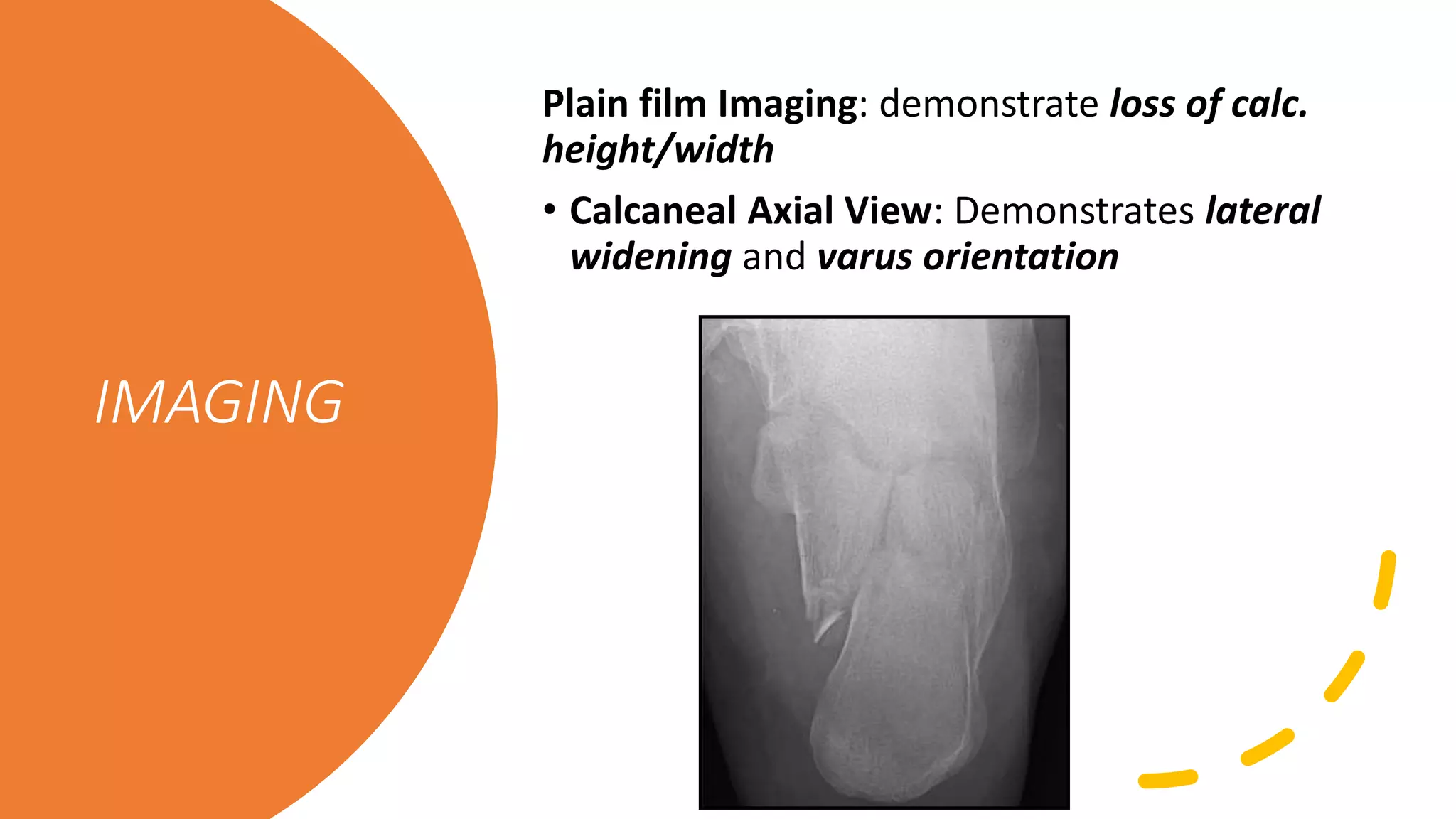
Plain film Imaging: demonstrate loss of calc.
height/width
• Calcaneal Axial View: Demonstrates lateral
widening and varus orientation
IMAGING
• Bohler’s Angle: Normally 25-40 degrees.
[Decreased with fracture]
• Critical Angle of Gissane: Normally 125-140
degrees [Increased with fracture]
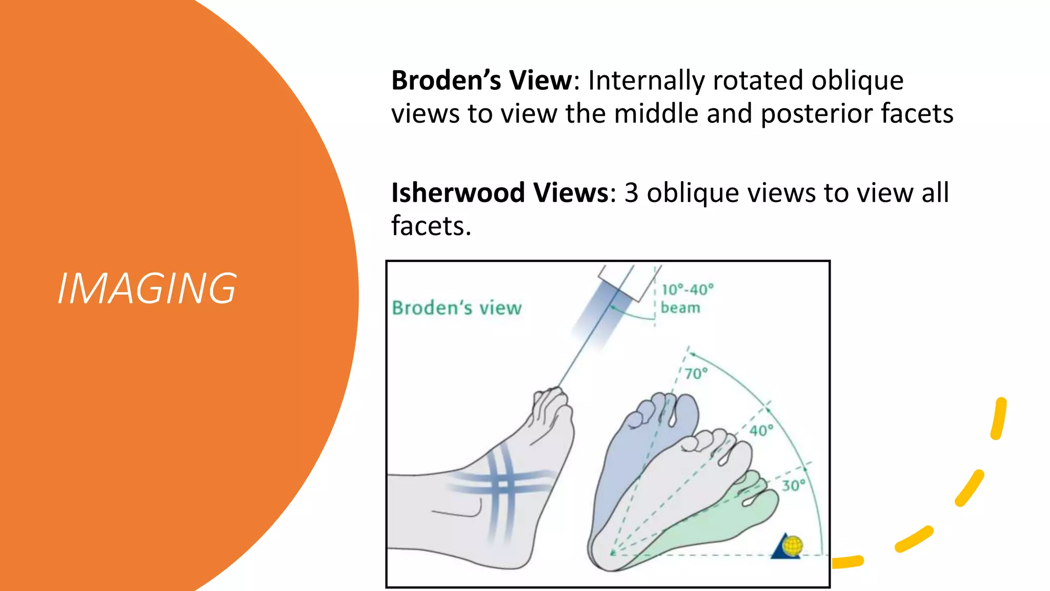
Broden’s View: Internally rotated oblique
views to view the middle and posterior facets
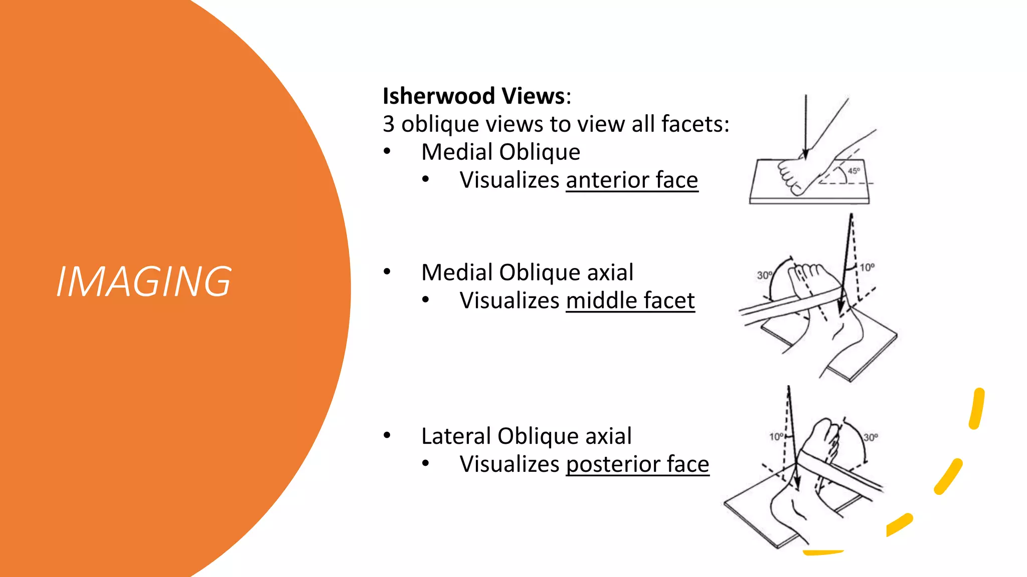
Isherwood Views: 3 oblique views to view all
facets.
IMAGING
Isherwood Views:
3 oblique views to view all facets:
• Medial Oblique
• Visualizes anterior face
• Medial Oblique axial
• Visualizes middle facet
• Lateral Oblique axial
• Visualizes posterior face
IMAGING
IMAGING
CT Scan:
Gold standard for evaluation and
surgical planning
The coronal view forms the basis of
the Sanders
Calcaneal
Fracture
classifications and
descriptions
1. Sanders Classification
2. Rowe Classification of calcaneal fractures
3. Essex-Lopresti Classification
4. Zwipp Classification
Sanders
Classification
Type “number” describes the # fragments formed
with fracture
A, B and C represent the location of fracture lines
A– Lateral
B – Center
C— Medial
Associated readings:
[Koval KJ, Sanders R. The radiographic evaluation of calcaneal fractures.
CORR. 1993 May; 290: 41-6.]
[Sanders R. Displaced intra-articular fractures of the calcaneus. JBJS-Am.
2000 Feb; 82(2): 225-50.]
Essex-Lopresti
Classification
Extra-articular (~25%)
Intra-articular (~75%)
• Tongue-type
• Joint depression
• fractures
Both intra-articular fractures
Have the same primary force, but different secondary
exit points.
[Essex-Lopresti P. The mechanism, reduction technique, and results in fractures of the os calcis.
Br J Surg 1952; 39: 395-419.]
Zwipp Classification
Assigns 2-12 points based
on:
• Number of fragments
• Number of involved
joints
• Open fracture or high
soft tissue injury
• Highly comminuted
nature, or associated
talar, cuboid, navicular
fractures [Rammelt S, Zwipp H. Calcaneus fractures: facts, controversies and
recent developments. Injury 2004; 35(5): 443-61.]
Treatment of
calcaneal
fractures
Goals of therapy are to:
• Restore calcaneal height
• Decrease calcaneal body widening (reduce
lateral wall blow-out)
• Take calcaneus out of varus
• Articular reduction.
AJM Sheet:
Appreciate the debate in the literature
between cast immobilization vs.
percutaneous reduction vs. ORIF vs.
primary arthrodesis. Possible use of
delta frame to allow for closed reduction
and balancing of soft tissue swelling pre-
operatively.
[Barei DP, et al. Fractures of the calcaneus. Orthop Clin North Am. 2002 Jan;
33(1): 263-85.]
Review the lateral extensile surgical approach
[Benirschke SK, Sangeorzan BJ. Extensive intraarticular fractures of the foot. Surgical
management of calcaneal fractures. CORR. 1993 Jul; 292: 128-134.]
AJM Sheet:
COMPLICATIONS
• Wound healing,
• Arthritis,
• Lateral ankle impingement,
• Malunion,
• Non-union, etc.
[Benirschke SK, Kramer PA. Wound healing complication in closed and open calc fractures. J
Orthop Trauma. 2004; 18(1): 1-6.]
[Cavadas PC, Landin L. Management of soft-tissue complications of the lateral approach for
calcaneal fractures. Plast Reconstr Surg. 2007; 120(2): 459-466.]
QUESTIONS AND ANSWERS
How often do
intra-articular
fractures occur?
• Approximately 75% are intra-articular
What three factors
determine the
pattern of
comminution and
location of the
fracture lines?
Position of the foot at impact
Force at impact
Bone quality
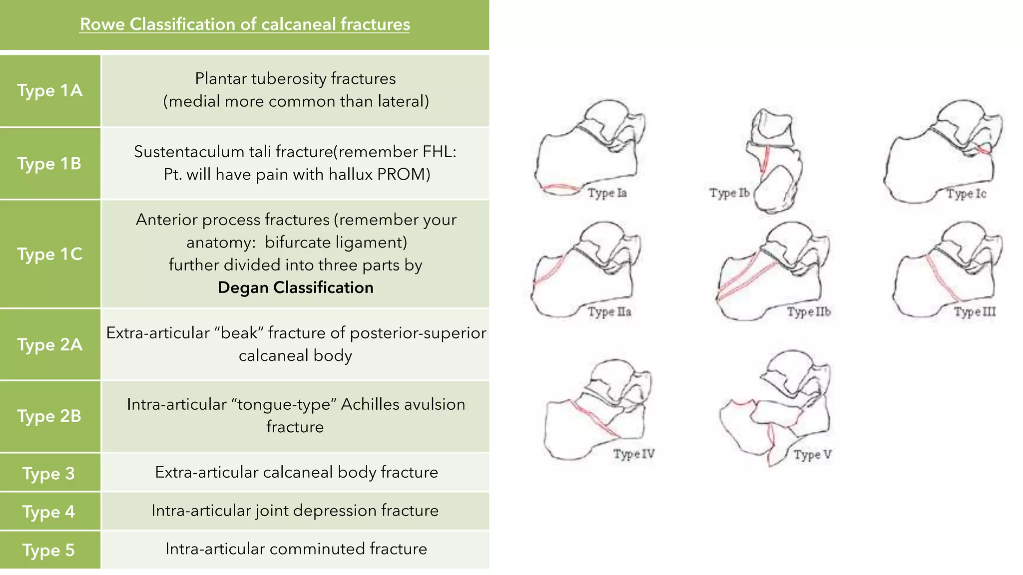
Describe the
Rowe
classification.
• Ia: plantar tuberosity fracture
• Ib: sustentaculum tali fracture
• Ic: anterior process fracture
• IIa: fracture of the posterior aspect of the
calcaneus not involving the Achilles tendon;
‘beak fracture’
IIb: avulsion fracture of the posterior aspect
of the calcaneus
• III: fracture of the body without STJ
involvement
• IV: fracture of the body with STJ involvement
• V: comminution of the body of the calcaneus
Describe the
Essex-Lopresti
classification?
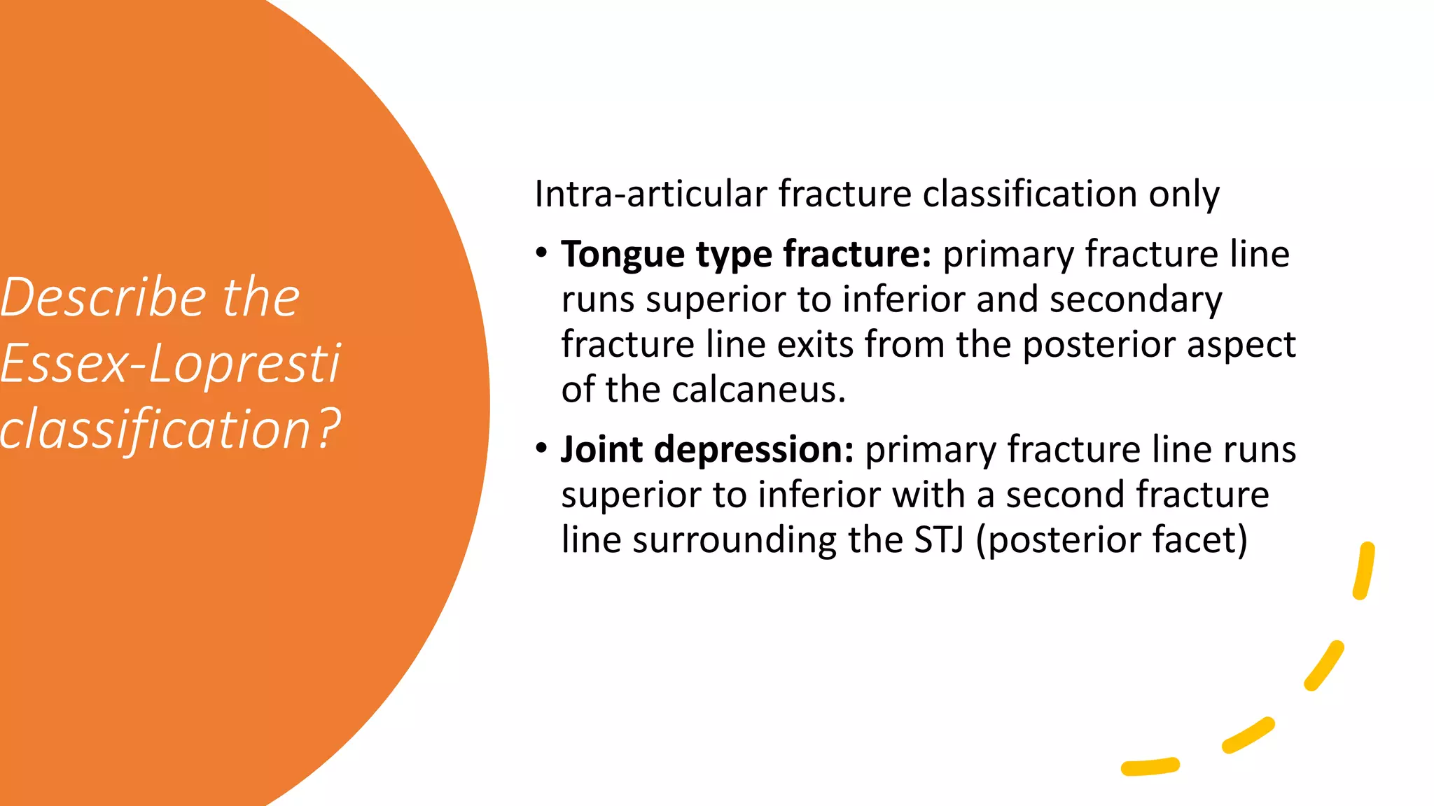
Intra-articular fracture classification only
• Tongue type fracture: primary fracture line
runs superior to inferior and secondary
fracture line exits from the posterior aspect
of the calcaneus.
• Joint depression: primary fracture line runs
superior to inferior with a second fracture
line surrounding the STJ (posterior facet)
Describe
Sanders
classification.
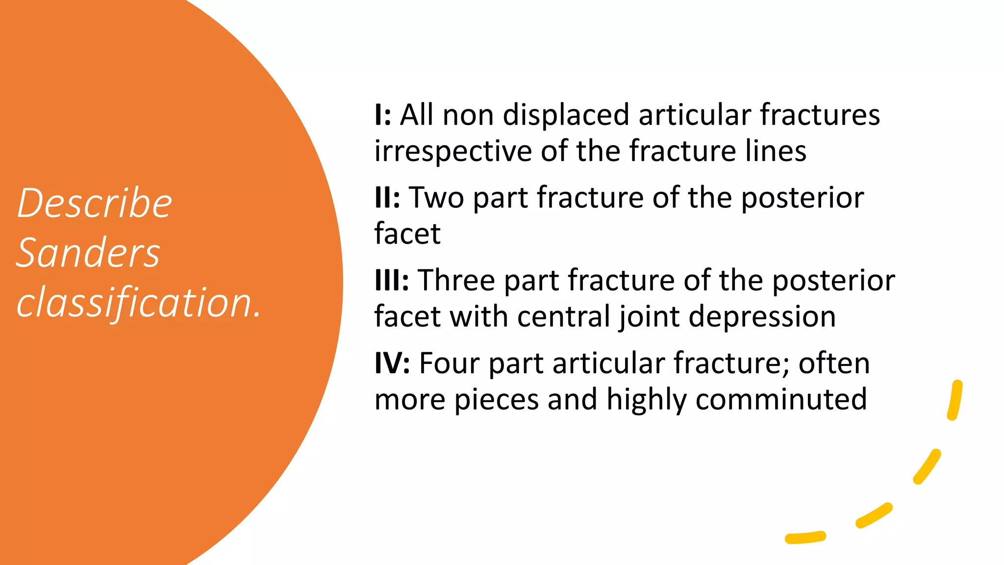
I: All non displaced articular fractures
irrespective of the fracture lines
II: Two part fracture of the posterior
facet
III: Three part fracture of the posterior
facet with central joint depression
IV: Four part articular fracture; often
more pieces and highly comminuted
How many
stages are in the
Sander’s
classification
(including
subtypes)?
Eight (I, IIA, IIB, IIC, IIIAB, IIIAC, IIIAB,
IIIAC, IV)
On which imaging
modality is the
Sanders
classification
based and what
slice is used?
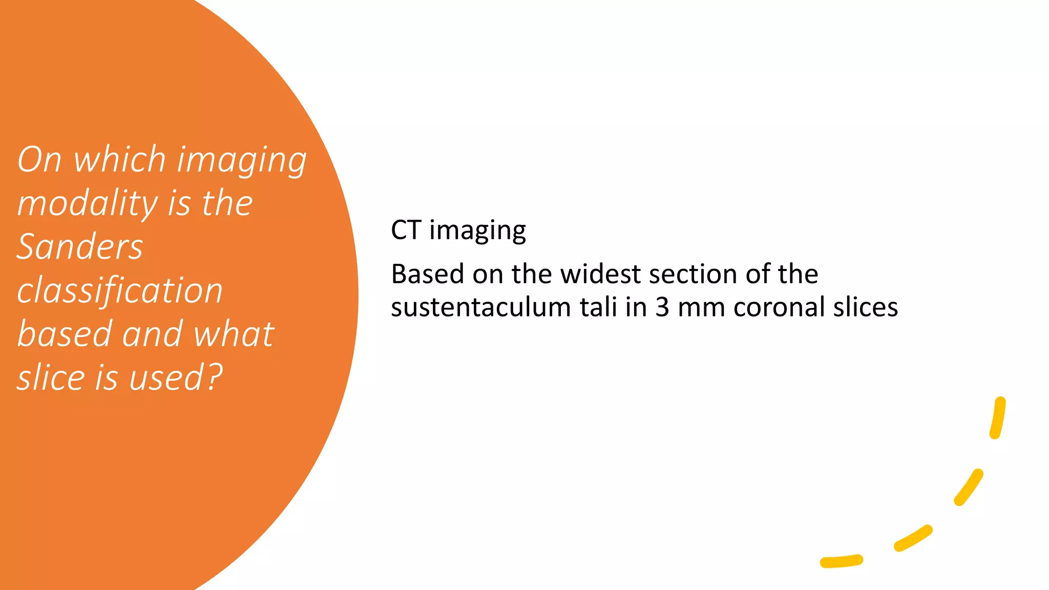
CT imaging
Based on the widest section of the
sustentaculum tali in 3 mm coronal slices
In the Sander’s
line
classification, is
line A medial or
lateral?
Lateral
The ecchymosis
seen in calcaneal
fractures is known
as what?
Mondour’s sign; this usually occurs
plantarly but can also occur distal to
both malleoli
Where are fracture
blisters most
commonly located
in calcaneal
fractures?
The medial side because during the
fracture there is predominantly
shearing and stretching of the soft
tissues on the medial side of the
foot.
What two
important angles
are associated
with calcaneal
fractures?
Bohler’s angle: Normally 20 – 40 degrees;
decreases with depression of the posterior STJ
Gissane’s angle: Normally 120 – 140 degrees;
will increase with the depression of the joint
What plain film
views would you
order and what
would you see on
each?
Lateral foot: see joint depression; evaluate
the two angles in the question above; check
for loss of height of the posterior STJ
AP foot: to evaluate all other foot bones for
additional fractures/pathology
Harris-Beath and/or Broden view: to evaluate
the posterior facet of the STJ
Lateral oblique: anterior process of the
calcaneus to check for CC joint involvement
What is a
Broden’s view
and how is it
taken? (Broden
projection I)
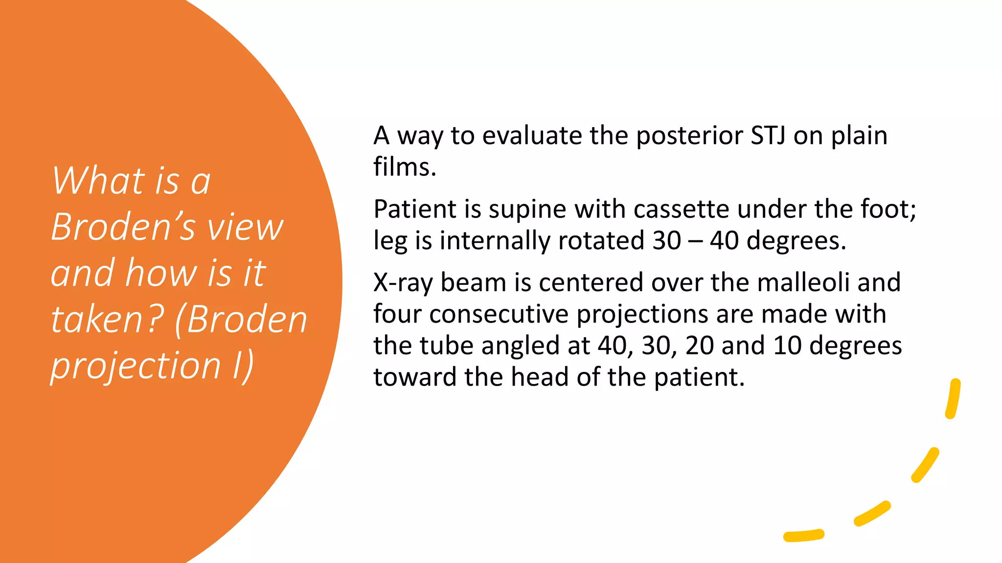
A way to evaluate the posterior STJ on plain
films.
Patient is supine with cassette under the foot;
leg is internally rotated 30 – 40 degrees.
X-ray beam is centered over the malleoli and
four consecutive projections are made with
the tube angled at 40, 30, 20 and 10 degrees
toward the head of the patient.
What are the
fragments
usually seen in
calcaneal
fractures?
• Superomedial fragment (Constant or
sustentacular fragment)
• Posterior facet fragment
(superolateral, semilunar or comet
fragment)
• Tuberosity fragment (main fragment)
• Anterior process fragment Anterior STJ
fragment
• The three important fragments that
must be reduced.
What are the
goals of ORIF
with calcaneal
fractures?
1. Restoration of length, width and
height of the calcaneus
2. Anatomic reduction of all involved
joint surfaces
3. Restitution of function by stable
osteosynthesis without joint trans-
fixation.
PEARL
• In July 2000 in the Journal of Orthopedic
Trauma there was a report of using
injectable bone cement for augmentation of
ORIF of calcaneal fractures. The authors
report using an injectable cement in the area
of the neutral triangle under the posterior
facet. This will allow for good resistance
from compression that ORIF alone can’t give.
At the end of their study, they were having
patients fully weight bear at 3 weeks post
op. This is about 10 weeks earlier than some
authors report. This could potentially be a
great tool for augmentation or internal
fixation in these fractures.
What are the
four ways to
treat calcaneal
fractures?
•Non-operative
•ORIF
•Ex-fix
•Primary STJ arthrodesis
What else should
be evaluated
when dealing
with calcaneal
fractures?
1. Proximal injuries (lower
back, spine, neck and head)
2. Bladder rupture
Which
vertebrae are
most commonly
injured?
L1, L2
• Wong (1966) found that 11% of males
with calcaneal fractures also had
vertebral
• Compression fractures
What is the
‘wrinkle test’?
A way to evaluate if the soft tissue
swelling has reduced enough for
surgical intervention
Dorsiflex and evert the foot and the
skin on the lateral side of the foot
will wrinkle
What are the
locations for the
incisions of ORIF
and the
advantages of
each?
• Lateral extensile, Modified Ollier
• Easy visualization of posterior facet and
calcaneocuboid joint
• Avoids neurovascular bundle
• Medial
• Initially popularized by McReynolds
• Easy reduction of the sustentacular fragment
• Seligson’s lateral extensile
Described by Giouild (F&A, 1984)
Some authors use both approaches so each
fragment can be adequately visualized Many
authors also use the lateral approach for which
there are many variations.
When should
primary
arthrodesis be
done?
In a highly comminuted intra
articular fracture (i.e. Sander’s type
IV)
Name ten
complications of
treatment of
calcaneal
fractures.
1. Nerve damage
2. Post traumatic arthritis
3. RSD
4. Compartment syndrome
5. Nerve entrapment
6. Wound dehiscence (with or without
calcaneal osteomyelitis)
7. Malposition after fixation
Name ten
complications
of treatment of
calcaneal
fractures.
8. Calcaneal malunion
Classified by Stephens and Sanders
Type I: large lateral exostosis with or without
extremely lateral arthrosis of the STJ
Type II: a lateral exostosis combined with major
arthrosis across the width of the STJ
Type III: a lateral exostosis, severe arthrosis of the
STJ and malunion of the calcaneal body with the
hindfoot in varus or valgus angulation
9. Peroneal tendonitis/subluxation
10. Heel pad pain, Damage to the fatty plantar heel
pad
What is the most
frequent post-op
complication with
ORIF of calcaneal
fractures?
• Wound dehiscence (cited numerous
places in the literature)
NOTE
There seems to be a large discrepancy in
outcomes following treatment of intra-
articular calcaneal fractures. This is seen
between those injuries suffered while at work
and those that are not. Since this injury
frequently occurs in the working population, it
is difficult not to include these subjects in
studies. Recently, there have been reports
alluding to this idea so hopefully in the future
we will see studies on injuries that are not
sustained at work.

More Related Content

What's hot

Fracture of Distal End Humerus.
Fracture of Distal End Humerus.Fracture of Distal End Humerus.
Fracture of Distal End Humerus.
Dr.Anshu Sharma
 
Calcaneal fractures --sito--29th aug 2015
Calcaneal fractures --sito--29th aug 2015Calcaneal fractures --sito--29th aug 2015
Calcaneal fractures --sito--29th aug 2015
Uday Bangalore
 
Talus fructures classification and managment
Talus fructures classification and managment Talus fructures classification and managment
Talus fructures classification and managment
drhakim90
 
Coronoid fracture
Coronoid fractureCoronoid fracture
Coronoid fracture
Mohamed Eldeeb
 
Talus anatomy, blood supply & fractures
Talus anatomy, blood supply & fracturesTalus anatomy, blood supply & fractures
Talus anatomy, blood supply & fractures
Hiren Divecha
 
Floating Knee
Floating KneeFloating Knee
Floating Knee
Dr Rohil Singh Kakkar
 
Calcaneal fractures
Calcaneal fracturesCalcaneal fractures
Calcaneal fractures
aviralchalise
 
TALUS FRACTURE AND MANAGEMENT.
TALUS FRACTURE AND MANAGEMENT.TALUS FRACTURE AND MANAGEMENT.
TALUS FRACTURE AND MANAGEMENT.
Dr. Anshu Sharma
 
39. tibial plafond (pilon) fractures
39. tibial plafond (pilon) fractures39. tibial plafond (pilon) fractures
39. tibial plafond (pilon) fractures
Muhammad Abdelghani
 
Principles of lock plate fixation AO
Principles of lock plate fixation AOPrinciples of lock plate fixation AO
Principles of lock plate fixation AO
Ahmad Sulong
 
Tips, tricks and pitfalls of proximal femoral nailing (PFN)
Tips, tricks and pitfalls of proximal femoral nailing (PFN)Tips, tricks and pitfalls of proximal femoral nailing (PFN)
Tips, tricks and pitfalls of proximal femoral nailing (PFN)
Puneeth Pai
 
Non union neck of femur
Non union neck of femurNon union neck of femur
Non union neck of femur
Jose Austine
 
Posterolateral corner injuries of knee joint
Posterolateral corner injuries of knee joint Posterolateral corner injuries of knee joint
Posterolateral corner injuries of knee joint
Samir Dwidmuthe
 
PROXIMAL TIBIAL FRACTURE.pptx
PROXIMAL TIBIAL FRACTURE.pptxPROXIMAL TIBIAL FRACTURE.pptx
PROXIMAL TIBIAL FRACTURE.pptx
RmsRms6
 
Calcaneal fractures
Calcaneal fracturesCalcaneal fractures
Calcaneal fractures
Ahmad Jafar
 
minimally invasive percutaneous plate osteosynthesis
minimally invasive percutaneous plate osteosynthesisminimally invasive percutaneous plate osteosynthesis
minimally invasive percutaneous plate osteosynthesisSagar Tomar
 
Fracture calcaneum and talus by dr ashutosh
Fracture calcaneum and talus by dr ashutoshFracture calcaneum and talus by dr ashutosh
Fracture calcaneum and talus by dr ashutosh
Ashutosh Kumar
 
Acetabular fracture ppt 4th sem
Acetabular fracture ppt 4th semAcetabular fracture ppt 4th sem
Acetabular fracture ppt 4th sem
ROSHAN YADAV
 
Complex elbow injury 2013
Complex elbow injury 2013Complex elbow injury 2013
Complex elbow injury 2013
Sumroeng Neti
 

What's hot (20)

pilon fractures or " plafond "
pilon fractures or  " plafond "pilon fractures or  " plafond "
pilon fractures or " plafond "
 
Fracture of Distal End Humerus.
Fracture of Distal End Humerus.Fracture of Distal End Humerus.
Fracture of Distal End Humerus.
 
Calcaneal fractures --sito--29th aug 2015
Calcaneal fractures --sito--29th aug 2015Calcaneal fractures --sito--29th aug 2015
Calcaneal fractures --sito--29th aug 2015
 
Talus fructures classification and managment
Talus fructures classification and managment Talus fructures classification and managment
Talus fructures classification and managment
 
Coronoid fracture
Coronoid fractureCoronoid fracture
Coronoid fracture
 
Talus anatomy, blood supply & fractures
Talus anatomy, blood supply & fracturesTalus anatomy, blood supply & fractures
Talus anatomy, blood supply & fractures
 
Floating Knee
Floating KneeFloating Knee
Floating Knee
 
Calcaneal fractures
Calcaneal fracturesCalcaneal fractures
Calcaneal fractures
 
TALUS FRACTURE AND MANAGEMENT.
TALUS FRACTURE AND MANAGEMENT.TALUS FRACTURE AND MANAGEMENT.
TALUS FRACTURE AND MANAGEMENT.
 
39. tibial plafond (pilon) fractures
39. tibial plafond (pilon) fractures39. tibial plafond (pilon) fractures
39. tibial plafond (pilon) fractures
 
Principles of lock plate fixation AO
Principles of lock plate fixation AOPrinciples of lock plate fixation AO
Principles of lock plate fixation AO
 
Tips, tricks and pitfalls of proximal femoral nailing (PFN)
Tips, tricks and pitfalls of proximal femoral nailing (PFN)Tips, tricks and pitfalls of proximal femoral nailing (PFN)
Tips, tricks and pitfalls of proximal femoral nailing (PFN)
 
Non union neck of femur
Non union neck of femurNon union neck of femur
Non union neck of femur
 
Posterolateral corner injuries of knee joint
Posterolateral corner injuries of knee joint Posterolateral corner injuries of knee joint
Posterolateral corner injuries of knee joint
 
PROXIMAL TIBIAL FRACTURE.pptx
PROXIMAL TIBIAL FRACTURE.pptxPROXIMAL TIBIAL FRACTURE.pptx
PROXIMAL TIBIAL FRACTURE.pptx
 
Calcaneal fractures
Calcaneal fracturesCalcaneal fractures
Calcaneal fractures
 
minimally invasive percutaneous plate osteosynthesis
minimally invasive percutaneous plate osteosynthesisminimally invasive percutaneous plate osteosynthesis
minimally invasive percutaneous plate osteosynthesis
 
Fracture calcaneum and talus by dr ashutosh
Fracture calcaneum and talus by dr ashutoshFracture calcaneum and talus by dr ashutosh
Fracture calcaneum and talus by dr ashutosh
 
Acetabular fracture ppt 4th sem
Acetabular fracture ppt 4th semAcetabular fracture ppt 4th sem
Acetabular fracture ppt 4th sem
 
Complex elbow injury 2013
Complex elbow injury 2013Complex elbow injury 2013
Complex elbow injury 2013
 

Similar to AJM Sheet: Calc Fracture

Tibial plateau fractures
Tibial plateau fracturesTibial plateau fractures
Tibial plateau fracturesYasser Alwabli
 
Proximal femoral fractures
Proximal femoral fracturesProximal femoral fractures
Proximal femoral fractures
Dr Souvik Paul
 
Inter trochanteric femoral fractures
Inter trochanteric femoral fracturesInter trochanteric femoral fractures
Inter trochanteric femoral fractures
Ihab El-Desouky
 
Pelvic fracture classification
Pelvic fracture classificationPelvic fracture classification
Pelvic fracture classification
Zahid Askar
 
L01_Hip-dislocatinos-femoral-head.ppt
L01_Hip-dislocatinos-femoral-head.pptL01_Hip-dislocatinos-femoral-head.ppt
L01_Hip-dislocatinos-femoral-head.ppt
toto798365
 
Principles of fractures on orthopedic field
Principles of fractures on orthopedic fieldPrinciples of fractures on orthopedic field
Principles of fractures on orthopedic field
ganaadyaksa
 
Fractures around elbow lateral condyle and intercondylar fractures
Fractures around elbow lateral condyle and intercondylar fracturesFractures around elbow lateral condyle and intercondylar fractures
Fractures around elbow lateral condyle and intercondylar fractures
Siddhartha Sinha
 
Ankle fracture
Ankle fracture Ankle fracture
Ankle fracture
AashishYadav35
 
Ankle and foot injuries
Ankle and foot injuriesAnkle and foot injuries
Ankle and foot injuries
Amardeep kaur
 
Fractures around elbow lateral condyle and intercondylar fractures
 Fractures around elbow lateral condyle and intercondylar fractures Fractures around elbow lateral condyle and intercondylar fractures
Fractures around elbow lateral condyle and intercondylar fractures
Siddhartha Sinha
 
THORACOLUMBAR SPINE INJURIES
THORACOLUMBAR SPINE INJURIESTHORACOLUMBAR SPINE INJURIES
THORACOLUMBAR SPINE INJURIES
Suman Subedi
 
Presentation1.pptx, radiological imaging of spinal trauma and spinal cord inj...
Presentation1.pptx, radiological imaging of spinal trauma and spinal cord inj...Presentation1.pptx, radiological imaging of spinal trauma and spinal cord inj...
Presentation1.pptx, radiological imaging of spinal trauma and spinal cord inj...Abdellah Nazeer
 
Thoracolumbar fracture for mbbs
Thoracolumbar fracture for mbbsThoracolumbar fracture for mbbs
Thoracolumbar fracture for mbbs
Dr Mizan
 
upper cervical trauma.pptx
upper cervical trauma.pptxupper cervical trauma.pptx
upper cervical trauma.pptx
Luckylogo Huang
 
pediatric fracture around elbow.pptx
pediatric fracture around elbow.pptxpediatric fracture around elbow.pptx
pediatric fracture around elbow.pptx
bishwabandhuniraula
 
Upper extremity fractures.pptx
Upper extremity fractures.pptxUpper extremity fractures.pptx
Upper extremity fractures.pptx
NathnaelCherinet
 
Distal radius fractures
Distal radius fracturesDistal radius fractures
Distal radius fractures
Dr Padma C Anand
 
Ankle Sprain
Ankle SprainAnkle Sprain
Ankle Sprain
Abdulla Kamal
 
ankle sprain presentation.pdf
ankle sprain presentation.pdfankle sprain presentation.pdf
ankle sprain presentation.pdf
AshrafHussein36
 
proximalfemoralfractures-190716152524.pptx
proximalfemoralfractures-190716152524.pptxproximalfemoralfractures-190716152524.pptx
proximalfemoralfractures-190716152524.pptx
gufp
 

Similar to AJM Sheet: Calc Fracture (20)

Tibial plateau fractures
Tibial plateau fracturesTibial plateau fractures
Tibial plateau fractures
 
Proximal femoral fractures
Proximal femoral fracturesProximal femoral fractures
Proximal femoral fractures
 
Inter trochanteric femoral fractures
Inter trochanteric femoral fracturesInter trochanteric femoral fractures
Inter trochanteric femoral fractures
 
Pelvic fracture classification
Pelvic fracture classificationPelvic fracture classification
Pelvic fracture classification
 
L01_Hip-dislocatinos-femoral-head.ppt
L01_Hip-dislocatinos-femoral-head.pptL01_Hip-dislocatinos-femoral-head.ppt
L01_Hip-dislocatinos-femoral-head.ppt
 
Principles of fractures on orthopedic field
Principles of fractures on orthopedic fieldPrinciples of fractures on orthopedic field
Principles of fractures on orthopedic field
 
Fractures around elbow lateral condyle and intercondylar fractures
Fractures around elbow lateral condyle and intercondylar fracturesFractures around elbow lateral condyle and intercondylar fractures
Fractures around elbow lateral condyle and intercondylar fractures
 
Ankle fracture
Ankle fracture Ankle fracture
Ankle fracture
 
Ankle and foot injuries
Ankle and foot injuriesAnkle and foot injuries
Ankle and foot injuries
 
Fractures around elbow lateral condyle and intercondylar fractures
 Fractures around elbow lateral condyle and intercondylar fractures Fractures around elbow lateral condyle and intercondylar fractures
Fractures around elbow lateral condyle and intercondylar fractures
 
THORACOLUMBAR SPINE INJURIES
THORACOLUMBAR SPINE INJURIESTHORACOLUMBAR SPINE INJURIES
THORACOLUMBAR SPINE INJURIES
 
Presentation1.pptx, radiological imaging of spinal trauma and spinal cord inj...
Presentation1.pptx, radiological imaging of spinal trauma and spinal cord inj...Presentation1.pptx, radiological imaging of spinal trauma and spinal cord inj...
Presentation1.pptx, radiological imaging of spinal trauma and spinal cord inj...
 
Thoracolumbar fracture for mbbs
Thoracolumbar fracture for mbbsThoracolumbar fracture for mbbs
Thoracolumbar fracture for mbbs
 
upper cervical trauma.pptx
upper cervical trauma.pptxupper cervical trauma.pptx
upper cervical trauma.pptx
 
pediatric fracture around elbow.pptx
pediatric fracture around elbow.pptxpediatric fracture around elbow.pptx
pediatric fracture around elbow.pptx
 
Upper extremity fractures.pptx
Upper extremity fractures.pptxUpper extremity fractures.pptx
Upper extremity fractures.pptx
 
Distal radius fractures
Distal radius fracturesDistal radius fractures
Distal radius fractures
 
Ankle Sprain
Ankle SprainAnkle Sprain
Ankle Sprain
 
ankle sprain presentation.pdf
ankle sprain presentation.pdfankle sprain presentation.pdf
ankle sprain presentation.pdf
 
proximalfemoralfractures-190716152524.pptx
proximalfemoralfractures-190716152524.pptxproximalfemoralfractures-190716152524.pptx
proximalfemoralfractures-190716152524.pptx
 

More from Podiatry Town

Ao fixation review questions
Ao fixation   review questionsAo fixation   review questions
Ao fixation review questions
Podiatry Town
 
Pod trivia part 1
Pod trivia part 1Pod trivia part 1
Pod trivia part 1
Podiatry Town
 
Met head avn q's
Met head avn  q'sMet head avn  q's
Met head avn q's
Podiatry Town
 
Pre op labs- elective surgery
Pre op labs- elective surgeryPre op labs- elective surgery
Pre op labs- elective surgery
Podiatry Town
 
AJM Sheet: Navicular fx
AJM Sheet: Navicular fxAJM Sheet: Navicular fx
AJM Sheet: Navicular fx
Podiatry Town
 
AJM Sheet: Lisfranc Trauma
AJM Sheet: Lisfranc TraumaAJM Sheet: Lisfranc Trauma
AJM Sheet: Lisfranc Trauma
Podiatry Town
 
AJM Sheet: Digital fracture
AJM Sheet: Digital fractureAJM Sheet: Digital fracture
AJM Sheet: Digital fracture
Podiatry Town
 
AJM Sheet: bone healing
AJM Sheet: bone healingAJM Sheet: bone healing
AJM Sheet: bone healing
Podiatry Town
 
AJM Sheet: Peri-op
AJM Sheet: Peri-opAJM Sheet: Peri-op
AJM Sheet: Peri-op
Podiatry Town
 
Ajm sheet dfi
Ajm sheet  dfiAjm sheet  dfi
Ajm sheet dfi
Podiatry Town
 
AJM Sheet: Ankle Fracture
AJM Sheet:  Ankle FractureAJM Sheet:  Ankle Fracture
AJM Sheet: Ankle Fracture
Podiatry Town
 
AJM Sheet: Talar fracture
AJM Sheet: Talar fractureAJM Sheet: Talar fracture
AJM Sheet: Talar fracture
Podiatry Town
 
Dvt work up
Dvt work upDvt work up
Dvt work up
Podiatry Town
 
Charcot case study and review questions
Charcot case study and review questionsCharcot case study and review questions
Charcot case study and review questions
Podiatry Town
 
Bunion work - up
Bunion work - upBunion work - up
Bunion work - up
Podiatry Town
 
AJM Sheet: 5 Podiatric Emergencies
AJM Sheet: 5 Podiatric EmergenciesAJM Sheet: 5 Podiatric Emergencies
AJM Sheet: 5 Podiatric Emergencies
Podiatry Town
 
AJM Sheet: pes cavus
AJM Sheet: pes cavusAJM Sheet: pes cavus
AJM Sheet: pes cavus
Podiatry Town
 
AJM Sheet: Digital Deformity Work-up
AJM Sheet: Digital Deformity Work-upAJM Sheet: Digital Deformity Work-up
AJM Sheet: Digital Deformity Work-up
Podiatry Town
 
AJM Sheet: HAV Work-up
AJM Sheet: HAV Work-upAJM Sheet: HAV Work-up
AJM Sheet: HAV Work-up
Podiatry Town
 
AJM Sheet: Ankle Equinus
AJM Sheet:  Ankle EquinusAJM Sheet:  Ankle Equinus
AJM Sheet: Ankle Equinus
Podiatry Town
 

More from Podiatry Town (20)

Ao fixation review questions
Ao fixation   review questionsAo fixation   review questions
Ao fixation review questions
 
Pod trivia part 1
Pod trivia part 1Pod trivia part 1
Pod trivia part 1
 
Met head avn q's
Met head avn  q'sMet head avn  q's
Met head avn q's
 
Pre op labs- elective surgery
Pre op labs- elective surgeryPre op labs- elective surgery
Pre op labs- elective surgery
 
AJM Sheet: Navicular fx
AJM Sheet: Navicular fxAJM Sheet: Navicular fx
AJM Sheet: Navicular fx
 
AJM Sheet: Lisfranc Trauma
AJM Sheet: Lisfranc TraumaAJM Sheet: Lisfranc Trauma
AJM Sheet: Lisfranc Trauma
 
AJM Sheet: Digital fracture
AJM Sheet: Digital fractureAJM Sheet: Digital fracture
AJM Sheet: Digital fracture
 
AJM Sheet: bone healing
AJM Sheet: bone healingAJM Sheet: bone healing
AJM Sheet: bone healing
 
AJM Sheet: Peri-op
AJM Sheet: Peri-opAJM Sheet: Peri-op
AJM Sheet: Peri-op
 
Ajm sheet dfi
Ajm sheet  dfiAjm sheet  dfi
Ajm sheet dfi
 
AJM Sheet: Ankle Fracture
AJM Sheet:  Ankle FractureAJM Sheet:  Ankle Fracture
AJM Sheet: Ankle Fracture
 
AJM Sheet: Talar fracture
AJM Sheet: Talar fractureAJM Sheet: Talar fracture
AJM Sheet: Talar fracture
 
Dvt work up
Dvt work upDvt work up
Dvt work up
 
Charcot case study and review questions
Charcot case study and review questionsCharcot case study and review questions
Charcot case study and review questions
 
Bunion work - up
Bunion work - upBunion work - up
Bunion work - up
 
AJM Sheet: 5 Podiatric Emergencies
AJM Sheet: 5 Podiatric EmergenciesAJM Sheet: 5 Podiatric Emergencies
AJM Sheet: 5 Podiatric Emergencies
 
AJM Sheet: pes cavus
AJM Sheet: pes cavusAJM Sheet: pes cavus
AJM Sheet: pes cavus
 
AJM Sheet: Digital Deformity Work-up
AJM Sheet: Digital Deformity Work-upAJM Sheet: Digital Deformity Work-up
AJM Sheet: Digital Deformity Work-up
 
AJM Sheet: HAV Work-up
AJM Sheet: HAV Work-upAJM Sheet: HAV Work-up
AJM Sheet: HAV Work-up
 
AJM Sheet: Ankle Equinus
AJM Sheet:  Ankle EquinusAJM Sheet:  Ankle Equinus
AJM Sheet: Ankle Equinus
 

Recently uploaded

How to Make a Field invisible in Odoo 17
How to Make a Field invisible in Odoo 17How to Make a Field invisible in Odoo 17
How to Make a Field invisible in Odoo 17
Celine George
 
Welcome to TechSoup New Member Orientation and Q&A (May 2024).pdf
Welcome to TechSoup   New Member Orientation and Q&A (May 2024).pdfWelcome to TechSoup   New Member Orientation and Q&A (May 2024).pdf
Welcome to TechSoup New Member Orientation and Q&A (May 2024).pdf
TechSoup
 
A Strategic Approach: GenAI in Education
A Strategic Approach: GenAI in EducationA Strategic Approach: GenAI in Education
A Strategic Approach: GenAI in Education
Peter Windle
 
Guidance_and_Counselling.pdf B.Ed. 4th Semester
Guidance_and_Counselling.pdf B.Ed. 4th SemesterGuidance_and_Counselling.pdf B.Ed. 4th Semester
Guidance_and_Counselling.pdf B.Ed. 4th Semester
Atul Kumar Singh
 
1.4 modern child centered education - mahatma gandhi-2.pptx
1.4 modern child centered education - mahatma gandhi-2.pptx1.4 modern child centered education - mahatma gandhi-2.pptx
1.4 modern child centered education - mahatma gandhi-2.pptx
JosvitaDsouza2
 
Sha'Carri Richardson Presentation 202345
Sha'Carri Richardson Presentation 202345Sha'Carri Richardson Presentation 202345
Sha'Carri Richardson Presentation 202345
beazzy04
 
CLASS 11 CBSE B.St Project AIDS TO TRADE - INSURANCE
CLASS 11 CBSE B.St Project AIDS TO TRADE - INSURANCECLASS 11 CBSE B.St Project AIDS TO TRADE - INSURANCE
CLASS 11 CBSE B.St Project AIDS TO TRADE - INSURANCE
BhavyaRajput3
 
Adversarial Attention Modeling for Multi-dimensional Emotion Regression.pdf
Adversarial Attention Modeling for Multi-dimensional Emotion Regression.pdfAdversarial Attention Modeling for Multi-dimensional Emotion Regression.pdf
Adversarial Attention Modeling for Multi-dimensional Emotion Regression.pdf
Po-Chuan Chen
 
Polish students' mobility in the Czech Republic
Polish students' mobility in the Czech RepublicPolish students' mobility in the Czech Republic
Polish students' mobility in the Czech Republic
Anna Sz.
 
The basics of sentences session 5pptx.pptx
The basics of sentences session 5pptx.pptxThe basics of sentences session 5pptx.pptx
The basics of sentences session 5pptx.pptx
heathfieldcps1
 
Acetabularia Information For Class 9 .docx
Acetabularia Information For Class 9  .docxAcetabularia Information For Class 9  .docx
Acetabularia Information For Class 9 .docx
vaibhavrinwa19
 
678020731-Sumas-y-Restas-Para-Colorear.pdf
678020731-Sumas-y-Restas-Para-Colorear.pdf678020731-Sumas-y-Restas-Para-Colorear.pdf
678020731-Sumas-y-Restas-Para-Colorear.pdf
CarlosHernanMontoyab2
 
June 3, 2024 Anti-Semitism Letter Sent to MIT President Kornbluth and MIT Cor...
June 3, 2024 Anti-Semitism Letter Sent to MIT President Kornbluth and MIT Cor...June 3, 2024 Anti-Semitism Letter Sent to MIT President Kornbluth and MIT Cor...
June 3, 2024 Anti-Semitism Letter Sent to MIT President Kornbluth and MIT Cor...
Levi Shapiro
 
The Roman Empire A Historical Colossus.pdf
The Roman Empire A Historical Colossus.pdfThe Roman Empire A Historical Colossus.pdf
The Roman Empire A Historical Colossus.pdf
kaushalkr1407
 
Unit 2- Research Aptitude (UGC NET Paper I).pdf
Unit 2- Research Aptitude (UGC NET Paper I).pdfUnit 2- Research Aptitude (UGC NET Paper I).pdf
Unit 2- Research Aptitude (UGC NET Paper I).pdf
Thiyagu K
 
Supporting (UKRI) OA monographs at Salford.pptx
Supporting (UKRI) OA monographs at Salford.pptxSupporting (UKRI) OA monographs at Salford.pptx
Supporting (UKRI) OA monographs at Salford.pptx
Jisc
 
Palestine last event orientationfvgnh .pptx
Palestine last event orientationfvgnh .pptxPalestine last event orientationfvgnh .pptx
Palestine last event orientationfvgnh .pptx
RaedMohamed3
 
Lapbook sobre os Regimes Totalitários.pdf
Lapbook sobre os Regimes Totalitários.pdfLapbook sobre os Regimes Totalitários.pdf
Lapbook sobre os Regimes Totalitários.pdf
Jean Carlos Nunes Paixão
 
Overview on Edible Vaccine: Pros & Cons with Mechanism
Overview on Edible Vaccine: Pros & Cons with MechanismOverview on Edible Vaccine: Pros & Cons with Mechanism
Overview on Edible Vaccine: Pros & Cons with Mechanism
DeeptiGupta154
 
Additional Benefits for Employee Website.pdf
Additional Benefits for Employee Website.pdfAdditional Benefits for Employee Website.pdf
Additional Benefits for Employee Website.pdf
joachimlavalley1
 

Recently uploaded (20)

How to Make a Field invisible in Odoo 17
How to Make a Field invisible in Odoo 17How to Make a Field invisible in Odoo 17
How to Make a Field invisible in Odoo 17
 
Welcome to TechSoup New Member Orientation and Q&A (May 2024).pdf
Welcome to TechSoup   New Member Orientation and Q&A (May 2024).pdfWelcome to TechSoup   New Member Orientation and Q&A (May 2024).pdf
Welcome to TechSoup New Member Orientation and Q&A (May 2024).pdf
 
A Strategic Approach: GenAI in Education
A Strategic Approach: GenAI in EducationA Strategic Approach: GenAI in Education
A Strategic Approach: GenAI in Education
 
Guidance_and_Counselling.pdf B.Ed. 4th Semester
Guidance_and_Counselling.pdf B.Ed. 4th SemesterGuidance_and_Counselling.pdf B.Ed. 4th Semester
Guidance_and_Counselling.pdf B.Ed. 4th Semester
 
1.4 modern child centered education - mahatma gandhi-2.pptx
1.4 modern child centered education - mahatma gandhi-2.pptx1.4 modern child centered education - mahatma gandhi-2.pptx
1.4 modern child centered education - mahatma gandhi-2.pptx
 
Sha'Carri Richardson Presentation 202345
Sha'Carri Richardson Presentation 202345Sha'Carri Richardson Presentation 202345
Sha'Carri Richardson Presentation 202345
 
CLASS 11 CBSE B.St Project AIDS TO TRADE - INSURANCE
CLASS 11 CBSE B.St Project AIDS TO TRADE - INSURANCECLASS 11 CBSE B.St Project AIDS TO TRADE - INSURANCE
CLASS 11 CBSE B.St Project AIDS TO TRADE - INSURANCE
 
Adversarial Attention Modeling for Multi-dimensional Emotion Regression.pdf
Adversarial Attention Modeling for Multi-dimensional Emotion Regression.pdfAdversarial Attention Modeling for Multi-dimensional Emotion Regression.pdf
Adversarial Attention Modeling for Multi-dimensional Emotion Regression.pdf
 
Polish students' mobility in the Czech Republic
Polish students' mobility in the Czech RepublicPolish students' mobility in the Czech Republic
Polish students' mobility in the Czech Republic
 
The basics of sentences session 5pptx.pptx
The basics of sentences session 5pptx.pptxThe basics of sentences session 5pptx.pptx
The basics of sentences session 5pptx.pptx
 
Acetabularia Information For Class 9 .docx
Acetabularia Information For Class 9  .docxAcetabularia Information For Class 9  .docx
Acetabularia Information For Class 9 .docx
 
678020731-Sumas-y-Restas-Para-Colorear.pdf
678020731-Sumas-y-Restas-Para-Colorear.pdf678020731-Sumas-y-Restas-Para-Colorear.pdf
678020731-Sumas-y-Restas-Para-Colorear.pdf
 
June 3, 2024 Anti-Semitism Letter Sent to MIT President Kornbluth and MIT Cor...
June 3, 2024 Anti-Semitism Letter Sent to MIT President Kornbluth and MIT Cor...June 3, 2024 Anti-Semitism Letter Sent to MIT President Kornbluth and MIT Cor...
June 3, 2024 Anti-Semitism Letter Sent to MIT President Kornbluth and MIT Cor...
 
The Roman Empire A Historical Colossus.pdf
The Roman Empire A Historical Colossus.pdfThe Roman Empire A Historical Colossus.pdf
The Roman Empire A Historical Colossus.pdf
 
Unit 2- Research Aptitude (UGC NET Paper I).pdf
Unit 2- Research Aptitude (UGC NET Paper I).pdfUnit 2- Research Aptitude (UGC NET Paper I).pdf
Unit 2- Research Aptitude (UGC NET Paper I).pdf
 
Supporting (UKRI) OA monographs at Salford.pptx
Supporting (UKRI) OA monographs at Salford.pptxSupporting (UKRI) OA monographs at Salford.pptx
Supporting (UKRI) OA monographs at Salford.pptx
 
Palestine last event orientationfvgnh .pptx
Palestine last event orientationfvgnh .pptxPalestine last event orientationfvgnh .pptx
Palestine last event orientationfvgnh .pptx
 
Lapbook sobre os Regimes Totalitários.pdf
Lapbook sobre os Regimes Totalitários.pdfLapbook sobre os Regimes Totalitários.pdf
Lapbook sobre os Regimes Totalitários.pdf
 
Overview on Edible Vaccine: Pros & Cons with Mechanism
Overview on Edible Vaccine: Pros & Cons with MechanismOverview on Edible Vaccine: Pros & Cons with Mechanism
Overview on Edible Vaccine: Pros & Cons with Mechanism
 
Additional Benefits for Employee Website.pdf
Additional Benefits for Employee Website.pdfAdditional Benefits for Employee Website.pdf
Additional Benefits for Employee Website.pdf
 

AJM Sheet: Calc Fracture

  • 2. AJM Sheet: The standard trauma work-up again applies with primary and secondary surveys. The following describes unique subjective findings, objective findings, diagnostic classifications and treatment considerations.
  • 3. SUBJECTIVE Demographics: • Men > Women: Age range generally 30-60 • Account for ~2% of all fractures: 2-10% are bilateral 10% associated with vertebral fracture (most commonly L1) 1% associated with pelvic fracture and urethral trauma.
  • 4. Common mechanisms of injury Direct axial load Vertical shear force/fall from height MVC Gastroc contraction Stress fracture Ballistics Iatrogenic surgical fracture
  • 5. OBJECTIVE Physical Exam: Pain with palpation to heel Short, wide heel Mondor’s Sign: Characteristic ecchymosis extending into plantar medial foot Hoffa’s sign: Less taut Achilles tendon on involved side Inability to bear weight Must rule out compartment syndrome
  • 6. IMAGING Plain film Imaging: demonstrate loss of calc. height/width • Calcaneal Axial View: Demonstrates lateral widening and varus orientation
  • 7. IMAGING • Bohler’s Angle: Normally 25-40 degrees. [Decreased with fracture] • Critical Angle of Gissane: Normally 125-140 degrees [Increased with fracture]
  • 8. Broden’s View: Internally rotated oblique views to view the middle and posterior facets Isherwood Views: 3 oblique views to view all facets. IMAGING
  • 9. Isherwood Views: 3 oblique views to view all facets: • Medial Oblique • Visualizes anterior face • Medial Oblique axial • Visualizes middle facet • Lateral Oblique axial • Visualizes posterior face IMAGING
  • 10. IMAGING CT Scan: Gold standard for evaluation and surgical planning The coronal view forms the basis of the Sanders
  • 11. Calcaneal Fracture classifications and descriptions 1. Sanders Classification 2. Rowe Classification of calcaneal fractures 3. Essex-Lopresti Classification 4. Zwipp Classification
  • 12. Sanders Classification Type “number” describes the # fragments formed with fracture A, B and C represent the location of fracture lines A– Lateral B – Center C— Medial Associated readings: [Koval KJ, Sanders R. The radiographic evaluation of calcaneal fractures. CORR. 1993 May; 290: 41-6.] [Sanders R. Displaced intra-articular fractures of the calcaneus. JBJS-Am. 2000 Feb; 82(2): 225-50.]
  • 13.
  • 14.
  • 15.
  • 16. Essex-Lopresti Classification Extra-articular (~25%) Intra-articular (~75%) • Tongue-type • Joint depression • fractures Both intra-articular fractures Have the same primary force, but different secondary exit points. [Essex-Lopresti P. The mechanism, reduction technique, and results in fractures of the os calcis. Br J Surg 1952; 39: 395-419.]
  • 17. Zwipp Classification Assigns 2-12 points based on: • Number of fragments • Number of involved joints • Open fracture or high soft tissue injury • Highly comminuted nature, or associated talar, cuboid, navicular fractures [Rammelt S, Zwipp H. Calcaneus fractures: facts, controversies and recent developments. Injury 2004; 35(5): 443-61.]
  • 18. Treatment of calcaneal fractures Goals of therapy are to: • Restore calcaneal height • Decrease calcaneal body widening (reduce lateral wall blow-out) • Take calcaneus out of varus • Articular reduction.
  • 19. AJM Sheet: Appreciate the debate in the literature between cast immobilization vs. percutaneous reduction vs. ORIF vs. primary arthrodesis. Possible use of delta frame to allow for closed reduction and balancing of soft tissue swelling pre- operatively. [Barei DP, et al. Fractures of the calcaneus. Orthop Clin North Am. 2002 Jan; 33(1): 263-85.] Review the lateral extensile surgical approach [Benirschke SK, Sangeorzan BJ. Extensive intraarticular fractures of the foot. Surgical management of calcaneal fractures. CORR. 1993 Jul; 292: 128-134.]
  • 21. COMPLICATIONS • Wound healing, • Arthritis, • Lateral ankle impingement, • Malunion, • Non-union, etc. [Benirschke SK, Kramer PA. Wound healing complication in closed and open calc fractures. J Orthop Trauma. 2004; 18(1): 1-6.] [Cavadas PC, Landin L. Management of soft-tissue complications of the lateral approach for calcaneal fractures. Plast Reconstr Surg. 2007; 120(2): 459-466.]
  • 23. How often do intra-articular fractures occur? • Approximately 75% are intra-articular
  • 24. What three factors determine the pattern of comminution and location of the fracture lines? Position of the foot at impact Force at impact Bone quality
  • 25. Describe the Rowe classification. • Ia: plantar tuberosity fracture • Ib: sustentaculum tali fracture • Ic: anterior process fracture • IIa: fracture of the posterior aspect of the calcaneus not involving the Achilles tendon; ‘beak fracture’ IIb: avulsion fracture of the posterior aspect of the calcaneus • III: fracture of the body without STJ involvement • IV: fracture of the body with STJ involvement • V: comminution of the body of the calcaneus
  • 26. Describe the Essex-Lopresti classification? Intra-articular fracture classification only • Tongue type fracture: primary fracture line runs superior to inferior and secondary fracture line exits from the posterior aspect of the calcaneus. • Joint depression: primary fracture line runs superior to inferior with a second fracture line surrounding the STJ (posterior facet)
  • 27. Describe Sanders classification. I: All non displaced articular fractures irrespective of the fracture lines II: Two part fracture of the posterior facet III: Three part fracture of the posterior facet with central joint depression IV: Four part articular fracture; often more pieces and highly comminuted
  • 28. How many stages are in the Sander’s classification (including subtypes)? Eight (I, IIA, IIB, IIC, IIIAB, IIIAC, IIIAB, IIIAC, IV)
  • 29. On which imaging modality is the Sanders classification based and what slice is used? CT imaging Based on the widest section of the sustentaculum tali in 3 mm coronal slices
  • 30. In the Sander’s line classification, is line A medial or lateral? Lateral
  • 31. The ecchymosis seen in calcaneal fractures is known as what? Mondour’s sign; this usually occurs plantarly but can also occur distal to both malleoli
  • 32. Where are fracture blisters most commonly located in calcaneal fractures? The medial side because during the fracture there is predominantly shearing and stretching of the soft tissues on the medial side of the foot.
  • 33. What two important angles are associated with calcaneal fractures? Bohler’s angle: Normally 20 – 40 degrees; decreases with depression of the posterior STJ Gissane’s angle: Normally 120 – 140 degrees; will increase with the depression of the joint
  • 34. What plain film views would you order and what would you see on each? Lateral foot: see joint depression; evaluate the two angles in the question above; check for loss of height of the posterior STJ AP foot: to evaluate all other foot bones for additional fractures/pathology Harris-Beath and/or Broden view: to evaluate the posterior facet of the STJ Lateral oblique: anterior process of the calcaneus to check for CC joint involvement
  • 35. What is a Broden’s view and how is it taken? (Broden projection I) A way to evaluate the posterior STJ on plain films. Patient is supine with cassette under the foot; leg is internally rotated 30 – 40 degrees. X-ray beam is centered over the malleoli and four consecutive projections are made with the tube angled at 40, 30, 20 and 10 degrees toward the head of the patient.
  • 36. What are the fragments usually seen in calcaneal fractures? • Superomedial fragment (Constant or sustentacular fragment) • Posterior facet fragment (superolateral, semilunar or comet fragment) • Tuberosity fragment (main fragment) • Anterior process fragment Anterior STJ fragment • The three important fragments that must be reduced.
  • 37. What are the goals of ORIF with calcaneal fractures? 1. Restoration of length, width and height of the calcaneus 2. Anatomic reduction of all involved joint surfaces 3. Restitution of function by stable osteosynthesis without joint trans- fixation.
  • 38. PEARL • In July 2000 in the Journal of Orthopedic Trauma there was a report of using injectable bone cement for augmentation of ORIF of calcaneal fractures. The authors report using an injectable cement in the area of the neutral triangle under the posterior facet. This will allow for good resistance from compression that ORIF alone can’t give. At the end of their study, they were having patients fully weight bear at 3 weeks post op. This is about 10 weeks earlier than some authors report. This could potentially be a great tool for augmentation or internal fixation in these fractures.
  • 39. What are the four ways to treat calcaneal fractures? •Non-operative •ORIF •Ex-fix •Primary STJ arthrodesis
  • 40. What else should be evaluated when dealing with calcaneal fractures? 1. Proximal injuries (lower back, spine, neck and head) 2. Bladder rupture
  • 41. Which vertebrae are most commonly injured? L1, L2 • Wong (1966) found that 11% of males with calcaneal fractures also had vertebral • Compression fractures
  • 42. What is the ‘wrinkle test’? A way to evaluate if the soft tissue swelling has reduced enough for surgical intervention Dorsiflex and evert the foot and the skin on the lateral side of the foot will wrinkle
  • 43. What are the locations for the incisions of ORIF and the advantages of each? • Lateral extensile, Modified Ollier • Easy visualization of posterior facet and calcaneocuboid joint • Avoids neurovascular bundle • Medial • Initially popularized by McReynolds • Easy reduction of the sustentacular fragment • Seligson’s lateral extensile Described by Giouild (F&A, 1984) Some authors use both approaches so each fragment can be adequately visualized Many authors also use the lateral approach for which there are many variations.
  • 44. When should primary arthrodesis be done? In a highly comminuted intra articular fracture (i.e. Sander’s type IV)
  • 45. Name ten complications of treatment of calcaneal fractures. 1. Nerve damage 2. Post traumatic arthritis 3. RSD 4. Compartment syndrome 5. Nerve entrapment 6. Wound dehiscence (with or without calcaneal osteomyelitis) 7. Malposition after fixation
  • 46. Name ten complications of treatment of calcaneal fractures. 8. Calcaneal malunion Classified by Stephens and Sanders Type I: large lateral exostosis with or without extremely lateral arthrosis of the STJ Type II: a lateral exostosis combined with major arthrosis across the width of the STJ Type III: a lateral exostosis, severe arthrosis of the STJ and malunion of the calcaneal body with the hindfoot in varus or valgus angulation 9. Peroneal tendonitis/subluxation 10. Heel pad pain, Damage to the fatty plantar heel pad
  • 47. What is the most frequent post-op complication with ORIF of calcaneal fractures? • Wound dehiscence (cited numerous places in the literature)
  • 48. NOTE There seems to be a large discrepancy in outcomes following treatment of intra- articular calcaneal fractures. This is seen between those injuries suffered while at work and those that are not. Since this injury frequently occurs in the working population, it is difficult not to include these subjects in studies. Recently, there have been reports alluding to this idea so hopefully in the future we will see studies on injuries that are not sustained at work.