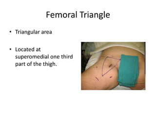
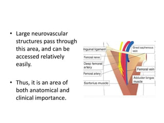
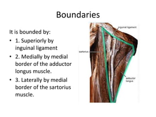
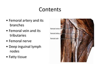
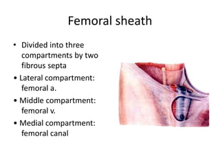
Femoral Triangle
Femoral Triangle
• Triangular area
• Located at
superomedial one third
part of the thigh.
• Large neurovascular
structures pass through
this area, and can be
accessed relatively
easily.
• Thus, it is an area of
both anatomical and
clinical importance.
Boundaries
It is bounded by:
• 1. Superiorly by
inguinal ligament
• 2. Medially by medial
border of the adductor
longus muscle.
• 3. Laterally by medial
border of the sartorius
muscle.
Boundaries
• The floor of the femoral
triangle is not flat but
gutter – shaped
• Its formed from medial
to lateral:
1. Adductor longus
2. Pectineus
3. Iliopsoas
• Its roof is formed by the
fascia lata (deep fascia)
Contents
• Femoral artery and its
branches
• Femoral vein and its
tributaries
• Femoral nerve
• Deep inguinal lymph
nodes
• Fatty tissue
Femoral sheath
• A funnel- shaped sheath
• Derived from transversalis fascia anteriorly
and iliac fascia posteriorly
• It surrounds the femoral vessels and lymphatic
about 2.5cm below the inguinal ligament.
Femoral sheath
• Divided into three
compartments by two
fibrous septa
• Lateral compartment:
femoral a.
• Middle compartment:
femoral v.
• Medial compartment:
femoral canal
Femoral canal
• The femoral canal is located in most medial part of the
femoral sheath.
• It is approximately 1.3cm long.
• The femoral ring is closed by a connective tissue layer –
the femoral septum.
• This septum is pierced by the lymphatic vessels exiting
the canal. Cloquet’s node is a lymph node situated in
the femoral canal.
Femoral canal
• The boundaries of the femoral ring
– Anteriorly: the inguinal ligament
– Medially: the lacunar ligament
– Posteriorly: the pecten of pubis
– Laterally: the femoral vein
Applied Anatomy
• stab wound at the apex of the femoral triangle
may cut all the large vessels of the lower limb ,
injury to femoral vessels results in fatal
hemorrhage.
• Heavy bleeding in the leg can be stopped by
applying pressure to points in the femoral triangle
• femoral vein is commonly used for intravenous
infusions in infants
Clinical Relevance of
Femoral Triangle
Femoral Pulse
• Just inferior to where the femoral artery
crosses the inguinal ligament, it can be
palpated to measure the femoral pulse.
• The femoral artery crosses midway between
the pubis symphysis and anterior superior iliac
spine.
• The femoral pulse can be
palpated in the femoral
triangle, 2 to 3 cm inferior
to the midpoint of the
inguinal ligament.
• The head of the femur
lies posterior to the
femoral artery at this site,
making compression of
the vessel easy.
Clinical Relevance of
Femoral Triangle
Access to the Femoral Artery
• The femoral artery is located superficially within the femoral
triangle, and is thus easy to access. This makes it suitable for
coronary angiography.
• In coronary angiography, the femoral artery is catheterised
with a long thin tube.
• This tube is navigated up the external iliac artery, common iliac
artery, aorta, and into the coronary vessels.
• A radio-opaque dye is then injected into the coronary vessels,
and any wall thickening or blockages can be visualised via x-ray.
Femoral venous catheterization
Indications
• Emergency venous access during
cardiopulmonary resuscitation (CPR), in that it
provides a rapid and reliable route for the
administration of drugs to the central
circulation of the patient in cardiac arrest
• In hypotensive trauma patients
• Urgent or emergency hemodialysis access
• Hemoperfusion access in patients with severe
drug overdose
• Central venous pressure monitoring
Saphena varix
• A saphena varix is a
dilation of
the saphenous vein at
its junction with the
femoral vein in the
groin due to valvular
incompetence.
The varix is:
• soft and compressible
• disappears immediately
on lying down
• exhibits an expansile
cough impulse
• demonstrates a fluid
thrill
Percussion over the
saphenous vein in the
thigh produces a
transmitted impulse in
the varix above
Inguinal lymphadenopathy
• The horizontal group
receive lymph vessels from
lower abdomen,urethra,
external genitalia and
lower half of anal canal.
• The vertical group
receives most of the
superficial lymph vessels
of the lower limb.
• Lymph node swellings are common
• Benign causes include sexually transmitted
infections such as herpes simplex,
lymphogranuloma venereum, chancroid, and
syphilis, and lower extremity skin infections
• Lymphadenopathy associated malignancies are
penile and vulvar squamous cell carcinomas and
melanoma.
Femoral hernia
• If a loop of intestine is
forced into the femoral
ring, it expands to form a
swelling in the upper part
of the thigh. Such a
condition is known as a
femoral hernia .
• A femoral hernia is more
common in women than in
men (possibly because their
wider pelvis and femoral
canal )
Richter’s hernia
• Richter’s hernia is a
variant of strangulated
hernia when only part of
the circumference of the
bowel is entrapped and is
common as a
complication of femoral
hernia.
• Part of the circumference
of the bowel is entrapped

Femoral Triangle.pptx

  • 1.
  • 2.
    Femoral Triangle • Triangulararea • Located at superomedial one third part of the thigh.
  • 3.
    • Large neurovascular structurespass through this area, and can be accessed relatively easily. • Thus, it is an area of both anatomical and clinical importance.
  • 4.
    Boundaries It is boundedby: • 1. Superiorly by inguinal ligament • 2. Medially by medial border of the adductor longus muscle. • 3. Laterally by medial border of the sartorius muscle.
  • 5.
    Boundaries • The floorof the femoral triangle is not flat but gutter – shaped • Its formed from medial to lateral: 1. Adductor longus 2. Pectineus 3. Iliopsoas • Its roof is formed by the fascia lata (deep fascia)
  • 6.
    Contents • Femoral arteryand its branches • Femoral vein and its tributaries • Femoral nerve • Deep inguinal lymph nodes • Fatty tissue
  • 7.
    Femoral sheath • Afunnel- shaped sheath • Derived from transversalis fascia anteriorly and iliac fascia posteriorly • It surrounds the femoral vessels and lymphatic about 2.5cm below the inguinal ligament.
  • 8.
    Femoral sheath • Dividedinto three compartments by two fibrous septa • Lateral compartment: femoral a. • Middle compartment: femoral v. • Medial compartment: femoral canal
  • 9.
    Femoral canal • Thefemoral canal is located in most medial part of the femoral sheath. • It is approximately 1.3cm long. • The femoral ring is closed by a connective tissue layer – the femoral septum. • This septum is pierced by the lymphatic vessels exiting the canal. Cloquet’s node is a lymph node situated in the femoral canal.
  • 10.
    Femoral canal • Theboundaries of the femoral ring – Anteriorly: the inguinal ligament – Medially: the lacunar ligament – Posteriorly: the pecten of pubis – Laterally: the femoral vein
  • 11.
    Applied Anatomy • stabwound at the apex of the femoral triangle may cut all the large vessels of the lower limb , injury to femoral vessels results in fatal hemorrhage. • Heavy bleeding in the leg can be stopped by applying pressure to points in the femoral triangle • femoral vein is commonly used for intravenous infusions in infants
  • 12.
    Clinical Relevance of FemoralTriangle Femoral Pulse • Just inferior to where the femoral artery crosses the inguinal ligament, it can be palpated to measure the femoral pulse. • The femoral artery crosses midway between the pubis symphysis and anterior superior iliac spine.
  • 13.
    • The femoralpulse can be palpated in the femoral triangle, 2 to 3 cm inferior to the midpoint of the inguinal ligament. • The head of the femur lies posterior to the femoral artery at this site, making compression of the vessel easy.
  • 14.
    Clinical Relevance of FemoralTriangle Access to the Femoral Artery • The femoral artery is located superficially within the femoral triangle, and is thus easy to access. This makes it suitable for coronary angiography. • In coronary angiography, the femoral artery is catheterised with a long thin tube. • This tube is navigated up the external iliac artery, common iliac artery, aorta, and into the coronary vessels. • A radio-opaque dye is then injected into the coronary vessels, and any wall thickening or blockages can be visualised via x-ray.
  • 15.
    Femoral venous catheterization Indications •Emergency venous access during cardiopulmonary resuscitation (CPR), in that it provides a rapid and reliable route for the administration of drugs to the central circulation of the patient in cardiac arrest • In hypotensive trauma patients
  • 16.
    • Urgent oremergency hemodialysis access • Hemoperfusion access in patients with severe drug overdose • Central venous pressure monitoring
  • 17.
    Saphena varix • Asaphena varix is a dilation of the saphenous vein at its junction with the femoral vein in the groin due to valvular incompetence.
  • 18.
    The varix is: •soft and compressible • disappears immediately on lying down • exhibits an expansile cough impulse • demonstrates a fluid thrill Percussion over the saphenous vein in the thigh produces a transmitted impulse in the varix above
  • 19.
    Inguinal lymphadenopathy • Thehorizontal group receive lymph vessels from lower abdomen,urethra, external genitalia and lower half of anal canal. • The vertical group receives most of the superficial lymph vessels of the lower limb.
  • 20.
    • Lymph nodeswellings are common • Benign causes include sexually transmitted infections such as herpes simplex, lymphogranuloma venereum, chancroid, and syphilis, and lower extremity skin infections • Lymphadenopathy associated malignancies are penile and vulvar squamous cell carcinomas and melanoma.
  • 21.
    Femoral hernia • Ifa loop of intestine is forced into the femoral ring, it expands to form a swelling in the upper part of the thigh. Such a condition is known as a femoral hernia . • A femoral hernia is more common in women than in men (possibly because their wider pelvis and femoral canal )
  • 22.
    Richter’s hernia • Richter’shernia is a variant of strangulated hernia when only part of the circumference of the bowel is entrapped and is common as a complication of femoral hernia. • Part of the circumference of the bowel is entrapped