2. Fat Emboli: Fat particles or droplets that
travel through the circulation
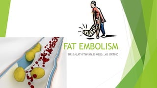
Fat Embolism: A process by which fat
emboli passes into the bloodstream and
lodges within a blood vessel.
Fat Embolism Syndrome (FES): serious
manifestation of fat embolism occasionally
causes multi system dysfunction, the lungs
are always involved and next is brain
3. FE vs. FES
Fat embolization is a well-known complication of skeletal
trauma and surgery involving instrumentation of the
femoral medullary canal.
Fat embolism syndrome (FES) is a physiological response to
fat within the systemic circulation.
Fat embolization and FES are not synonymus.
The embolization of fat can be detected in almost all
patients who sustain a pelvic or femoral fracture, but the
incidence of FES is less than 1%.
4. Fat Embolism Syndrome
Mortality: 10-20%
Clinical diagnosis, No specific laboratory test is
diagnostic.
Mostly associated with long bone and pelvic fractures, and
more frequent in closed fractures.
Single long bone fracture has 1-3% chance of developing
FES, and increases with number of fractures.
Onset is 24-72 hours from initial insult.
5. Causes of fat embolism
TRAUMA RELATED:
Blunt trauma: Long bone (Femur, tibia, pelvic)
factures orthopedic procedures
Soft tissue injury(chest compression with or
without rib fracture)
Burn
Liposuction
Bone marrow harvesting and transplant.
6. NON TRAUMA RELATED
Pancreatitis
Diabetes mellitus
Osteomyelitis and panniculitis
Bone tumor lysis
Steroid therapy
Sickle cell hemoglobinopathy
Alcoholic liver disease
Fat infusion
7. Most common cause of FES is blunt trauma.
90 % occurs after blunt trauma complicated by
long- bone fractures
Closed fractures had higher incidence compared
to open fractures. The intramedullary bone
pressure is lower in case of open fractures, which
reduces the bulk of fat emboli propelled into the
blood stream.
8. Non-traumatic fat embolism
It occurs due to the process of fat or marrow necrosis or
by the increased concentration of lipids in the blood.
It may be caused by agglutination of chylomicrons and
VLDL by high levels of plasma CRP.
As in Acute pancreatitis in patients with types I, IV, and V
hyperlipidaemia and avascular necrosis of bone in patients
with corticosteroid-induced hyperlipidaemia.
9. Drug-related causes of FES
Infusion of lipids at rates greater than the normal
clearance capacity of lipids.
Agglutination of lipid emulsion particles with fibrin.
Agglutination of endogenous or infused exogenous fat such
as Intra lipid.
FES can occur in SC crisis.
Bone marrow necrosis as a result of hypoxia may release
fat.
11. Pathophysiology of FES
Exact mechanism unknown, but
two main hypothesis
1. Mechanical Hypothesis
2.Biochemical Hypothesis
12. Mechanical Hypothesis
Obstruction of vessels and capillaries
Increase in inter medullary pressure forces fat and marrow into blood stream.
Bone marrow contents enter the venous system and lodge in the
lungs as emboli.
Smaller fat droplets travel through the pulmonary capillaries into the
systemic circulation
Embolization to cerebral vessels or renal vessels also leads to central nervous
system and renal dysfunction
13. Biochemical Hypothesis
Toxicity of free fatty acids
Circulating free fatty acids directly affect the pneumocytes,
producing abnormalities in gas exchange.
Coexisting shock, hypovolemia and sepsis impair liver function and
augment toxic effects of free fatty acids.
Hormonal changes caused by trauma or sepsis induce systemic
release of free fatty acids as chylomicrons.
Acute-phase reactants( C-reactive proteins) cause chylomicrons to
coalesce.
It explains non traumatic forms of fat embolism syndrome and why
symptoms take 12 hours to develop
14. FE in ARDS
Fat emboli obstructs lung vessel
(20microns), platelets and fibrin adhere
to it Lipase increases FFA
Inflammatory changes- >endothelial
damage- >ARDS
15. CLINICAL FEATURES
Asymptomatic for the first 12-48 hours
Pulmonary Dysfunction
Neurological (nonspecific)
Dermatological Signs
16. Pulmonary
Hypoxia, rales, pleural friction rub
ARDS may develop.
CXR usually normal early on, later may show
‘snowstorm’ pattern- diffuse bilateral infiltrates
CT chest: ground glass opacification with
interlobular septal thickening.
17. Neurological findings
Usually occur after respiratory symptoms
Incidence- 80% patients with FES
Minor global dysfunction is most common-ranges from mild delirium to
coma.
Seizures/focal deficits
Transient and reversible in most cases.
CT Head: general edema, usually nonspecific
MRI brain: Low density on T1, and high intensity T2 signal, correlates
to degree of impairment.